Pathology Image Detail
Caption |
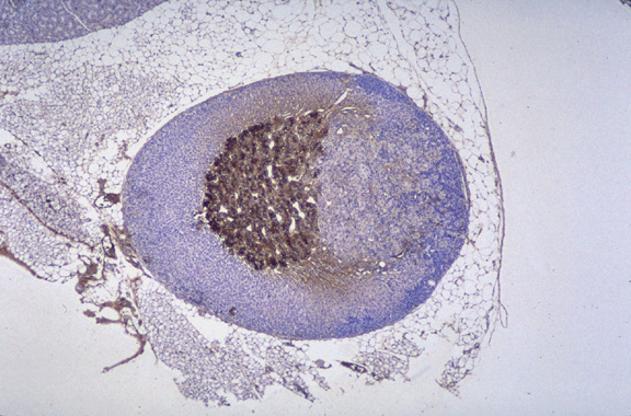
Adrenocortical adenoma. The kidney is in the upper left corner of this image. The adrenal gland has been subjected to immunohistochemistry for detection of expression of tyrosine hydroxylase. The center of the gland (adrenal medulla) is positive for this enzyme (dark brown). The cortex is blue due to the counterstain (hematoxylin). To the right of the medulla is an round mass that compresses the cortex. The cells resemble the cortex and do not express tyrosine hydroxylase and therefore are most likely of adrenal cortex origin. |
Contributor |
Sundberg J (J:94307) |
Pathologist |
Sundberg J (J:94307) |
Method |
IHC for tyrosine hydroxylase |
Type: antibody |
|
Model |
|
Strain |
|