Pathology Image Detail
Caption |
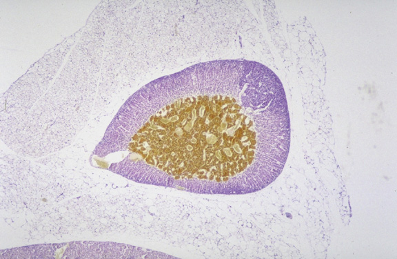
Adrenocortical adenoma. The classical histochemical approach to evaluating the adrenal medulla is to fix it in chromium based fixatives as shown here. The medulla stains brown while the remainder of the gland, the cortex, is blue due to the counterstain. In the upper right quadrant of the cortex is a dark blue nodule that is an adrenal cortical adenoma since it does not stain brown, is located within the adrenal cortex, and has cellular features consistent with the cortical cells. |
Contributor |
Sundberg J (J:94307) |
Pathologist |
Sundberg J (J:94307) |
Method |
chromium fixation method for adrenal medulla |
Model |
|
Strain |
|