Caption |
This image was submitted by JM Ward. Image is from study set created by Herbert C. Morse III (NIAID, NIH), Torgny Fredrickson (NIAID, NIH), and Jerrold M. Ward (NCI, NIH) in 2001. A whole-slide scan image cane be viewed at https://images.jax.org/webclient/img_detail/15575. |
Description |
immunoblastic lymphoma |
Notes |
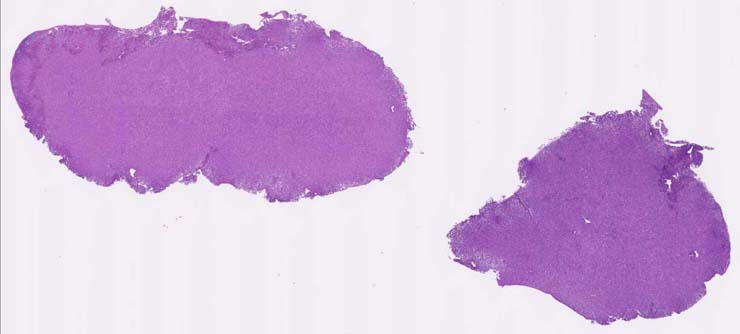
Immunoblastic lymphoma (diffuse large B-cell lymphoma), spleen. The spleen is infiltrated by a mixed population of lymphoid cells (immunoblastic, centroblastic) with nuclei of variable size and shape and with a moderate amount of eosinophilic cytoplasm. Nuclei vary from large and central, acentric along the nuclear membrane. Mitotic figures are common. Some plasmacytoid cells can be seen. Tumor cells were CD45R+ and infiltrating CD3+ cells were seen. Repoterd CD45R and CD3 staining patterns have no submitted images. |
Contributor |
Ward JM (J:107304) |
Pathologist |
Ward JM (J:107304) |
Copyright |
This work was supported in part with funds from the Mouse Model of Cancers Consortium, National Cancer Institute, National Institutes of health, and by contract N01-C056000 from the National cancer Institute. |
Method |
H&E |