Caption |
This image was submitted by JM Ward. Image is from study set created by Herbert C. Morse III (NIAID, NIH), Torgny Fredrickson (NIAID, NIH), and Jerrold M. Ward (NCI, NIH) in 2001. A whole-slide scan image cane be viewed at https://images.jax.org/webclient/img_detail/15602/. |
Description |
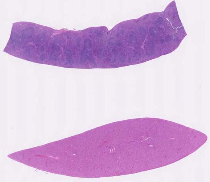
splenic marginal zone hyperplasia |
Notes |
Marginal zone hyperplasia, spleen. The marginal zones are increased in thickness for this line of mice and contain a mixed population of lymphoid cells with variable amount of eosinophilic cytoplasm. The cells vary from large lymphoblastic cells with round to irregular nuclei and medium-sized nucleoli, to smaller lymphoid cells including mature lymphocytes. These cells may have also infiltrated the red pulp diffusely but only IHC can be used to show that fact. The white pulp is normal in structure with some germinal centers present. |
Contributor |
Ward JM (J:107304) |
Pathologist |
Ward JM (J:107304) |
Copyright |
This work was supported in part with funds from the Mouse Model of Cancers Consortium, National Cancer Institute, National Institutes of health, and by contract N01-C056000 from the National cancer Institute. |
Method |
H&E |