Caption |
This image was submitted by JM Ward. Image is from study set created by Herbert C. Morse III (NIAID, NIH), Torgny Fredrickson (NIAID, NIH), and Jerrold M. Ward (NCI, NIH) in 2001. A whole-slide scan image cane be viewed at https://images.jax.org/webclient/img_detail/15605. |
Description |
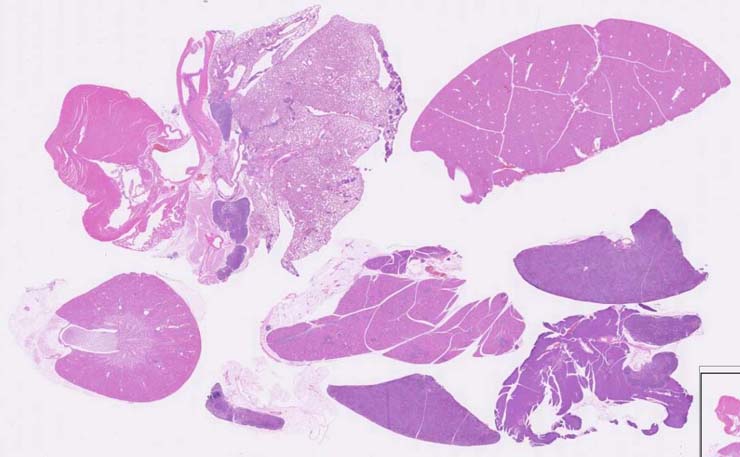
splenic marginal zone lymphoma |
Notes |
Marginal zone lymphoma, spleen. Only small remnants of the normal white pulp can be seen. On the edges of the atrophied white pulp areas seen in a location of the marginal zone, there are a population of lymphoid cells that are also seen in the red pulp. Tumor cells replace most of the white pulp areas, sometimes in a nodular pattern and infiltrate the red pulp. Tumor cells are variable in size and shape but are mostly round cells with a nucleolus and some eosinophilic cytoplasm with mixtures of better differentiated lymphoid cells. Mitotic figures are seen. Tumor cells are CD45R+ and some infiltrating non-neoplastic CD3+ cells are observed. |
Contributor |
Ward JM (J:107304) |
Pathologist |
Ward JM (J:107304) |
Copyright |
This work was supported in part with funds from the Mouse Model of Cancers Consortium, National Cancer Institute, National Institutes of health, and by contract N01-C056000 from the National cancer Institute. |
Method |
H&E |