Caption |
This image was submitted by JM Ward. Image is from study set created by Herbert C. Morse III (NIAID, NIH), Torgny Fredrickson (NIAID, NIH), and Jerrold M. Ward (NCI, NIH) in 2001. A whole-slide scan image cane be viewed at https://images.jax.org/webclient/img_detail/15905. |
Description |
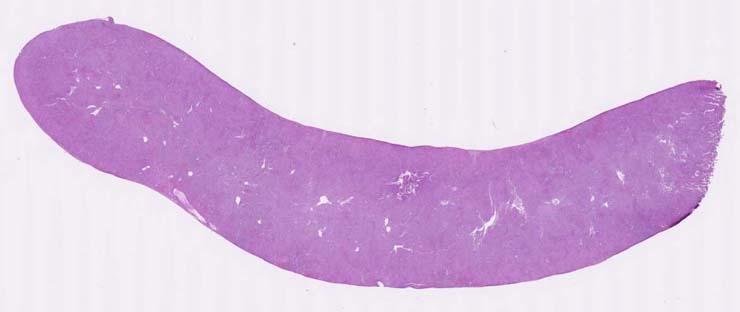
splenic marginal zone lymphoma |
Notes |
Marginal zone lymphoma, spleen, advanced. The marginal zones and red pulp are infiltrated by a population of lymphoid cells with irregular to round nuclei with prominent nuclei and a moderate amount of eosinophilic cytoplasm. Small remnants of the white pulp can be seen. Foci of small basophilic cells (erythroid islands) are found throughout the red pulp. Metastases were not found in other tissues. Bouin?s fixation. From a Trp53 null mouse. |
Contributor |
Ward JM (J:107304) |
Pathologist |
Ward JM (J:107304) |
Copyright |
This work was supported in part with funds from the Mouse Model of Cancers Consortium, National Cancer Institute, National Institutes of health, and by contract N01-C056000 from the National cancer Institute. |
Method |
H&E |