Caption |
This image was submitted by JM Ward. Image is from study set created by Herbert C. Morse III (NIAID, NIH), Torgny Fredrickson (NIAID, NIH), and Jerrold M. Ward (NCI, NIH) in 2001. A whole-slide scan image cane be viewed at https://images.jax.org/webclient/img_detail/15638. |
Description |
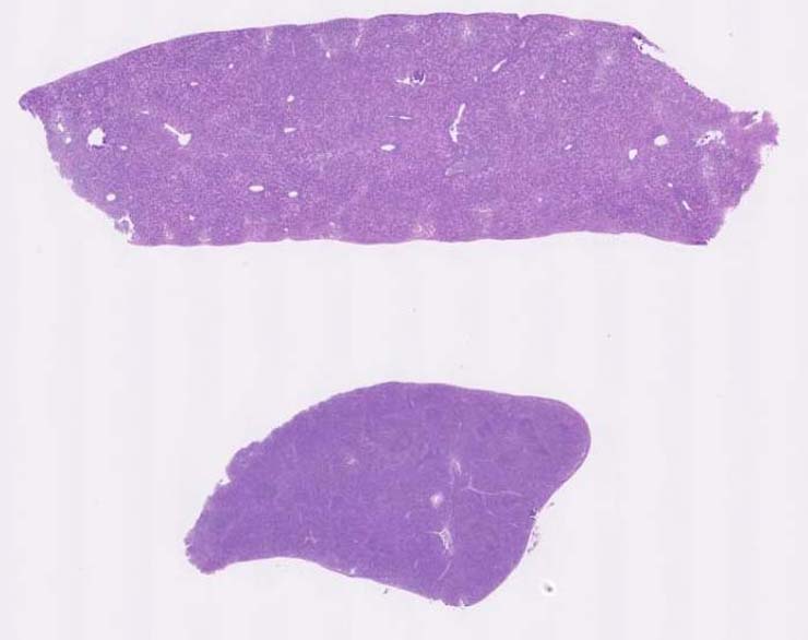
erythroid leukemia |
Notes |
Erythroid Leukemia, spleen, liver. The spleen and liver are diffusely infiltrated by tumor cells that have round nuclei, prominent nucleoli and a moderate amount of eosinophilic cytoplasm. Many mitotic figures are noted. Apoptotic tumor cells are seen in the spleen. Remnants of splenic white pulp areas are seen. In the liver the tumor cells form round nodular lesions within sinusoids. In both liver and spleen, some smaller darker cells are noted which probably represent maturing nucleated erythrocytes. The tumor cells are CD45R-, CD3- and F4/80-. No IHC slides provided or shown. |
Contributor |
Ward JM (J:107304) |
Pathologist |
Ward JM (J:107304) |
Copyright |
This work was supported in part with funds from the Mouse Model of Cancers Consortium, National Cancer Institute, National Institutes of health, and by contract N01-C056000 from the National cancer Institute. |
Method |
H&E |