Caption |
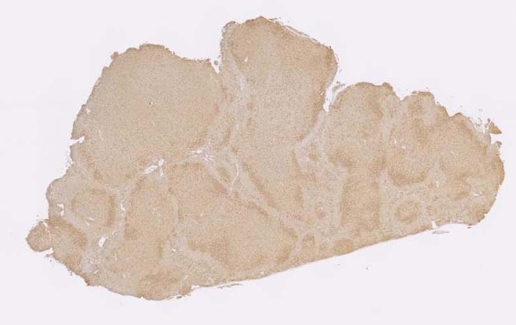
This image was submitted by JM Ward. Image is from study set created by Herbert C. Morse III (NIAID, NIH), Torgny Fredrickson (NIAID, NIH), and Jerrold M. Ward (NCI, NIH) in 2001. This slide was immunostained for CD3. A whole-slide scan of this image can be viewed at https://images.jax.org/webclient/img_detail/15545. |
Description |
follicular center cell lymphoma |
Notes |
Follicular lymphoma, spleen. The enlarged spleen contains white pulp areas are expanded to various degrees and with loss of normal white pulp structure. No marginal zone, PALS, follicles or germinal centers can be seen. A rim of normal small lymphocytes is seen at the edge of the expanded abnormal white pulp. The white pulp contains a mixture of immature blast cells (centroblasts) with large nucleoli, medium sized lymphocytes with inconspicuous nucleoli, and small mature lymphocytes. Some cells have pale eosinophilic cytoplasm. Little red pulp is seen. The lymphoma cells are CD45R positive, some non-neoplastic tumor infiltrating CD3+ lymphocytes are also seen. |
Contributor |
Ward JM (J:107304) |
Pathologist |
Ward JM (J:107304) |
Copyright |
This work was supported in part with funds from the Mouse Model of Cancers Consortium, National Cancer Institute, National Institutes of health, and by contract N01-C056000 from the National cancer Institute. |