Caption |
This image was submitted by JM Ward. Image is from study set created by Herbert C. Morse III (NIAID, NIH), Torgny Fredrickson (NIAID, NIH), and Jerrold M. Ward (NCI, NIH) in 2001. A whole-slide scan image can be viewed at https://images.jax.org/webclient/img_detail/15896. |
Description |
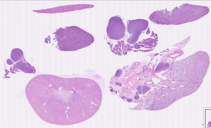
Marginal zone lymphoma, spleen, with metastases |
Notes |
Marginal zone lymphoma, spleen, with metastases. The spleen has small (atrophied) white pulp areas. In the marginal zone and red pulp are marginal zone lymphoma cells that are variable in size, shape and amount of eosinophilic cytoplasm. Nuclei are round, oval and irregular with nucleoli. The pale eosinophilic cytoplasm is abundant in some tumor cells. The mediastinal and pancreatic-duodenal lymph nodes and mediastinum are infiltrated with a uniform population of immature lymphoid cells with much less cytoplasm. They are presumed to be metastatic splenic marginal zone lymphoma cells. Tumor cells are CD45R+, more so in the lymph node. CD45R slide not provided or shown. |
Contributor |
Ward JM (J:107304) |
Pathologist |
Ward JM (J:107304) |
Copyright |
This work was supported in part with funds from the Mouse Model of Cancers Consortium, National Cancer Institute, National Institutes of health, and by contract N01-C056000 from the National cancer Institute. |
Method |
H&E |