|
| MTB ID |
Tumor Name |
Organ(s) Affected |
Treatment Type |
Agents |
Strain Name |
Strain Sex |
Reproductive Status |
Tumor Frequency |
Age at Necropsy |
Description |
Reference |
| MTB:88546 |
Blood vessel hemangiosarcoma |
Skin |
None (spontaneous) |
|
|
Male |
reproductive status not specified |
observed |
30 months |
dermal hemangiosarcoma |
J:236843 |
|
Image Caption:Fig. 5 "G. Dermal hemangiosarcoma from a 30-month-old male CB6F1."
|

|
|
Image ID:6343 |
|
Source of Image:Pettan-Brewer C |
|
Pathologist:Pettan-Brewer C |
|
|
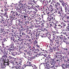
Image Caption:Fig. 5 "H. Higher magnification of G. Irregular vascular channels are formed by atypical and anisokaryotic endothelial cells."
|
|
|
Image ID:6344 |
|
Source of Image:Pettan-Brewer C |
|
Pathologist:Pettan-Brewer C |
|
|
|
| MTB ID |
Tumor Name |
Organ(s) Affected |
Treatment Type |
Agents |
Strain Name |
Strain Sex |
Reproductive Status |
Tumor Frequency |
Age at Necropsy |
Description |
Reference |
| MTB:88543 |
Eye - Harderian gland adenoma |
Eye - Harderian gland |
None (spontaneous) |
|
|
Male |
reproductive status not specified |
observed |
24 months |
Harderian gland adenoma |
J:236843 |
|
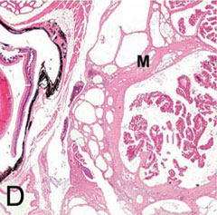
Image Caption:Fig. 3 "D. Hematoxylin and eosin-stained section of the Harderian gland adenoma from the CB6F1 mouse in panel 3B. The mutliobulated and expansible mass (M) is located in the retro-orbital space compresses the eye (E)."
|
|
|
Image ID:6342 |
|
Source of Image:Pettan-Brewer C |
|
Pathologist:Pettan-Brewer C |
|
Method / Stain:H&E |
|
|
|
| MTB ID |
Tumor Name |
Organ(s) Affected |
Treatment Type |
Agents |
Strain Name |
Strain Sex |
Reproductive Status |
Tumor Frequency |
Age at Necropsy |
Description |
Reference |
| MTB:88599 |
Leukocyte lymphoma |
Spleen |
None (spontaneous) |
|
|
Female |
reproductive status not specified |
observed |
30 months |
pleomorphic/follicular malignant lymphoma in the spleen |
J:236843 |
|
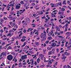
Image Caption:Fig. "L. Higher magnification of K. Pleomorphic lymphoma can resemble HSA. Anatomic distribution, morphology, and immunohistochemistry can aid in differentiation."
|
|
|
Image ID:6349 |
|
Source of Image:Pettan-Brewer C |
|
Pathologist:Pettan-Brewer C |
|
|
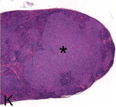
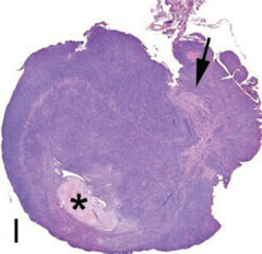
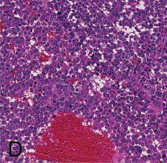
Image Caption:Fig. 5 "K. Spleen with pleomorphic/follicular malignant lymphoma (*) from a 30-month-old CB6F1 female."
|
|
|
Image ID:6348 |
|
Source of Image:Pettan-Brewer C |
|
Pathologist:Pettan-Brewer C |
|
|
|
| MTB ID |
Tumor Name |
Organ(s) Affected |
Treatment Type |
Agents |
Strain Name |
Strain Sex |
Reproductive Status |
Tumor Frequency |
Age at Necropsy |
Description |
Reference |
| MTB:88522 |
Leukocyte - Monocyte - Macrophage - Histiocyte histiocytic sarcoma |
Uterus |
None (spontaneous) |
|
|
Female |
reproductive status not specified |
observed |
28 months |
histiocytic sarcoma (HSA) in the uterus |
J:236843 |
|
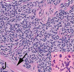
Image Caption:Fig. 5 "I. Uterus from a 28-month-old B6 mouse is effaced with histiocytic sarcoma (HSA). Broad ligament (arrow) and formed lumen (*) are indicated."
|
|
|
Image ID:6335 |
|
Source of Image:Pettan-Brewer C |
|
Pathologist:Pettan-Brewer C |
|
|
Image Caption:Fig. 5 "J. Higher magnification of HSA in I. Neoplastic histiocytes may have elongated oval to round nuclei or form multinucleated giant cells (inset). Myometrial smooth muscle (arrow) is indicated."
|
|
|
Image ID:6336 |
|
Source of Image:Pettan-Brewer C |
|
Pathologist:Pettan-Brewer C |
|
|
|
| MTB ID |
Tumor Name |
Organ(s) Affected |
Treatment Type |
Agents |
Strain Name |
Strain Sex |
Reproductive Status |
Tumor Frequency |
Age at Necropsy |
Description |
Reference |
| MTB:88578 |
Liver hepatocellular carcinoma |
Liver |
None (spontaneous) |
|
|
Female |
reproductive status not specified |
observed |
30 months |
solid and trabecular hepatocellular carcinoma (HCC) |
J:236843 |
|
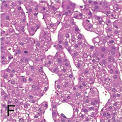
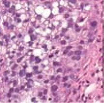
Image Caption:Fig. 5 "F. Higher magnification of trabecular HCC."
|
|
|
Image ID:6347 |
|
Source of Image:Pettan-Brewer C |
|
Pathologist:Pettan-Brewer C |
|
|
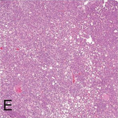
Image Caption:Fig. 5 "E. Liver with solid and trabecular hepatocellular carcinoma (HCC) from a 30-month-old female CB6F1."
|
|
|
Image ID:6346 |
|
Source of Image:Pettan-Brewer C |
|
Pathologist:Pettan-Brewer C |
|
|
|
| MTB ID |
Tumor Name |
Organ(s) Affected |
Treatment Type |
Agents |
Strain Name |
Strain Sex |
Reproductive Status |
Tumor Frequency |
Age at Necropsy |
Description |
Reference |
| MTB:88576 |
Lung adenocarcinoma - mucinous |
Lung |
None (spontaneous) |
|
|
Female |
reproductive status not specified |
observed |
30 months |
mucinous pulmonary adenocarcinoma |
J:236843 |
|
Image Caption:Fig. 5B "Inset: Mucinous pulmonary adenocarcinoma from lungs pictures in Fig. 4G."
|
|
|
Image ID:6345 |
|
Source of Image:Pettan-Brewer C |
|
Pathologist:Pettan-Brewer C |
|
|
|
| MTB ID |
Tumor Name |
Organ(s) Affected |
Treatment Type |
Agents |
Strain Name |
Strain Sex |
Reproductive Status |
Tumor Frequency |
Age at Necropsy |
Description |
Reference |
| MTB:88541 |
Peritoneum - Mesentery hyperplasia |
Peritoneum - Mesentery |
None (spontaneous) |
|
|
Unspecified |
reproductive status not specified |
observed |
16-36 months |
hyperplastic mesenteric milky spot |
J:236843 |
|
Image Caption:Fig. 6 "Histological presentations of subclinical chronic systemic inflammation-associated lesions seen in C57BL/6 mice aged 16-36 months. A. Hyperplastic mesenteric milky spot."
|
|
|
Image ID:6339 |
|
Source of Image:Pettan-Brewer C |
|
Pathologist:Pettan-Brewer C |
|
|
|
| MTB ID |
Tumor Name |
Organ(s) Affected |
Treatment Type |
Agents |
Strain Name |
Strain Sex |
Reproductive Status |
Tumor Frequency |
Age at Necropsy |
Description |
Reference |
| MTB:88530 |
Pituitary gland - Pars distalis adenocarcinoma |
Pituitary gland - Pars distalis |
None (spontaneous) |
|
|
Mixed Population |
reproductive status not specified |
1% |
28 months |
pituitary adenocarcinoma of the pars distalis in a female mouse |
J:236843 |
|
Image Caption:Fig. 5 "D. Higher magnification of the pituitary adenocarcinoma in C."
|
|
|
Image ID:6338 |
|
Source of Image:Pettan-Brewer C |
|
Pathologist:Pettan-Brewer C |
|
|
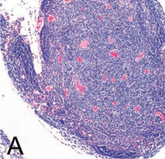
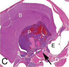
Image Caption:Fig. 5 "C. Decalcified cross-section of the head with a pituitary adenocarcinoma (P) of the pars distalis from mouse pictured in 2B. Pars distalis tumors typically are hemorrhagic and develop blood filled cystic spaces. Malignancy, in this case, was evident with invasion into the sphenoid bone (arrow). Brain (B), pituitary tumor (P) and ear (E) are indicated."
|
|
|
Image ID:6337 |
|
Source of Image:Pettan-Brewer C |
|
Pathologist:Pettan-Brewer C |
|
|
|
| MTB ID |
Tumor Name |
Organ(s) Affected |
Treatment Type |
Agents |
Strain Name |
Strain Sex |
Reproductive Status |
Tumor Frequency |
Age at Necropsy |
Description |
Reference |
| MTB:88542 |
Tooth dysplasia |
Tooth |
None (spontaneous) |
|
|
Unspecified |
reproductive status not specified |
observed |
28 months |
incisor dysplasia |
J:236843 |
|
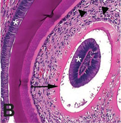
Image Caption:Fig. 7 "B. Higher magnification of the intrapulpal denticles. Denticles with an unusual presentation of a central column of ameloblasts (*) encircled with clear space, predentin (arrow, pink band) and odontoblasts (arrowhead). Denticles may represent dysplastic tooth development. Normal position of ameloblasts (*) and odontoblasts (arrow) are also indicated."
|
|
|
Image ID:6341 |
|
Source of Image:Pettan-Brewer C |
|
Pathologist:Pettan-Brewer C |
|
|
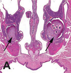
Image Caption:Fig. 7 "A. Decal cross-section of the nose and incisor roots (arrows) with unilateral multiple immature intrapulpal denticles from a 28-month-old B6 mouse."
|
|
|
Image ID:6340 |
|
Source of Image:Pettan-Brewer C |
|
Pathologist:Pettan-Brewer C |
|
|