|
| MTB ID |
Tumor Name |
Organ(s) Affected |
Treatment Type |
Agents |
Strain Name |
Strain Sex |
Reproductive Status |
Tumor Frequency |
Age at Necropsy |
Description |
Reference |
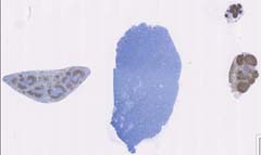
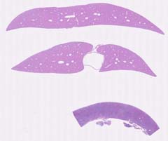
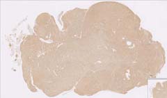
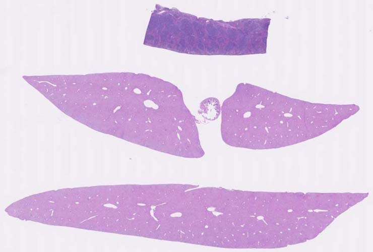
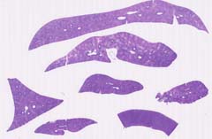
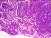
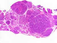
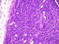
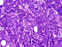
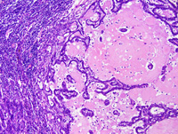
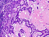
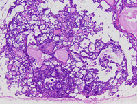
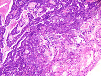
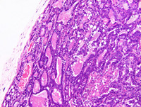
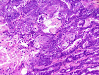
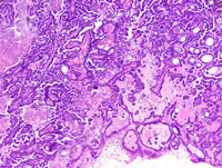
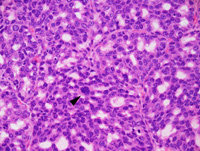
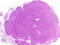
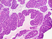
| MTB:69172 |
Erythrocyte leukemia - erythroleukemia |
Erythrocyte |
None (spontaneous) |
|
|
Unspecified |
reproductive status not specified |
observed |
|
erythroid leukemia |
J:107304 |
|
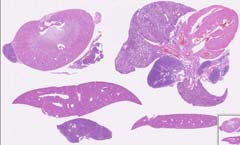
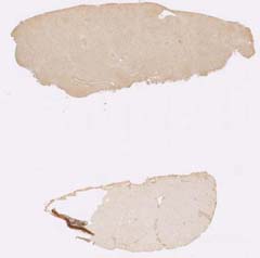
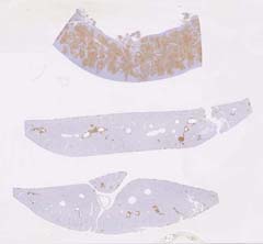
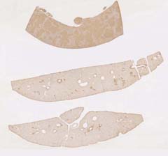
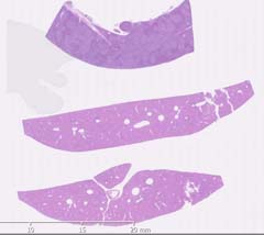
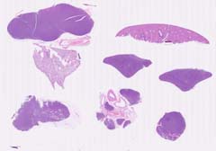
Image Caption:This image was submitted by JM Ward. Image is from study set created by Herbert C. Morse III (NIAID, NIH), Torgny Fredrickson (NIAID, NIH), and Jerrold M. Ward (NCI, NIH) in 2001. A whole-slide scan image cane be viewed at https://images.jax.org/webclient/img_detail/15638.
|
|
|
Image ID:5761 |
|
Source of Image:Ward JM |
|
Pathologist:Ward JM |
|
Method / Stain:H&E |
|
|
|
| MTB ID |
Tumor Name |
Organ(s) Affected |
Treatment Type |
Agents |
Strain Name |
Strain Sex |
Reproductive Status |
Tumor Frequency |
Age at Necropsy |
Description |
Reference |
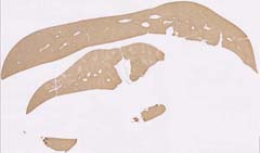
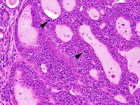
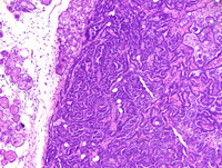
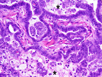
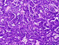
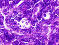
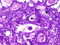
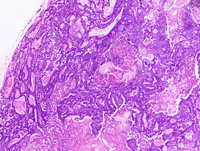
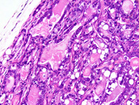
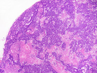
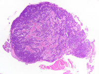
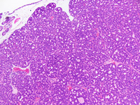
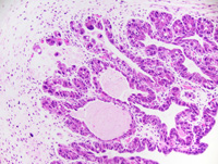
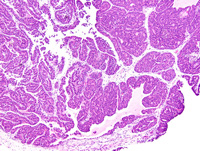
| MTB:69112 |
Leukocyte lymphoma - immunoblastic |
Leukocyte |
None (spontaneous) |
|
|
Unspecified |
reproductive status not specified |
observed |
|
immunoblastic lymphoma |
J:107304 |
|
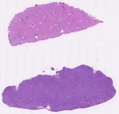
Image Caption:This image was submitted by JM Ward. Image is from study set created by Herbert C. Morse III (NIAID, NIH), Torgny Fredrickson (NIAID, NIH), and Jerrold M. Ward (NCI, NIH) in 2001. A whole-slide scan image cane be viewed at https://images.jax.org/webclient/img_detail/15575.
|
|
|
Image ID:5747 |
|
Source of Image:Ward JM |
|
Pathologist:Ward JM |
|
Method / Stain:H&E |
|
|
|
| MTB ID |
Tumor Name |
Organ(s) Affected |
Treatment Type |
Agents |
Strain Name |
Strain Sex |
Reproductive Status |
Tumor Frequency |
Age at Necropsy |
Description |
Reference |
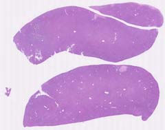
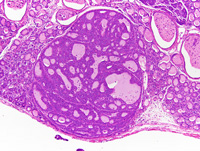
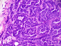
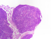
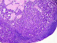
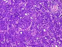
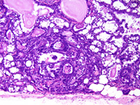
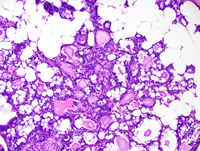
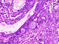
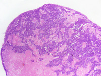
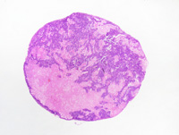
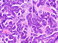
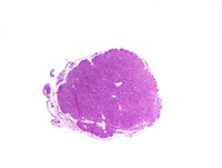
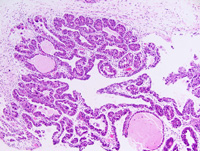
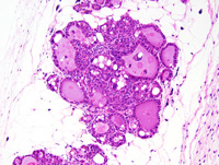
| MTB:69130 |
Leukocyte lymphoma - histiocytic |
Leukocyte |
None (spontaneous) |
|
|
Unspecified |
reproductive status not specified |
observed |
|
histiocyte-rich lymphoma |
J:107304 |
|
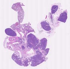
Image Caption:This image was submitted by JM Ward. Image is from study set created by Herbert C. Morse III (NIAID, NIH), Torgny Fredrickson (NIAID, NIH), and Jerrold M. Ward (NCI, NIH) in 2001. A whole-slide scan image cane be viewed at https://images.jax.org/webclient/img_detail/15587.
|
|
|
Image ID:5749 |
|
Source of Image:Ward JM |
|
Pathologist:Ward JM |
|
Method / Stain:H&E |
|
|
|
| MTB ID |
Tumor Name |
Organ(s) Affected |
Treatment Type |
Agents |
Strain Name |
Strain Sex |
Reproductive Status |
Tumor Frequency |
Age at Necropsy |
Description |
Reference |
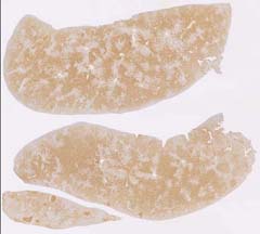
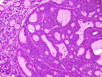
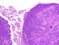
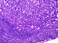
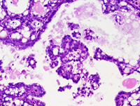
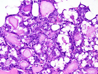
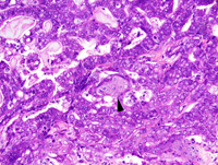
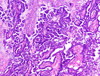
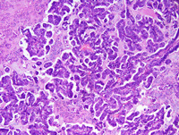
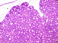
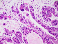
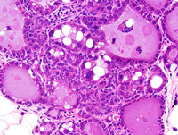
| MTB:69138 |
Leukocyte lymphoma - histiocytic |
Leukocyte |
None (spontaneous) |
|
|
Unspecified |
reproductive status not specified |
observed |
|
histiocyte-rich lymphoma |
J:107304 |
|
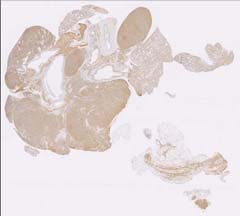
Image Caption:This image was submitted by JM Ward. Image is from study set created by Herbert C. Morse III (NIAID, NIH), Torgny Fredrickson (NIAID, NIH), and Jerrold M. Ward (NCI, NIH) in 2001. A whole-slide scan image cane be viewed at https://images.jax.org/webclient/img_detail/15590.
|
|
|
Image ID:5750 |
|
Source of Image:Ward JM |
|
Pathologist:Ward JM |
|
Method / Stain:H&E |
|
|
|
| MTB ID |
Tumor Name |
Organ(s) Affected |
Treatment Type |
Agents |
Strain Name |
Strain Sex |
Reproductive Status |
Tumor Frequency |
Age at Necropsy |
Description |
Reference |
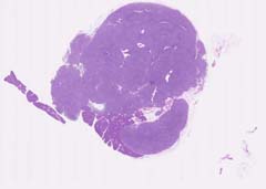
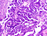
| MTB:69147 |
Leukocyte lymphoma - splenic marginal zone |
Spleen |
None (spontaneous) |
|
|
Unspecified |
reproductive status not specified |
observed |
|
splenic marginal zone lymphoma |
J:107304 |
|
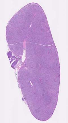
Image Caption:This image was submitted by JM Ward. Image is from study set created by Herbert C. Morse III (NIAID, NIH), Torgny Fredrickson (NIAID, NIH), and Jerrold M. Ward (NCI, NIH) in 2001. This slide was immunostained for CD45R. A whole-slide scan of this image can be viewed at https://images.jax.org/webclient/img_detail/15608.
|
|
|
Image ID:5777 |
|
Source of Image:Ward JM |
|
Pathologist:Ward JM |
|
|
Image Caption:This image was submitted by JM Ward. Image is from study set created by Herbert C. Morse III (NIAID, NIH), Torgny Fredrickson (NIAID, NIH), and Jerrold M. Ward (NCI, NIH) in 2001. This slide was immunostained for CD3. A whole-slide scan of this image can be viewed at https://images.jax.org/webclient/img_detail/15611/.
|
|
|
Image ID:5778 |
|
Source of Image:Ward JM |
|
Pathologist:Ward JM |
|
|
Image Caption:This image was submitted by JM Ward. Image is from study set created by Herbert C. Morse III (NIAID, NIH), Torgny Fredrickson (NIAID, NIH), and Jerrold M. Ward (NCI, NIH) in 2001. A whole-slide scan image cane be viewed at https://images.jax.org/webclient/img_detail/15605.
|
|
|
Image ID:5753 |
|
Source of Image:Ward JM |
|
Pathologist:Ward JM |
|
Method / Stain:H&E |
|
|
|
| MTB ID |
Tumor Name |
Organ(s) Affected |
Treatment Type |
Agents |
Strain Name |
Strain Sex |
Reproductive Status |
Tumor Frequency |
Age at Necropsy |
Description |
Reference |
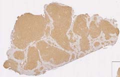
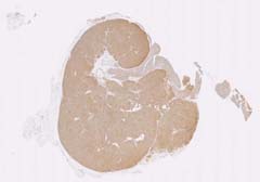
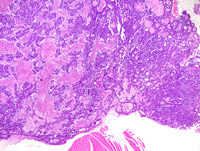
| MTB:69151 |
Leukocyte lymphoma - splenic marginal zone |
Spleen |
None (spontaneous) |
|
|
Unspecified |
reproductive status not specified |
observed |
|
Marginal zone lymphoma, spleen, with metastases |
J:107304 |
|
Image Caption:This image was submitted by JM Ward. Image is from study set created by Herbert C. Morse III (NIAID, NIH), Torgny Fredrickson (NIAID, NIH), and Jerrold M. Ward (NCI, NIH) in 2001. A whole-slide scan image can be viewed at https://images.jax.org/webclient/img_detail/15896.
|
|
|
Image ID:5835 |
|
Source of Image:Ward JM |
|
Pathologist:Ward JM |
|
Method / Stain:H&E |
|
|
|
| MTB ID |
Tumor Name |
Organ(s) Affected |
Treatment Type |
Agents |
Strain Name |
Strain Sex |
Reproductive Status |
Tumor Frequency |
Age at Necropsy |
Description |
Reference |
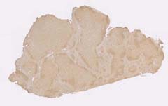
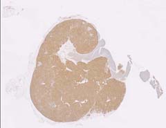
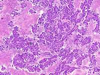
| MTB:69592 |
Leukocyte lymphoma - splenic marginal zone |
Spleen |
None (spontaneous) |
|
|
Unspecified |
reproductive status not specified |
observed |
|
splenic marginal zone lymphoma |
J:107304 |
|
Image Caption:This image was submitted by JM Ward. Image is from study set created by Herbert C. Morse III (NIAID, NIH), Torgny Fredrickson (NIAID, NIH), and Jerrold M. Ward (NCI, NIH) in 2001. A whole-slide scan image cane be viewed at https://images.jax.org/webclient/img_detail/15905.
|
|
|
Image ID:5754 |
|
Source of Image:Ward JM |
|
Pathologist:Ward JM |
|
Method / Stain:H&E |
|
|
|
| MTB ID |
Tumor Name |
Organ(s) Affected |
Treatment Type |
Agents |
Strain Name |
Strain Sex |
Reproductive Status |
Tumor Frequency |
Age at Necropsy |
Description |
Reference |
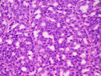
| MTB:69154 |
Leukocyte - Lymphoblast lymphoma - lymphoblastic |
Leukocyte - Lymphoblast |
None (spontaneous) |
|
|
Unspecified |
reproductive status not specified |
observed |
|
lymphoblastic lymphoma |
J:107304 |
|
Image Caption:This image was submitted by JM Ward. Image is from study set created by Herbert C. Morse III (NIAID, NIH), Torgny Fredrickson (NIAID, NIH), and Jerrold M. Ward (NCI, NIH) in 2001. A whole-slide scan image cane be viewed at https://images.jax.org/webclient/img_detail/15620.
|

|
|
Image ID:5755 |
|
Source of Image:Ward JM |
|
Pathologist:Ward JM |
|
Method / Stain:H&E |
|
|
|
| MTB ID |
Tumor Name |
Organ(s) Affected |
Treatment Type |
Agents |
Strain Name |
Strain Sex |
Reproductive Status |
Tumor Frequency |
Age at Necropsy |
Description |
Reference |
| MTB:69121 |
Leukocyte - Lymphocyte - B-lymphocyte lymphoma - Burkitt |
Leukocyte - Lymphocyte - B-lymphocyte |
None (spontaneous) |
|
|
Unspecified |
reproductive status not specified |
observed |
|
Diffuse large B-cell lymphoma, centroblastic, spleen |
J:107304 |
|
Image Caption:This image was submitted by JM Ward. Image is from study set created by Herbert C. Morse III (NIAID, NIH), Torgny Fredrickson (NIAID, NIH), and Jerrold M. Ward (NCI, NIH) in 2001. This slide was immunostained for CD45R. A whole-slide scan of this image can be viewed at https://images.jax.org/webclient/img_detail/15584.
|
|
|
Image ID:5774 |
|
Source of Image:Ward JM |
|
Pathologist:Ward JM |
|
|
Image Caption:This image was submitted by JM Ward. Image is from study set created by Herbert C. Morse III (NIAID, NIH), Torgny Fredrickson (NIAID, NIH), and Jerrold M. Ward (NCI, NIH) in 2001. This slide was immunostained for CD3. A whole-slide scan of this image can be viewed at https://images.jax.org/webclient/img_detail/15581.
|
|
|
Image ID:5773 |
|
Source of Image:Ward JM |
|
Pathologist:Ward JM |
|
|
Image Caption:This image was submitted by JM Ward. Image is from study set created by Herbert C. Morse III (NIAID, NIH), Torgny Fredrickson (NIAID, NIH), and Jerrold M. Ward (NCI, NIH) in 2001. A whole-slide scan image cane be viewed at https://images.jax.org/webclient/img_detail/15578.
|
|
|
Image ID:5748 |
|
Source of Image:Ward JM |
|
Pathologist:Ward JM |
|
Method / Stain:H&E |
|
|
|
| MTB ID |
Tumor Name |
Organ(s) Affected |
Treatment Type |
Agents |
Strain Name |
Strain Sex |
Reproductive Status |
Tumor Frequency |
Age at Necropsy |
Description |
Reference |
| MTB:69143 |
Leukocyte - Lymphocyte - B-lymphocyte lymphoma - lymphoblastic |
Leukocyte - Lymphocyte - B-lymphocyte |
None (spontaneous) |
|
|
Unspecified |
reproductive status not specified |
observed |
|
B-cell lymphoblastic lymphoma |
J:107304 |
|
Image Caption:This image was submitted by JM Ward. Image is from study set created by Herbert C. Morse III (NIAID, NIH), Torgny Fredrickson (NIAID, NIH), and Jerrold M. Ward (NCI, NIH) in 2001. A whole-slide scan image cane be viewed at https://images.jax.org/webclient/img_detail/15593.
|
|
|
Image ID:5751 |
|
Source of Image:Ward JM |
|
Pathologist:Ward JM |
|
Method / Stain:H&E |
|
|
Image Caption:This image was submitted by JM Ward. Image is from study set created by Herbert C. Morse III (NIAID, NIH), Torgny Fredrickson (NIAID, NIH), and Jerrold M. Ward (NCI, NIH) in 2001. This slide was immunostained for CD3. A whole-slide scan of this image can be viewed at https://images.jax.org/webclient/img_detail/15596.
|
|
|
Image ID:5775 |
|
Source of Image:Ward JM |
|
Pathologist:Ward JM |
|
|
Image Caption:This image was submitted by JM Ward. Image is from study set created by Herbert C. Morse III (NIAID, NIH), Torgny Fredrickson (NIAID, NIH), and Jerrold M. Ward (NCI, NIH) in 2001. This slide was immunostained for CD45R. A whole-slide scan of this image can be viewed at https://images.jax.org/webclient/img_detail/15599.
|
|
|
Image ID:5776 |
|
Source of Image:Ward JM |
|
Pathologist:Ward JM |
|
|
|
| MTB ID |
Tumor Name |
Organ(s) Affected |
Treatment Type |
Agents |
Strain Name |
Strain Sex |
Reproductive Status |
Tumor Frequency |
Age at Necropsy |
Description |
Reference |
| MTB:69100 |
Leukocyte - Lymphocyte - B-lymphocyte - Follicular center cell lymphoma - follicular |
Spleen |
None (spontaneous) |
|
|
Unspecified |
reproductive status not specified |
observed |
|
follicular center cell lymphoma |
J:107304 |
|
Image Caption:This image was submitted by JM Ward. Image is from study set created by Herbert C. Morse III (NIAID, NIH), Torgny Fredrickson (NIAID, NIH), and Jerrold M. Ward (NCI, NIH) in 2001. This slide was immunostained for CD45R. A whole-slide scan of this image can be viewed at https://images.jax.org/webclient/img_detail/15548.
|
|
|
Image ID:5765 |
|
Source of Image:Ward JM |
|
Pathologist:Ward JM |
|
|
Image Caption:This image was submitted by JM Ward. Image is from study set created by Herbert C. Morse III (NIAID, NIH), Torgny Fredrickson (NIAID, NIH), and Jerrold M. Ward (NCI, NIH) in 2001. This slide was immunostained for CD3. A whole-slide scan of this image can be viewed at https://images.jax.org/webclient/img_detail/15545.
|
|
|
Image ID:5764 |
|
Source of Image:Ward JM |
|
Pathologist:Ward JM |
|
|
Image Caption:This image was submitted by JM Ward. Image is from study set created by Herbert C. Morse III (NIAID, NIH), Torgny Fredrickson (NIAID, NIH), and Jerrold M. Ward (NCI, NIH) in 2001. A whole-slide scan of this image can be viewed at https://images.jax.org/webclient/img_detail/15893.
|
|
|
Image ID:5740 |
|
Source of Image:Ward JM |
|
Pathologist:Ward JM |
|
Method / Stain:H&E |
|
|
|
| MTB ID |
Tumor Name |
Organ(s) Affected |
Treatment Type |
Agents |
Strain Name |
Strain Sex |
Reproductive Status |
Tumor Frequency |
Age at Necropsy |
Description |
Reference |
| MTB:69101 |
Leukocyte - Lymphocyte - B-lymphocyte - Follicular center cell lymphoma - follicular |
Leukocyte - Lymphocyte - B-lymphocyte - Follicular center cell |
None (spontaneous) |
|
|
Unspecified |
reproductive status not specified |
observed |
|
follicular center cell lymphoma |
J:107304 |
|
Image Caption:This image was submitted by JM Ward. Image is from study set created by Herbert C. Morse III (NIAID, NIH), Torgny Fredrickson (NIAID, NIH), and Jerrold M. Ward (NCI, NIH) in 2001. This slide was immunostained for CD45R. A whole-slide scan of this image can be viewed at https://images.jax.org/webclient/img_detail/15890.
|
|
|
Image ID:5766 |
|
Source of Image:Ward JM |
|
Pathologist:Ward JM |
|
|
Image Caption:This image was submitted by JM Ward. Image is from study set created by Herbert C. Morse III (NIAID, NIH), Torgny Fredrickson (NIAID, NIH), and Jerrold M. Ward (NCI, NIH) in 2001. This slide was immunostained for CD3. A whole-slide scan of this image can be viewed at https://images.jax.org/webclient/img_detail/15899.
|
|
|
Image ID:5767 |
|
Source of Image:Ward JM |
|
Pathologist:Ward JM |
|
|
Image Caption:This image was submitted by JM Ward. Image is from study set created by Herbert C. Morse III (NIAID, NIH), Torgny Fredrickson (NIAID, NIH), and Jerrold M. Ward (NCI, NIH) in 2001. A whole-slide scan of this image can be viewed at https://images.jax.org/webclient/img_detail/16007/.
|
|
|
Image ID:5741 |
|
Source of Image:Ward JM |
|
Pathologist:Ward JM |
|
Method / Stain:H&E |
|
|
|
| MTB ID |
Tumor Name |
Organ(s) Affected |
Treatment Type |
Agents |
Strain Name |
Strain Sex |
Reproductive Status |
Tumor Frequency |
Age at Necropsy |
Description |
Reference |
| MTB:69102 |
Leukocyte - Lymphocyte - B-lymphocyte - Follicular center cell lymphoma - follicular |
Leukocyte - Lymphocyte - B-lymphocyte - Follicular center cell |
None (spontaneous) |
|
|
Unspecified |
reproductive status not specified |
observed |
|
follicular center cell lymphoma |
J:107304 |
|
Image Caption:This image was submitted by JM Ward. Image is from study set created by Herbert C. Morse III (NIAID, NIH), Torgny Fredrickson (NIAID, NIH), and Jerrold M. Ward (NCI, NIH) in 2001. A whole-slide scan of this image can be viewed at https://images.jax.org/webclient/img_detail/15845/.
|
|
|
Image ID:5742 |
|
Source of Image:Ward JM |
|
Pathologist:Ward JM |
|
Method / Stain:H&E |
|
|
|
| MTB ID |
Tumor Name |
Organ(s) Affected |
Treatment Type |
Agents |
Strain Name |
Strain Sex |
Reproductive Status |
Tumor Frequency |
Age at Necropsy |
Description |
Reference |
| MTB:69103 |
Leukocyte - Lymphocyte - B-lymphocyte - Follicular center cell lymphoma - follicular |
Leukocyte - Lymphocyte - B-lymphocyte - Follicular center cell |
None (spontaneous) |
|
|
Unspecified |
reproductive status not specified |
observed |
|
follicular center cell lymphoma |
J:107304 |
|
Image Caption:This image was submitted by JM Ward. Image is from study set created by Herbert C. Morse III (NIAID, NIH), Torgny Fredrickson (NIAID, NIH), and Jerrold M. Ward (NCI, NIH) in 2001. A whole-slide scan of this image can be viewed at https://images.jax.org/webclient/img_detail/15851.
|
|
|
Image ID:5744 |
|
Source of Image:Ward JM |
|
Pathologist:Ward JM |
|
Method / Stain:H&E |
|
|
Image Caption:This image was submitted by JM Ward. Image is from study set created by Herbert C. Morse III (NIAID, NIH), Torgny Fredrickson (NIAID, NIH), and Jerrold M. Ward (NCI, NIH) in 2001. This slide was immunostained for CD3. A whole-slide scan of this image can be viewed at https://images.jax.org/webclient/img_detail/15566.
|
|
|
Image ID:5768 |
|
Source of Image:Ward JM |
|
Pathologist:Ward JM |
|
|
|
| MTB ID |
Tumor Name |
Organ(s) Affected |
Treatment Type |
Agents |
Strain Name |
Strain Sex |
Reproductive Status |
Tumor Frequency |
Age at Necropsy |
Description |
Reference |
| MTB:69105 |
Leukocyte - Lymphocyte - B-lymphocyte - Follicular center cell lymphoma - follicular |
Leukocyte - Lymphocyte - B-lymphocyte - Follicular center cell |
None (spontaneous) |
|
|
Unspecified |
reproductive status not specified |
observed |
|
follicular center cell lymphoma |
J:107304 |
|
Image Caption:This image was submitted by JM Ward. Image is from study set created by Herbert C. Morse III (NIAID, NIH), Torgny Fredrickson (NIAID, NIH), and Jerrold M. Ward (NCI, NIH) in 2001. A whole-slide scan image cane be viewed at https://images.jax.org/webclient/img_detail/15848.
|
|
|
Image ID:5743 |
|
Source of Image:Ward JM |
|
Pathologist:Ward JM |
|
Method / Stain:H&E |
|
|
Image Caption:This image was submitted by JM Ward. Image is from study set created by Herbert C. Morse III (NIAID, NIH), Torgny Fredrickson (NIAID, NIH), and Jerrold M. Ward (NCI, NIH) in 2001. This slide was immunostained for CD45R. A whole-slide scan of this image can be viewed at https://images.jax.org/webclient/img_detail/15560
|
|
|
Image ID:5769 |
|
Source of Image:Ward JM |
|
Pathologist:Ward JM |
|
|
Image Caption:This image was submitted by JM Ward. Image is from study set created by Herbert C. Morse III (NIAID, NIH), Torgny Fredrickson (NIAID, NIH), and Jerrold M. Ward (NCI, NIH) in 2001. This slide was immunostained for CD3. A whole-slide scan of this image can be viewed at https://images.jax.org/webclient/img_detail/15563.
|
|
|
Image ID:5770 |
|
Source of Image:Ward JM |
|
Pathologist:Ward JM |
|
|
|
| MTB ID |
Tumor Name |
Organ(s) Affected |
Treatment Type |
Agents |
Strain Name |
Strain Sex |
Reproductive Status |
Tumor Frequency |
Age at Necropsy |
Description |
Reference |
| MTB:69109 |
Leukocyte - Lymphocyte - B-lymphocyte - Follicular center cell lymphoma - follicular |
Leukocyte - Lymphocyte - B-lymphocyte - Follicular center cell |
None (spontaneous) |
|
|
Unspecified |
reproductive status not specified |
observed |
|
follicular center cell lymphoma |
J:107304 |
|
Image Caption:This image was submitted by JM Ward. Image is from study set created by Herbert C. Morse III (NIAID, NIH), Torgny Fredrickson (NIAID, NIH), and Jerrold M. Ward (NCI, NIH) in 2001. A whole-slide scan of this image can be viewed at https://images.jax.org/webclient/img_detail/15854/.
|
|
|
Image ID:5745 |
|
Source of Image:Ward JM |
|
Pathologist:Ward JM |
|
Method / Stain:H&E |
|
|
Image Caption:This image was submitted by JM Ward. Image is from study set created by Herbert C. Morse III (NIAID, NIH), Torgny Fredrickson (NIAID, NIH), and Jerrold M. Ward (NCI, NIH) in 2001. This slide was immunostained for CD3. A whole-slide scan of this image can be viewed at https://images.jax.org/webclient/img_detail/15908.
|
|
|
Image ID:5772 |
|
Source of Image:Ward JM |
|
Pathologist:Ward JM |
|
|
Image Caption:This image was submitted by JM Ward. Image is from study set created by Herbert C. Morse III (NIAID, NIH), Torgny Fredrickson (NIAID, NIH), and Jerrold M. Ward (NCI, NIH) in 2001. This slide was immunostained for CD45R. A whole-slide scan of this image can be viewed at https://images.jax.org/webclient/img_detail/15902.
|
|
|
Image ID:5771 |
|
Source of Image:Ward JM |
|
Pathologist:Ward JM |
|
|
|
| MTB ID |
Tumor Name |
Organ(s) Affected |
Treatment Type |
Agents |
Strain Name |
Strain Sex |
Reproductive Status |
Tumor Frequency |
Age at Necropsy |
Description |
Reference |
| MTB:71233 |
Leukocyte - Lymphocyte - B-lymphocyte - Follicular center cell lymphoma - follicular |
Spleen |
None (spontaneous) |
|
|
Unspecified |
reproductive status not specified |
observed |
|
follicular lymphoma, spleen |
J:107304 |
|
Image Caption:This image was submitted by JM Ward. Image is from study set created by Herbert C. Morse III (NIAID, NIH), Torgny Fredrickson (NIAID, NIH), and Jerrold M. Ward (NCI, NIH) in 2001. A whole-slide scan image cane be viewed at https://images.jax.org/webclient/img_detail/15857/.
|
|
|
Image ID:5834 |
|
Source of Image:Ward JM |
|
Pathologist:Ward JM |
|
Method / Stain:H&E |
|
|
|
| MTB ID |
Tumor Name |
Organ(s) Affected |
Treatment Type |
Agents |
Strain Name |
Strain Sex |
Reproductive Status |
Tumor Frequency |
Age at Necropsy |
Description |
Reference |
| MTB:69111 |
Leukocyte - Lymphocyte - B-lymphocyte - Plasma cell lymphoma |
Leukocyte - Lymphocyte - B-lymphocyte - Plasma cell |
None (spontaneous) |
|
|
Unspecified |
reproductive status not specified |
observed |
|
plasma cell lymphoma |
J:107304 |
|
Image Caption:This image was submitted by JM Ward. Image is from study set created by Herbert C. Morse III (NIAID, NIH), Torgny Fredrickson (NIAID, NIH), and Jerrold M. Ward (NCI, NIH) in 2001. A whole-slide scan image can be viewed at https://images.jax.org/webclient/img_detail/15860/
|
|
|
Image ID:5746 |
|
Source of Image:Ward JM |
|
Pathologist:Ward JM |
|
Method / Stain:H&E |
|
|
|
| MTB ID |
Tumor Name |
Organ(s) Affected |
Treatment Type |
Agents |
Strain Name |
Strain Sex |
Reproductive Status |
Tumor Frequency |
Age at Necropsy |
Description |
Reference |
| MTB:69593 |
Leukocyte - Lymphocyte - Pre-T-lymphocyte lymphoma - lymphoblastic |
Thymus |
None (spontaneous) |
|
|
Unspecified |
reproductive status not specified |
observed |
|
lymphoblastic lymphoma, thymic origin, early |
J:107304 |
|
Image Caption:This image was submitted by JM Ward. Image is from study set created by Herbert C. Morse III (NIAID, NIH), Torgny Fredrickson (NIAID, NIH), and Jerrold M. Ward (NCI, NIH) in 2001. This slide was immunostained for CD3. A whole-slide scan of this image can be viewed at https://images.jax.org/webclient/img_detail/15863/.
|
|
|
Image ID:5781 |
|
Source of Image:Ward JM |
|
Pathologist:Ward JM |
|
|
Image Caption:This image was submitted by JM Ward. Image is from study set created by Herbert C. Morse III (NIAID, NIH), Torgny Fredrickson (NIAID, NIH), and Jerrold M. Ward (NCI, NIH) in 2001. A whole-slide scan image cane be viewed at https://images.jax.org/webclient/img_detail/15623.
|
|
|
Image ID:5756 |
|
Source of Image:Ward JM |
|
Pathologist:Ward JM |
|
Method / Stain:H&E |
|
|
|
| MTB ID |
Tumor Name |
Organ(s) Affected |
Treatment Type |
Agents |
Strain Name |
Strain Sex |
Reproductive Status |
Tumor Frequency |
Age at Necropsy |
Description |
Reference |
| MTB:69594 |
Leukocyte - Lymphocyte - Pre-T-lymphocyte lymphoma - mixed |
Thymus |
None (spontaneous) |
|
|
Unspecified |
reproductive status not specified |
observed |
|
Lymphoblastic lymphoma, thymic T-cell origin and marginal zone lymphoma, spleen |
J:107304 |
|
Image Caption:This image was submitted by JM Ward. Image is from study set created by Herbert C. Morse III (NIAID, NIH), Torgny Fredrickson (NIAID, NIH), and Jerrold M. Ward (NCI, NIH) in 2001. This slide was immunostained for CD3. A whole-slide scan of this image can be viewed at https://images.jax.org/webclient/img_detail/15878.
|
|
|
Image ID:5788 |
|
Source of Image:Ward JM |
|
Pathologist:Ward JM |
|
|
Image Caption:This image was submitted by JM Ward. Image is from study set created by Herbert C. Morse III (NIAID, NIH), Torgny Fredrickson (NIAID, NIH), and Jerrold M. Ward (NCI, NIH) in 2001. This slide was immunostained for CD45R. A whole-slide scan of this image can be viewed at https://images.jax.org/webclient/img_detail/15881.
|
|
|
Image ID:5789 |
|
Source of Image:Ward JM |
|
Pathologist:Ward JM |
|
|
Image Caption:This image was submitted by JM Ward. Image is from study set created by Herbert C. Morse III (NIAID, NIH), Torgny Fredrickson (NIAID, NIH), and Jerrold M. Ward (NCI, NIH) in 2001. A whole-slide scan image cane be viewed at https://images.jax.org/webclient/img_detail/15641.
|
|
|
Image ID:5762 |
|
Source of Image:Ward JM |
|
Pathologist:Ward JM |
|
Method / Stain:H&E |
|
|
Image Caption:This image was submitted by JM Ward. Image is from study set created by Herbert C. Morse III (NIAID, NIH), Torgny Fredrickson (NIAID, NIH), and Jerrold M. Ward (NCI, NIH) in 2001. This slide was immunostained for TDT. A whole-slide scan of this image can be viewed at https://images.jax.org/webclient/img_detail/15884.
|
|
|
Image ID:5790 |
|
Source of Image:Ward JM |
|
Pathologist:Ward JM |
|
|
|
| MTB ID |
Tumor Name |
Organ(s) Affected |
Treatment Type |
Agents |
Strain Name |
Strain Sex |
Reproductive Status |
Tumor Frequency |
Age at Necropsy |
Description |
Reference |
| MTB:69155 |
Leukocyte - Lymphocyte - T-lymphocyte lymphoma - lymphoblastic |
Thymus |
Chemical/Drug |
dimethylbenzanthracene (DMBA) |
|
Unspecified |
reproductive status not specified |
observed |
|
thymic origin lymphoblastic lymphoma |
J:107304 |
|
Image Caption:This image was submitted by JM Ward. Image is from study set created by Herbert C. Morse III (NIAID, NIH), Torgny Fredrickson (NIAID, NIH), and Jerrold M. Ward (NCI, NIH) in 2001. A whole-slide scan image cane be viewed at https://images.jax.org/webclient/img_detail/15626/.
|
|
|
Image ID:5757 |
|
Source of Image:Ward JM |
|
Pathologist:Ward JM |
|
Method / Stain:H&E |
|
|
Image Caption:This image was submitted by JM Ward. Image is from study set created by Herbert C. Morse III (NIAID, NIH), Torgny Fredrickson (NIAID, NIH), and Jerrold M. Ward (NCI, NIH) in 2001. This slide was immunostained for CD3. A whole-slide scan of this image can be viewed at https://images.jax.org/webclient/img_detail/15866.
|
|
|
Image ID:5782 |
|
Source of Image:Ward JM |
|
Pathologist:Ward JM |
|
|
|
| MTB ID |
Tumor Name |
Organ(s) Affected |
Treatment Type |
Agents |
Strain Name |
Strain Sex |
Reproductive Status |
Tumor Frequency |
Age at Necropsy |
Description |
Reference |
| MTB:69156 |
Leukocyte - Monocyte - Macrophage - Histiocyte histiocytic sarcoma |
Leukocyte - Monocyte - Macrophage - Histiocyte |
None (spontaneous) |
|
|
Unspecified |
reproductive status not specified |
observed |
|
histiocytic sarcoma |
J:107304 |
|
Image Caption:This image was submitted by JM Ward. Image is from study set created by Herbert C. Morse III (NIAID, NIH), Torgny Fredrickson (NIAID, NIH), and Jerrold M. Ward (NCI, NIH) in 2001. A whole-slide scan image cane be viewed at https://images.jax.org/webclient/img_detail/15629/.
|
|
|
Image ID:5758 |
|
Source of Image:Ward JM |
|
Pathologist:Ward JM |
|
Method / Stain:H&E |
|
|
Image Caption:This image was submitted by JM Ward. Image is from study set created by Herbert C. Morse III (NIAID, NIH), Torgny Fredrickson (NIAID, NIH), and Jerrold M. Ward (NCI, NIH) in 2001. This slide was immunostained for CD68. A whole-slide scan of this image can be viewed at https://images.jax.org/webclient/img_detail/15869/.
|
|
|
Image ID:5783 |
|
Source of Image:Ward JM |
|
Pathologist:Ward JM |
|
|
Image Caption:This image was submitted by JM Ward. Image is from study set created by Herbert C. Morse III (NIAID, NIH), Torgny Fredrickson (NIAID, NIH), and Jerrold M. Ward (NCI, NIH) in 2001. This slide was immunostained for F4/80. A whole-slide scan of this image can be viewed at https://images.jax.org/webclient/img_detail/15872/.
|
|
|
Image ID:5784 |
|
Source of Image:Ward JM |
|
Pathologist:Ward JM |
|
|
|
| MTB ID |
Tumor Name |
Organ(s) Affected |
Treatment Type |
Agents |
Strain Name |
Strain Sex |
Reproductive Status |
Tumor Frequency |
Age at Necropsy |
Description |
Reference |
| MTB:69158 |
Leukocyte - Myelocyte (Granulocyte) leukemia - myelocytic |
Leukocyte - Myelocyte (Granulocyte) |
None (spontaneous) |
|
|
Unspecified |
reproductive status not specified |
observed |
|
Myeloid leukemia, undifferentiated, without maturation, spleen |
J:107304 |
|
Image Caption:This image was submitted by JM Ward. Image is from study set created by Herbert C. Morse III (NIAID, NIH), Torgny Fredrickson (NIAID, NIH), and Jerrold M. Ward (NCI, NIH) in 2001. A whole-slide scan image cane be viewed at https://images.jax.org/webclient/img_detail/15635.
|
|
|
Image ID:5760 |
|
Source of Image:Ward JM |
|
Pathologist:Ward JM |
|
Method / Stain:H&E |
|
|
|
| MTB ID |
Tumor Name |
Organ(s) Affected |
Treatment Type |
Agents |
Strain Name |
Strain Sex |
Reproductive Status |
Tumor Frequency |
Age at Necropsy |
Description |
Reference |
| MTB:71024 |
Leukocyte - Myelocyte (Granulocyte) leukemia - myelocytic |
Spleen |
None (spontaneous) |
|
|
Unspecified |
reproductive status not specified |
observed |
|
Myeloid leukemia, preleukemic, spleen |
J:107304 |
|
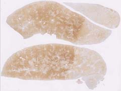
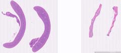
Image Caption:Image was submitted by Jerrald Ward. Image is from study set created by Herbert C. Morse III (NIAID, NIH), Torgny Fredrickson (NIAID, NIH), and Jerrold M. Ward (NCI, NIH) in 2001. Whole-slide scan of this image is available at https://images.jax.org/webclient/img_detail/15632.
|
|
|
Image ID:5832 |
|
Source of Image:Ward JM |
|
Pathologist:Ward JM |
|
Method / Stain:H&E |
|
|
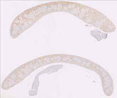
Image Caption:Image was submitted by Jerrald Ward. This slide was stained for INOS. Image is from study set created by Herbert C. Morse III (NIAID, NIH), Torgny Fredrickson (NIAID, NIH), and Jerrold M. Ward (NCI, NIH) in 2001. Whole-slide scan of this image is available at https://images.jax.org/webclient/img_detail/15875.
|
|
|
Image ID:5833 |
|
Source of Image:Ward JM |
|
Pathologist:Ward JM |
|
|
|
| MTB ID |
Tumor Name |
Organ(s) Affected |
Treatment Type |
Agents |
Strain Name |
Strain Sex |
Reproductive Status |
Tumor Frequency |
Age at Necropsy |
Description |
Reference |
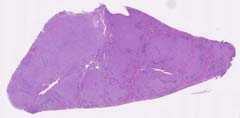
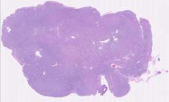
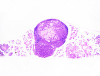
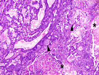
| MTB:28197 |
Mammary gland adenocarcinoma |
Mammary gland |
None (spontaneous) |
|
|
Female |
reproductive status not specified |
observed |
120 days |
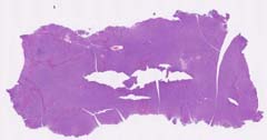
Adenocarcinoma, glandular, circumscribed, mammary gland |
J:94320 |
|
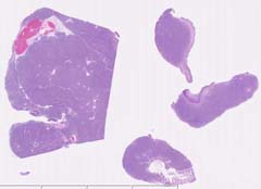
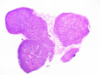
Image Caption:Mammary gland: there is, in the same field, a solid mammary adenocarcinoma (upper right corner of the field) with an invasive growth pattern, and a small circumscribed adenocarcinoma (lower left corner of the field) separated by areas of atypical hyperplasia and in situ carcinoma.
|
|
|
Image ID:1393 |
|
Source of Image:Ward JM |
|
Pathologist:Mikaelian I |
|
Method / Stain:H&E |
|
|
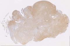
Image Caption:Circumscribed adenocarcinoma: neoplastic cells multifocally pile-up in a disorderly fashion. Neoplastic cells are polygonal to columnar, with indistinct cell borders and a moderate to large amount of strongly amphophilic cytoplasm. The nucleus is central, round to oval, hypochromatic, and with a large basophilic nucleolus. Mitoses are numerous(arrowheads).
|
|
|
Image ID:1397 |
|
Source of Image:Ward JM |
|
Pathologist:Mikaelian I |
|
Method / Stain:H&E |
|
|
Image Caption:Circumscribed adenocarcinoma: the mammary parenchyma is effaced by a nodular, expensile, unencapsulated, densely cellular, polycystic mass. This mass is composed of glands that show various degrees of ectasia, contain an amorphous acidophilic material, and are lined by a one cell-thick cuboidal to columnar epithelium supported by a scant amount of fibrovascular stroma.
|
|
|
Image ID:1395 |
|
Source of Image:Ward JM |
|
Pathologist:Mikaelian I |
|
Method / Stain:H&E |
|
|
Image Caption:Circumscribed adenocarcinoma: the mammary parenchyma is effaced by a nodular, expensile, unencapsulated, densely cellular, polycystic mass. This mass is composed of glands that show various degrees of ectasia, contain an amorphous acidophilic material, and are lined by a one cell-thick cuboidal to columnar epithelium supported by a scant amount of fibrovascular stroma. Neoplastic cells multifocally pile-up in a disorderly fashion. Neoplastic cells are polygonal to columnar, with indistinct cell borders and a moderate to large amount of strongly amphophilic cytoplasm. The nucleus is central, round to oval, hypochromatic, and with a large basophilic nucleolus. Mitoses are numerous.
|
|
|
Image ID:1396 |
|
Source of Image:Ward JM |
|
Pathologist:Mikaelian I |
|
Method / Stain:H&E |
|
|
Image Caption:Mammary gland: the mammary gland is markedly expanded and its' architecture is distorted by numerous hyperplastic and neoplastic nodules. Some neoplasms are expensile while some are invasive.
|
|
|
Image ID:1394 |
|
Source of Image:Ward JM |
|
Pathologist:Mikaelian I |
|
Method / Stain:H&E |
|
|
|
| MTB ID |
Tumor Name |
Organ(s) Affected |
Treatment Type |
Agents |
Strain Name |
Strain Sex |
Reproductive Status |
Tumor Frequency |
Age at Necropsy |
Description |
Reference |
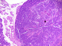
| MTB:29169 |
Mammary gland adenocarcinoma - papillary |
Mammary gland |
None (spontaneous) |
|
|
Female |
reproductive status not specified |
observed |
unknown |
Adenocarcinoma, papillary, mammary gland |
J:94320 |
|
Image Caption:Mammary gland: this neoplasm is nodular, unencapsulated, densely cellular, and locally invasive. It is composed of thin branched papillae and tubules lined by a one cell-thick columnar epithelium and supported by a small to moderate amount of fibrovascular stroma. Neoplastic cells are columnar, with indistinct cell borders and a moderate of amphophilic cytoplasm. The nucleus is central, oval, oriented at right angle with the basement membrane, medium-sized, and hyperchromatic.
|
|
|
Image ID:1418 |
|
Source of Image:Ward JM |
|
Pathologist:Mikaelian I |
|
Method / Stain:H&E |
|
|
Image Caption:Mammary gland: the is composed of thin branched papillae and tubules lined by a one cell-thick columnar epithelium and supported by a small to moderate amount of fibrovascular stroma. Neoplastic cells are columnar, with indistinct cell borders and a moderate of amphophilic cytoplasm. The nucleus is central, oval, oriented at right angle with the basement membrane, medium-sized, and hyperchromatic. Anisokaryosis and anisocytosis are mild. Mitoses are numerous.
|
|
|
Image ID:1420 |
|
Source of Image:Ward JM |
|
Pathologist:Mikaelian I |
|
Method / Stain:H&E |
|
|
Image Caption:Mammary gland: this neoplasm is nodular, unencapsulated, densely cellular, and locally invasive. It is composed of thin branched papillae and tubules lined by a one cell-thick columnar epithelium and supported by a small to moderate amount of fibrovascular stroma. Large areas of liquefactive necrosis (*) are present at the center of the tumor.
|
|
|
Image ID:1416 |
|
Source of Image:Ward JM |
|
Pathologist:Mikaelian I |
|
Method / Stain:H&E |
|
|
Image Caption:Mammary gland: this is composed of thin branched papillae and tubules lined by a one cell-thick columnar epithelium and supported by a small to moderate amount of fibrovascular stroma. Neoplastic cells are columnar, with indistinct cell borders and a moderate of amphophilic cytoplasm. The nucleus is central, oval, oriented at right angle with the basement membrane, medium-sized, and hyperchromatic.
|
|
|
Image ID:1419 |
|
Source of Image:Ward JM |
|
Pathologist:Mikaelian I |
|
Method / Stain:H&E |
|
|
Image Caption:Mammary gland: this photomicrograph illustrates the central portions of the neoplasm. At the center of the neoplasm, the extracellular space between neoplastic papillae was often packed with macrophages (*), the cytoplasm of which was foamy.
|
|
|
Image ID:1421 |
|
Source of Image:Ward JM |
|
Pathologist:Mikaelian I |
|
Method / Stain:H&E |
|
|
Image Caption:Mammary gland: this neoplasm is nodular, unencapsulated, densely cellular, and locally invasive. It is composed of thin branched papillae and tubules lined by a one cell-thick columnar epithelium and supported by a small to moderate amount of fibrovascular stroma.
|
|
|
Image ID:1415 |
|
Source of Image:Ward JM |
|
Pathologist:Mikaelian I |
|
Method / Stain:H&E |
|
|
Image Caption:Mammary gland: this neoplasm is nodular, unencapsulated, densely cellular, and locally invasive. It is composed of thin branched papillae and tubules lined by a one cell-thick columnar epithelium and supported by a small to moderate amount of fibrovascular stroma. Large areas of liquefactive necrosis (*) are present at the center of the tumor.
|
|
|
Image ID:1417 |
|
Source of Image:Ward JM |
|
Pathologist:Mikaelian I |
|
Method / Stain:H&E |
|
|
Image Caption:Mammary gland: multiple nodular, densely cellular, unencapsulated, locally invasive, well-delineated neoplasms almost entirely replace the mammary gland.
|
|
|
Image ID:1414 |
|
Source of Image:Ward JM |
|
Pathologist:Mikaelian I |
|
Method / Stain:H&E |
|
|
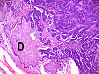
Image Caption:Papillary mammary adenocarcinoma: this photomicrograph illustrates a peripheral portion of the neoplasm. The neoplasm is continuous with the epithelium of an interlobular mammary duct (D).
|
|
|
Image ID:1422 |
|
Source of Image:Ward JM |
|
Pathologist:Mikaelian I |
|
Method / Stain:H&E |
|
|
|
| MTB ID |
Tumor Name |
Organ(s) Affected |
Treatment Type |
Agents |
Strain Name |
Strain Sex |
Reproductive Status |
Tumor Frequency |
Age at Necropsy |
Description |
Reference |
| MTB:29170 |
Mammary gland adenocarcinoma in situ |
Mammary gland |
None (spontaneous) |
|
|
Female |
reproductive status not specified |
observed |
unknown |
In situ papillary adenocarcinoma, mammary gland |
J:94320 |
|
Image Caption:Mammary gland: all alveoli and ducts are moderately ectatic and contain various proportions of inspissated proteins, a proteinaceous fluid, and lipid droplets. In addition, a few ducts and alveoli contain small papillary projections lined by a one cell-thick attenuated to cuboidal epithelium.
|
|
|
Image ID:1423 |
|
Source of Image:Ward JM |
|
Pathologist:Mikaelian I |
|
Method / Stain:H&E |
|
|
Image Caption:Mammary gland: a few ducts and alveoli contain small papillary projections lined by a one cell-thick attenuated to cuboidal epithelium. Mitoses (arrowhead) are detected in the epithelium lining these papillae. Most of the epithelium lining acini and ducts is atrophic, except in a few areas (arrow) where cells have large nuclei.
|
|
|
Image ID:1424 |
|
Source of Image:Ward JM |
|
Pathologist:Mikaelian I |
|
Method / Stain:H&E |
|
|
|
| MTB ID |
Tumor Name |
Organ(s) Affected |
Treatment Type |
Agents |
Strain Name |
Strain Sex |
Reproductive Status |
Tumor Frequency |
Age at Necropsy |
Description |
Reference |
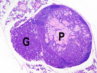
| MTB:29171 |
Mammary gland adenocarcinoma - mixed |
Mammary gland |
None (spontaneous) |
|
|
Female |
reproductive status not specified |
observed |
unknown |
Adenocarcinoma, papillary and glandular, secretory, with prominent lymphocytic and plasmacytic inflammation, mammary gland |
J:94320 |
|
Image Caption:Mammary adenocarcinoma: this photomicrograph illustrates a portion of the neoplasm where the papillary pattern is less obvious and is intermingled with areas with a glandular pattern. Degenerated neoplastic cells and neutrophils are present between neoplastic papillae.
|
|
|
Image ID:1433 |
|
Source of Image:Ward JM |
|
Pathologist:Mikaelian I |
|
Method / Stain:H&E |
|
|
Image Caption:Mammary adenocarcinoma: a very prominent lymphocytic and plasmacytic infiltration is present in the stroma of the neoplasm at its' periphery. The neoplasm is composed, in this area, of closely-packed papillae supported by a small amount of fibrovascular stroma. Neoplastic structures are lined by a one cell-thick cuboidal to columnar epithelium.
|
|
|
Image ID:1426 |
|
Source of Image:Ward JM |
|
Pathologist:Mikaelian I |
|
Method / Stain:H&E |
|
|
Image Caption:Mammary adenocarcinoma: a very prominent lymphocytic and plasmacytic infiltration is present in the stroma of the neoplasm at its' periphery. The neoplasm is composed, in this area, of closely-packed papillae supported by a small amount of fibrovascular stroma. Neoplastic structures are lined by a one cell-thick cuboidal to columnar epithelium.
|
|
|
Image ID:1427 |
|
Source of Image:Ward JM |
|
Pathologist:Mikaelian I |
|
Method / Stain:H&E |
|
|
Image Caption:Mammary gland: the mammary parenchyma is effaced by a multilobular, unencapsulated, densely cellular, neoplasm. Most of the neoplasm has a papillary pattern (P), while one neoplastic lobule has a glandular pattern (G).
|
|
|
Image ID:1425 |
|
Source of Image:Ward JM |
|
Pathologist:Mikaelian I |
|
Method / Stain:H&E |
|
|
Image Caption:Mammary adenocarcinoma: this photomicrograph illustrates a portion of the neoplasm where the papillary pattern is prominent. The papillae are separated by a large amount of amorphous acidophilic material that contains a small number of desquamated and degenerated neoplastic cells intermingled with a few macrophages.
|
|
|
Image ID:1430 |
|
Source of Image:Ward JM |
|
Pathologist:Mikaelian I |
|
Method / Stain:H&E |
|
|
Image Caption:Mammary adenocarcinoma: the neoplasm is composed, in this area, of closely-packed papillae supported by a small amount of fibrovascular stroma. Neoplastic structures are lined by a one cell-thick cuboidal to columnar epithelium. Neoplastic cells have distinct cell borders and a moderate amount of strongly amphophilic cytoplasm that occasionally contains small lipid vacuoles. The nucleus is large, central, oval, hyperchromatic, and with a coarsely-clumped chromatin. Numerous neoplastic cells undergo single cell necrosis and mitotic rate is elevated. Macrophages with a foamy cytoplasm are present in between neoplastic papillae.
|
|
|
Image ID:1428 |
|
Source of Image:Ward JM |
|
Pathologist:Mikaelian I |
|
Method / Stain:H&E |
|
|
Image Caption:Mammary adenocarcinoma: this photomicrograph illustrates a portion of the neoplasm where the papillary pattern is less obvious and is intermingled with areas with a glandular pattern.
|
|
|
Image ID:1432 |
|
Source of Image:Ward JM |
|
Pathologist:Mikaelian I |
|
Method / Stain:H&E |
|
|
Image Caption:Mammary gland: the mammary parenchyma is effaced by a multilobular, unencapsulated, densely cellular, neoplasm.
|
|
|
Image ID:1434 |
|
Source of Image:Ward JM |
|
Pathologist:Mikaelian I |
|
Method / Stain:H&E |
|
|
Image Caption:Mammary adenocarcinoma: this photomicrograph illustrates a portion of the neoplasm where the papillary pattern is prominent. The papillae are separated by a large amount of amorphous acidophilic material that contains a small number of desquamated and degenerated neoplastic cells intermingled with a moderate number of macrophages with a foamy cytoplasm.
|
|
|
Image ID:1431 |
|
Source of Image:Ward JM |
|
Pathologist:Mikaelian I |
|
Method / Stain:H&E |
|
|
Image Caption:Mammary adenocarcinoma: the lumen of some glands contains apoptotic bodies.
|
|
|
Image ID:1429 |
|
Source of Image:Ward JM |
|
Pathologist:Mikaelian I |
|
Method / Stain:H&E |
|
|
|
| MTB ID |
Tumor Name |
Organ(s) Affected |
Treatment Type |
Agents |
Strain Name |
Strain Sex |
Reproductive Status |
Tumor Frequency |
Age at Necropsy |
Description |
Reference |
| MTB:29172 |
Mammary gland carcinoma in situ |
Mammary gland |
None (spontaneous) |
|
|
Female |
reproductive status not specified |
observed |
unknown |
Carcinoma in situ, papillary, mammary gland |
J:94320 |
|
Image Caption:Mammary gland: the cellular area in this carcinoma in situ is characterized by epithelial cells piling-up and forming small papillae in a structure that resembles an intralobular duct. This change is associated with mild to moderate interstitial fibrosis and lymphoplasmacytic inflammation. Most neoplastic cells contain lipid vacuoles which are larger and more numerous in the cells lining the dysplastic acini surrounding the most advanced lesions.
|
|
|
Image ID:1436 |
|
Source of Image:Ward JM |
|
Pathologist:Mikaelian I |
|
Method / Stain:H&E |
|
|
Image Caption:In situ mammary carcinoma: this photomicrograp illustrates a portion of the neoplasm where neoplastic cells form discrete papillae that are located in a moderately dilated intralobular duct. Atypia in this area is mild.
|

|
|
Image ID:1438 |
|
Source of Image:Ward JM |
|
Pathologist:Mikaelian I |
|
Method / Stain:H&E |
|
|
Image Caption:In situ mammary carcinoma: this photomicrograp illustrates a portion of the neoplasm where neoplastic cells form discrete papillae that are located in a moderately dilated intralobular duct. Atypia in this area is mild.
|
|
|
Image ID:1439 |
|
Source of Image:Ward JM |
|
Pathologist:Mikaelian I |
|
Method / Stain:H&E |
|
|
Image Caption:Mammary gland: the mammary gland is focally expanded by a nodular, ill-defined, unencapsulated, moderately cellular mass. A more cellular area is present at the center of this mass.
|
|
|
Image ID:1435 |
|
Source of Image:Ward JM |
|
Pathologist:Mikaelian I |
|
Method / Stain:H&E |
|
|
Image Caption:Mammary gland: the cellular area in this carcinoma in situ is characterized by epithelial cells piling-up and forming small papillae in a structure that resembles an intralobular duct. This change is associated with mild to moderate interstitial fibrosis and lymphoplasmacytic inflammation. Most neoplastic cells contain lipid vacuoles which are larger and more numerous in the cells lining the dysplastic acini surrounding the most advanced lesions. A few minute areas of cornification (arroheads) are also present.
|
|
|
Image ID:1437 |
|
Source of Image:Ward JM |
|
Pathologist:Mikaelian I |
|
Method / Stain:H&E |
|
|
|
| MTB ID |
Tumor Name |
Organ(s) Affected |
Treatment Type |
Agents |
Strain Name |
Strain Sex |
Reproductive Status |
Tumor Frequency |
Age at Necropsy |
Description |
Reference |
| MTB:29173 |
Mammary gland carcinoma in situ |
Mammary gland |
None (spontaneous) |
|
|
Female |
reproductive status not specified |
observed |
unknown |
Carcinoma in situ (early stage), mammary gland |
J:94320 |
|
Image Caption:Early stages of in situ mammary carcinoma: this photomicrograph represents the appearance of the mammary gland of this mouse model in the early stages of neoplastic transformation. There is prominent lobular hyperplasia with accumulation of inspissated proteinaceous material in many alveoli and intralobular ducts. Alveoli and intralobular ducts are lined by a prominent epithelium that confers a "crowded" appearance to these structures.
|
|
|
Image ID:1440 |
|
Source of Image:Ward JM |
|
Pathologist:Mikaelian I |
|
Method / Stain:H&E |
|
|
Image Caption:Early stages of in situ mammary carcinoma: this photomicrograph represents the appearance of the mammary gland of this mouse model in the early stages of neoplastic transformation. There is prominent lobular hyperplasia with accumulation of inspissated proteinaceous material in many alveoli and intralobular ducts. Alveoli and intralobular ducts are lined by a prominent epithelium that confers a "crowded" appearance to these structures. Cells do not pile-up disorderly and there is no evidence of atypia. Apoptotic cells are numerous.
|
|
|
Image ID:1441 |
|
Source of Image:Ward JM |
|
Pathologist:Mikaelian I |
|
Method / Stain:H&E |
|
|
|
| MTB ID |
Tumor Name |
Organ(s) Affected |
Treatment Type |
Agents |
Strain Name |
Strain Sex |
Reproductive Status |
Tumor Frequency |
Age at Necropsy |
Description |
Reference |
| MTB:29174 |
Mammary gland adenocarcinoma - papillary |
Mammary gland |
None (spontaneous) |
|
|
Female |
reproductive status not specified |
observed |
unknown |
Adenocarcinoma, papillary, secretory, with squamous differentiation |
J:94320 |
|
Image Caption:Papillary and secretory mammary adenocarcinoma with squamous differentiation: this photomicrograph illustrates an area of squamous differentiation that results in the formation of squames. Neutrophils infiltrate this portion of the neoplasm.
|
|
|
Image ID:1450 |
|
Source of Image:Ward JM |
|
Pathologist:Mikaelian I |
|
Method / Stain:H&E |
|
|
Image Caption:Mammary gland: the mammary gland is replaced by a nodular, unencapsulated, well-delineated, densely cellular, polycystic neoplasm. The neoplasm is composed of closely-packed branched papillae and glands supported by a small to moderate amount of fibrovascular stroma. A large amount of coarsely granular material (degenerated neoplastic cells) separate neoplastic papillae and fill neoplastic glands.
|
|
|
Image ID:1445 |
|
Source of Image:Ward JM |
|
Pathologist:Mikaelian I |
|
Method / Stain:H&E |
|
|
Image Caption:Papillary and secretory mammary adenocarcinoma with squamous differentiation: this photomicrograph illustrates an area of squamous differentiation with the formation of nests of cornified cells.
|
|
|
Image ID:1449 |
|
Source of Image:Ward JM |
|
Pathologist:Mikaelian I |
|
Method / Stain:H&E |
|
|
Image Caption:Papillary and secretory mammary adenocarcinoma with squamous differentiation: this photomicrograph illustrates an area of squamous differentiation that results in the formation of nests of cornified cells (arrowhead). Cells in this area have distinct cell borders. Cells at various degrees of cornification are also present in the lower portions of the photomicrograph.
|
|
|
Image ID:1451 |
|
Source of Image:Ward JM |
|
Pathologist:Mikaelian I |
|
Method / Stain:H&E |
|
|
Image Caption:Papillary mammary adenocarcinoma: neoplastic glands are cuboidal to low columnar, with indistinct cell borders and a moderate amount of amphophilic cytoplasm which occasionally contains large lipid vacuoles. The nucleus is central, oval, medium-sized and normochromatic. Anisokaryosis and anisocytosis are mild. Neoplastic glands are filled with an inspissated proteinaceous material and desquamated neoplastic cells.
|
|
|
Image ID:1446 |
|
Source of Image:Ward JM |
|
Pathologist:Mikaelian I |
|
Method / Stain:H&E |
|
|
Image Caption:Papillary mammary adenocarcinoma: neoplastic glands are cuboidal to low columnar, with indistinct cell borders and a moderate amount of amphophilic cytoplasm which occasionally contains large lipid vacuoles. The nucleus is central, oval, medium-sized and normochromatic. Anisokaryosis and anisocytosis are mild. Neoplastic glands are filled with an inspissated proteinaceous material and desquamated neoplastic cells.
|
|
|
Image ID:1447 |
|
Source of Image:Ward JM |
|
Pathologist:Mikaelian I |
|
Method / Stain:H&E |
|
|
Image Caption:Mammary gland: the mammary gland is replaced by a nodular, unencapsulated, well-delineated, densely cellular, polycystic neoplasm. The neoplasm is composed of closely-packed branched papillae and glands supported by a small to moderate amount of fibrovascular stroma. A large amount of coarsely granular material (degenerated neoplastic cells) separate neoplastic papillae.
|
|
|
Image ID:1443 |
|
Source of Image:Ward JM |
|
Pathologist:Mikaelian I |
|
Method / Stain:H&E |
|
|
Image Caption:Papillary and secretory mammary adenocarcinoma with squamous differentiation: this photomicrograph illustrates an area of transition between a portion of the neoplasm with a glandular pattern (on the left of the photomicrograph) and a portion of the neoplasm with squamous differentiation (on the right of the photomicrophotograph). Squamous differentiation is associated with the formation of nests of cells undergoing cornification (arrowheads) and production of squames (*).
|
|
|
Image ID:1448 |
|
Source of Image:Ward JM |
|
Pathologist:Mikaelian I |
|
Method / Stain:H&E |
|
|
Image Caption:Papillary and secretory mammary adenocarcinoma with squamous differentiation: this photomicrograph illustrates an area of squamous differentiation that results in the formation of nests of cornified cells and squames.
|
|
|
Image ID:1452 |
|
Source of Image:Ward JM |
|
Pathologist:Mikaelian I |
|
Method / Stain:H&E |
|
|
Image Caption:Mammary gland: the mammary gland is replaced by a nodular, unencapsulated, well-delineated, densely cellular, polycystic neoplasm. The neoplasm is composed of closely-packed branched papillae and glands supported by a small to moderate amount of fibrovascular stroma. A large amount of coarsely granular material (degenerated neoplastic cells) separate neoplastic papillae.
|
|
|
Image ID:1444 |
|
Source of Image:Ward JM |
|
Pathologist:Mikaelian I |
|
Method / Stain:H&E |
|
|
Image Caption:Mammary gland: the mammary gland is replaced by a nodular, unencapsulated, well-delineated, densely cellular, polycystic neoplasm.
|
|
|
Image ID:1442 |
|
Source of Image:Ward JM |
|
Pathologist:Mikaelian I |
|
Method / Stain:H&E |
|
|
|
| MTB ID |
Tumor Name |
Organ(s) Affected |
Treatment Type |
Agents |
Strain Name |
Strain Sex |
Reproductive Status |
Tumor Frequency |
Age at Necropsy |
Description |
Reference |
| MTB:29175 |
Mammary gland adenocarcinoma - papillary |
Mammary gland |
None (spontaneous) |
|
|
Female |
reproductive status not specified |
observed |
unknown |
Adenocarcinoma, papillary, secretory, mammary gland |
J:94320 |
|
Image Caption:Papillary and secretory mammary adenocarcinoma: the neoplasm is composed of glands and papillae lined by a one cell-thick cuboidal to low columnar epithelium and supported by a small to moderate amount of fibrovascular stroma.
|
|
|
Image ID:1456 |
|
Source of Image:Ward JM |
|
Pathologist:Mikaelian I |
|
Method / Stain:H&E |
|
|
Image Caption:Papillary and secretory mammary adenocarcinoma: the neoplasm is composed of glands and papillae lined by a one cell-thick cuboidal to low columnar epithelium and supported by a small to moderate amount of fibrovascular stroma.
|
|
|
Image ID:1455 |
|
Source of Image:Ward JM |
|
Pathologist:Mikaelian I |
|
Method / Stain:H&E |
|
|
Image Caption:Mammary gland: the mammary gland is replaced by a nodular, unencapsulated, well-delineated, densely cellular, neoplasm that contains, in its' center, large areas composed of desquamated and degenerated neoplastic cells.
|
|
|
Image ID:1453 |
|
Source of Image:Ward JM |
|
Pathologist:Mikaelian I |
|
Method / Stain:H&E |
|
|
Image Caption:Papillary and secretory mammary adenocarcinoma: this photomicrograph illustrates the papillary pattern of this neoplasm. There are many mitotic figures.
|
|
|
Image ID:1460 |
|
Source of Image:Ward JM |
|
Pathologist:Mikaelian I |
|
Method / Stain:H&E |
|
|
Image Caption:Papillary and secretory mammary adenocarcinoma: this photomicrograph illustrates the papillary pattern of this neoplasm.
|
|
|
Image ID:1459 |
|
Source of Image:Ward JM |
|
Pathologist:Mikaelian I |
|
Method / Stain:H&E |
|
|
Image Caption:Papillary and secretory mammary adenocarcinoma: the neoplasm is composed of glands and papillae lined by a one cell-thick cuboidal to low columnar epithelium and supported by a small to moderate amount of collagen-rich fibrovascular stroma. Neoplastic cells are cuboidal to low columnar, with indistinct cell borders and a moderate amount of strongly amphophilic cytoplasm that occasionally contains a few small vacuoles of lipids. The nucleus is basal to central, medium-sized, oval, and normochromatic. The luminal spaces are filled with degenerated neoplastic cells. The stroma is infiltrated by a moderate number of neutrophils, lymphocytes and plasma cells.
|
|
|
Image ID:1457 |
|
Source of Image:Ward JM |
|
Pathologist:Mikaelian I |
|
Method / Stain:H&E |
|
|
Image Caption:Mammary gland: the mammary gland is replaced by a nodular, unencapsulated, well-delineated, densely cellular, neoplasm that contains, in its' center, large areas composed of desquamated and degenerated neoplastic cells. The neoplasm is composed of thin branched papillae and a few glands supported by a small to moderate amount of fibrovascular stroma.
|
|
|
Image ID:1454 |
|
Source of Image:Ward JM |
|
Pathologist:Mikaelian I |
|
Method / Stain:H&E |
|
|
Image Caption:Papillary and secretory mammary adenocarcinoma: this photomicrograph illustrates the papillary pattern of this neoplasm.
|
|
|
Image ID:1458 |
|
Source of Image:Ward JM |
|
Pathologist:Mikaelian I |
|
Method / Stain:H&E |
|
|
|
| MTB ID |
Tumor Name |
Organ(s) Affected |
Treatment Type |
Agents |
Strain Name |
Strain Sex |
Reproductive Status |
Tumor Frequency |
Age at Necropsy |
Description |
Reference |
| MTB:29176 |
Mammary gland adenoma |
Mammary gland |
None (spontaneous) |
|
|
Female |
reproductive status not specified |
observed |
300 days |
Adenoma, glandular, mammary gland |
J:94320 |
|
Image Caption:Mammary gland: the neoplasm is composed of large nest that have numerous small secondary lumens lined by a one cell-thick cuboidal epithelium. Neoplastic cells are cuboidal, with indistinct cell borders and a moderate amount of strongly acidophilic cytoplasm. The nucleus is central, oval, medium-sixed and normochromatic. Anisokaryosis is moderate. Anisocytosis is mild.
|
|
|
Image ID:1465 |
|
Source of Image:Ward JM |
|
Pathologist:Mikaelian I |
|
Method / Stain:H&E |
|
|
Image Caption:Glandular adenoma: a few neoplastic cells are multinucleated (arrowhead). This feature is consistent with aneuploidy and is often observed in tumors with alterations of p53.
|
|
|
Image ID:1466 |
|
Source of Image:Ward JM |
|
Pathologist:Mikaelian I |
|
Method / Stain:H&E |
|
|
Image Caption:Mammary gland: the mammary gland is effaced by a nodular, unencapsulated, densely cellular, expensile neoplasm. This neoplasm is composed of large nest that have numerous small secondary lumens.
|
|
|
Image ID:1463 |
|
Source of Image:Ward JM |
|
Pathologist:Mikaelian I |
|
Method / Stain:H&E |
|
|
Image Caption:Mammary gland: the mammary gland is effaced by a nodular, unencapsulated, densely cellular, expensile neoplasm.
|
|
|
Image ID:1461 |
|
Source of Image:Ward JM |
|
Pathologist:Mikaelian I |
|
Method / Stain:H&E |
|
|
Image Caption:Mammary gland: the mammary gland is effaced by a nodular, unencapsulated, densely cellular, expensile neoplasm. This neoplasm is composed of large nest that have numerous small secondary lumens lined by a one cell-thick cuboidal epithelium. Neoplastic cells are cuboidal, with indistinct cell borders and a moderate amount of strongly acidophilic cytoplasm. The nucleus is central, oval, medium-sixed and normochromatic. Anisokaryosis is moderate. Anisocytosis is mild.
|
|
|
Image ID:1464 |
|
Source of Image:Ward JM |
|
Pathologist:Mikaelian I |
|
Method / Stain:H&E |
|
|
Image Caption:Mammary gland: the mammary gland is effaced by a nodular, unencapsulated, densely cellular, expensile neoplasm. This neoplasm is composed of large nest that have numerous small secondary lumens.
|
|
|
Image ID:1462 |
|
Source of Image:Ward JM |
|
Pathologist:Mikaelian I |
|
Method / Stain:H&E |
|
|
|
| MTB ID |
Tumor Name |
Organ(s) Affected |
Treatment Type |
Agents |
Strain Name |
Strain Sex |
Reproductive Status |
Tumor Frequency |
Age at Necropsy |
Description |
Reference |
| MTB:29177 |
Mammary gland cyst - macrocyst |
Mammary gland |
None (spontaneous) |
|
|
Female |
reproductive status not specified |
observed |
300 days |
Mammary gland - Macrocyst, with atypia |
J:94320 |
|
Image Caption:Mammary gland: the mammary gland is expanded by a large polycystic mass lined by a one to two cells-thick cuboidal to tall columnar epithelium.
|
|
|
Image ID:1468 |
|
Source of Image:Ward JM |
|
Pathologist:Mikaelian I |
|
Method / Stain:H&E |
|
|
Image Caption:Mammary gland: the mammary gland is expanded by a large polycystic mass lined by a one to two cells-thick cuboidal to tall columnar epithelium.
|
|
|
Image ID:1467 |
|
Source of Image:Ward JM |
|
Pathologist:Mikaelian I |
|
Method / Stain:H&E |
|
|
Image Caption:Mammary gland: the mammary gland is expanded by a large polycystic mass lined by a one to two cells-thick cuboidal to tall columnar epithelium. The cells lining the cysts have distinct cell borders and a moderate to large amount of acidophilic cytoplasm that often contain lipid vacuoles of various sizes. The nucleus is randomly located in the cell, is large, oval, hyperchromatic, and with a coarsely clumped chromatin. Anisokaryosis and anisocytosis are prominent. A few mitoses are detected.
|
|
|
Image ID:1469 |
|
Source of Image:Ward JM |
|
Pathologist:Mikaelian I |
|
Method / Stain:H&E |
|
|
|
| MTB ID |
Tumor Name |
Organ(s) Affected |
Treatment Type |
Agents |
Strain Name |
Strain Sex |
Reproductive Status |
Tumor Frequency |
Age at Necropsy |
Description |
Reference |
| MTB:29178 |
Mammary gland adenocarcinoma - papillary |
Mammary gland |
None (spontaneous) |
|
|
Female |
reproductive status not specified |
observed |
unknown |
Adenocarcinoma, papillary, arising in a macrocyst, mammary gland |
J:94320 |
|
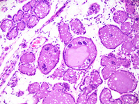
Image Caption:Mammary gland: the mammary gland is effaced by a nodular, unencapsulated, densely cellular, polycystic mass. This mass is composed of papillae and glands with a prominent branching pattern.
|
|
|
Image ID:1470 |
|
Source of Image:Ward JM |
|
Pathologist:Mikaelian I |
|
Method / Stain:H&E |
|
|
Image Caption:Mammary adenocarcinoma: this photomicrograph represents portions of the neoplasm characterized by the formation of solid areas with central coagulation necrosis and formation of small secondary lumens.
|
|
|
Image ID:1475 |
|
Source of Image:Ward JM |
|
Pathologist:Mikaelian I |
|
Method / Stain:H&E |
|
|
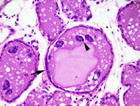
Image Caption:Mammary gland: neoplastic epithelial structures generally are several cells-thick (up to 10 cells-thick) and are characterized by the formation of secondary lumens. Neoplastic cells are large, cuboidal to polygonal, with distinct cell borders and a large amount of strongly acidophilic cytoplasm. The nucleus is central, oval, medium-sized, normochromatic, with a coarsely-clumped chromatin and 1-3 small basophilic nucleoli. Anisokaryosis and anisocytosis are mild.
|
|
|
Image ID:1473 |
|
Source of Image:Ward JM |
|
Pathologist:Mikaelian I |
|
Method / Stain:H&E |
|
|
Image Caption:Mammary adenocarcinoma: this photomicrograph represents portions of the neoplasm characterized by the formation of solid areas with central coagulation necrosis and formation of small secondary lumens. Nuclear palissading is prominent at the periphery of this neoplastic lobule.
|
|
|
Image ID:1476 |
|
Source of Image:Ward JM |
|
Pathologist:Mikaelian I |
|
Method / Stain:H&E |
|
|
Image Caption:Mammary gland: the mammary gland is effaced by a nodular, unencapsulated, densely cellular, polycystic mass. This mass is composed of papillae and glands with a prominent branching pattern. The epithelial structures generally are several cells-thick (up to 10 cells-thick) and are characterized by the formation of secondary lumens. Neoplastic cells are large, cuboidal to polygonal, with distinct cell borders and a large amount of strongly acidophilic cytoplasm. The nucleus is central, oval, and medium-sized.
|
|
|
Image ID:1472 |
|
Source of Image:Ward JM |
|
Pathologist:Mikaelian I |
|
Method / Stain:H&E |
|
|
Image Caption:Mammary gland: the mammary gland is effaced by a nodular, unencapsulated, densely cellular, polycystic mass. This mass is composed of papillae and glands with a prominent branching pattern. The morphology of this lesion is similar to that of macrocysts, except that the glandular and papillary areas located at the periphery of the cysts in this tumors are more cellular than in macrocysts.
|
|
|
Image ID:1471 |
|
Source of Image:Ward JM |
|
Pathologist:Mikaelian I |
|
Method / Stain:H&E |
|
|
Image Caption:Mammary adenocarcinoma: this photomicrograph represents portions of the neoplasm characterized by the formation of solid areas with central coagulation necrosis (*) and formation of small secondary lumens.
|

|
|
Image ID:1474 |
|
Source of Image:Ward JM |
|
Pathologist:Mikaelian I |
|
Method / Stain:H&E |
|
|
|
| MTB ID |
Tumor Name |
Organ(s) Affected |
Treatment Type |
Agents |
Strain Name |
Strain Sex |
Reproductive Status |
Tumor Frequency |
Age at Necropsy |
Description |
Reference |
| MTB:29179 |
Mammary gland hyperplasia - lobular |
Mammary gland |
None (spontaneous) |
|
|
Female |
reproductive status not specified |
observed |
unknown |
Lobular hyperplasia with atypia, mammary gland |
J:94320 |
|
Image Caption:Mammary gland: mammary lobules are prominent, are filled with an often inspissated proteinaceous fluid, and are lined by a prominent epithelium. Luminal cells of all alveoli contain lipid vacuoles and proteinaceous droplets. Anisokaryosis, a common feature of tumors with altered Trp53 expression, is prominent. There is also moderate interstitial lymphocytic and plasmacytic inflammation.
|
|
|
Image ID:1479 |
|
Source of Image:Ward JM |
|
Pathologist:Mikaelian I |
|
Method / Stain:H&E |
|
|
Image Caption:Mammary gland: mammary lobules are prominent, are filled with an often inspissated proteinaceous fluid, and are lined by a prominent epithelium. Luminal cells of all alveoli contain lipid vacuoles and proteinaceous droplets. Anisokaryosis, a common feature of tumors with altered Trp53 expression, is prominent. There is also moderate interstitial lymphocytic and plasmacytic inflammation.
|
|
|
Image ID:1480 |
|
Source of Image:Ward JM |
|
Pathologist:Mikaelian I |
|
Method / Stain:H&E |
|
|
Image Caption:Mammary gland: mammary lobules are prominent, are filled with an often inspissated proteinaceous fluid, and are lined by a prominent epithelium. Luminal cells of all alveoli contain lipid vacuoles and proteinaceous droplets. Anisokaryosis, a common feature of tumors with altered Trp53 expression, is prominent.
|
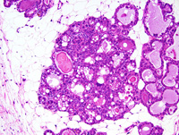
|
|
Image ID:1477 |
|
Source of Image:Ward JM |
|
Pathologist:Mikaelian I |
|
Method / Stain:H&E |
|
|
Image Caption:Mammary gland: mammary lobules are prominent, are filled with an often inspissated proteinaceous fluid, and are lined by a prominent epithelium. Luminal cells of all alveoli contain lipid vacuoles and proteinaceous droplets. Anisokaryosis, a common feature of tumors with altered Trp53 expression, is prominent.
|
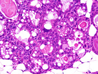
|
|
Image ID:1478 |
|
Source of Image:Ward JM |
|
Pathologist:Mikaelian I |
|
Method / Stain:H&E |
|
|
|
| MTB ID |
Tumor Name |
Organ(s) Affected |
Treatment Type |
Agents |
Strain Name |
Strain Sex |
Reproductive Status |
Tumor Frequency |
Age at Necropsy |
Description |
Reference |
| MTB:29180 |
Mammary gland hyperplasia - alveolar |
Mammary gland |
None (spontaneous) |
|
|
Unspecified |
reproductive status not specified |
observed |
unknown |
Hyperplastic alveolar nodule, mammary gland |
J:94320 |
|
Image Caption:Hyperplastic alveolar nodule: the nodule is unencapsulated. It is composed of closely-packed glands and ducts that show various degrees of ectasia.
|
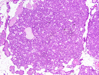
|
|
Image ID:1482 |
|
Source of Image:Ward JM |
|
Pathologist:Mikaelian I |
|
Method / Stain:H&E |
|
|
Image Caption:Hyperplastic alveolar nodule: the nodule is unencapsulated. It is composed of closely-packed glands and ducts that show various degrees of ectasia. Acini are lined by a one cell-thick cuboidal epithelium with prominent small lipid vacuoles and proteinaceous droplets. There is no evidence of atypia.
|
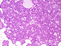
|
|
Image ID:1483 |
|
Source of Image:Ward JM |
|
Pathologist:Mikaelian I |
|
Method / Stain:H&E |
|
|
Image Caption:Mammary gland: two small, non-compressive, expensile, densely cellular nodules are present in a lactating mammary gland.
|
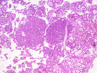
|
|
Image ID:1481 |
|
Source of Image:Ward JM |
|
Pathologist:Mikaelian I |
|
Method / Stain:H&E |
|
|
Image Caption:Hyperplastic alveolar nodule: the nodule is unencapsulated. It is composed of closely-packed glands and ducts that show various degrees of ectasia. Acini are lined by a one cell-thick cuboidal epithelium with prominent small lipid vacuoles and proteinaceous droplets. There is no evidence of atypia. A few mitoses are present.
|
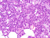
|
|
Image ID:1484 |
|
Source of Image:Ward JM |
|
Pathologist:Mikaelian I |
|
Method / Stain:H&E |
|
|
|
| MTB ID |
Tumor Name |
Organ(s) Affected |
Treatment Type |
Agents |
Strain Name |
Strain Sex |
Reproductive Status |
Tumor Frequency |
Age at Necropsy |
Description |
Reference |
| MTB:29181 |
Mammary gland adenoma |
Mammary gland |
None (spontaneous) |
|
|
Unspecified |
reproductive status not specified |
observed |
unknown |
Adenoma, glandular, secretory, mammary gland |
J:94320 |
|
Image Caption:Mammary gland: the mammary parenchyma is focally effaced by a small, nodular, densely cellular neoplastic mass surrounded by a thin fibrous capsule. This neoplasm is composed of closely packed glands lined by a one cell-thick cuboidal epithelium supported by a small amount of fibrovascular stroma. The glands are dilated by a small amount of proteinaceous material. The cells are cuboidal, with indistinct cell borders and a moderate amount of strongly acidophilic cytoplasm that often lipid vacuoles and proteinaceous droplets of various sizes. The nucleus is often basal, round to oval, and slightly hypochromatic. Anisokaryosis and anisocytosis are mild. A few mitoses are present.
|
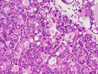
|
|
Image ID:1488 |
|
Source of Image:Ward JM |
|
Pathologist:Mikaelian I |
|
Method / Stain:H&E |
|
|
Image Caption:Mammary gland: the mammary parenchyma is focally effaced by a small, nodular, densely cellular neoplastic mass surrounded by a thin fibrous capsule. This neoplasm is composed of closely packed glands lined by a one cell-thick cuboidal epithelium supported by a small amount of fibrovascular stroma. The glands are dilated by a small amount of proteinaceous material. The cells are cuboidal, with indistinct cell borders and a moderate amount of strongly acidophilic cytoplasm that often lipid vacuoles and proteinaceous droplets of various sizes. The nucleus is ofetn basal, round to oval, and slightly hypochromatic. Anisokaryosis and anisocytosis are mild.
|
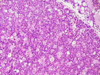
|
|
Image ID:1487 |
|
Source of Image:Ward JM |
|
Pathologist:Mikaelian I |
|
Method / Stain:H&E |
|
|
Image Caption:Mammary gland: the mammary parenchyma is focally effaced by a small, nodular, densely cellular neoplastic mass surrounded by a thin fibrous capsule. This neoplasm is composed of closely packed glands lined by a one cell-thick cuboidal epithelium supported by a small amount of fibrovascular stroma. The glands are dilated by a small amount of proteinaceous material. The cells are cuboidal, with indistinct cell borders and a moderate amount of strongly acidophilic cytoplasm that often lipid vacuoles and proteinaceous droplets of various sizes. The adjacent mammary gland is lactational.
|
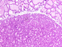
|
|
Image ID:1486 |
|
Source of Image:Ward JM |
|
Pathologist:Mikaelian I |
|
Method / Stain:H&E |
|
|
Image Caption:Mammary gland: the mammary parenchyma is focally effaced by a small, nodular, densely cellular neoplastic mass surrounded by a thin fibrous capsule. This neoplasm is composed of closely packed glands supported by a small amount of fibrovascular stroma. The adjacent mammary gland is lactational.
|
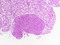
|
|
Image ID:1485 |
|
Source of Image:Ward JM |
|
Pathologist:Mikaelian I |
|
Method / Stain:H&E |
|
|
|
| MTB ID |
Tumor Name |
Organ(s) Affected |
Treatment Type |
Agents |
Strain Name |
Strain Sex |
Reproductive Status |
Tumor Frequency |
Age at Necropsy |
Description |
Reference |
| MTB:29182 |
Mammary gland hyperplasia |
Mammary gland |
None (spontaneous) |
|
|
Unspecified |
reproductive status not specified |
observed |
unknown |
Hyperplasia and atypia, mammary gland |
J:94320 |
|
Image Caption:Mammary gland: this photomicrograph illustrates a portion of the mammary gland that is lactational, hyperplastic, and shows prominent atypia. Many cells in this area are large and have a large hyperchromatic nucleus with a contoured nuclear membrane. These features are consistent with aneuploidy, a phenomenon that is commonly observed in tumors with altered expression of Trp53. There is also a mild interstitial lymphocytic inflammation.
|
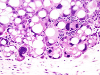
|
|
Image ID:1490 |
|
Source of Image:Ward JM |
|
Pathologist:Mikaelian I |
|
Method / Stain:H&E |
|
|
Image Caption:Mammary gland: this photomicrograph illustrates a portion of the mammary gland that is lactational, hyperplastic, and shows prominent atypia. A pentapolar mitosis is present.
|
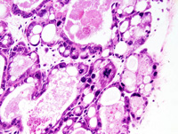
|
|
Image ID:1491 |
|
Source of Image:Ward JM |
|
Pathologist:Mikaelian I |
|
Method / Stain:H&E |
|
|
Image Caption:Mammary gland: this photomicrograph illustrates a portion of the mammary gland that is lactational, hyperplastic, and shows prominent atypia. Many cells in this area are large and have a large hyperchromatic nucleus with a contoured nuclear membrane. These features are consistent with aneuploidy, a phenomenon that is commonly observed in tumors with altered expression of Trp53.
|
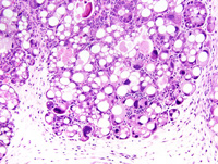
|
|
Image ID:1489 |
|
Source of Image:Ward JM |
|
Pathologist:Mikaelian I |
|
Method / Stain:H&E |
|
|
|
| MTB ID |
Tumor Name |
Organ(s) Affected |
Treatment Type |
Agents |
Strain Name |
Strain Sex |
Reproductive Status |
Tumor Frequency |
Age at Necropsy |
Description |
Reference |
| MTB:29183 |
Mammary gland adenoma |
Mammary gland |
None (spontaneous) |
|
|
Female |
reproductive status not specified |
observed |
unknown |
Adenoma, glandular, secretory, with atypia, mammary gland |
J:94320 |
|
Image Caption:Glandular adenoma of the mammary gland: this photomicrograph illustrates a portion of the neoplasm that is more cellular and shows more atypia than the rest of the neoplasm.
|
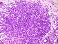
|
|
Image ID:1501 |
|
Source of Image:Ward JM |
|
Pathologist:Mikaelian I |
|
Method / Stain:H&E |
|
|
Image Caption:Glandular adenoma of the mammary gland: this photomicrograph illustrates a portion of the neoplasm that is surrounded by a capsule infiltrated by a small number of lymphocytes and plasma cells. Inflammation extends into the neoplasm which comprises a moderate amount of stroma.
|
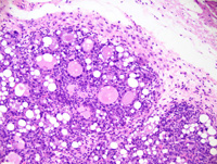
|
|
Image ID:1498 |
|
Source of Image:Ward JM |
|
Pathologist:Mikaelian I |
|
Method / Stain:H&E |
|
|
Image Caption:Glandular adenoma of the mammary gland: this photomicrograph illustrates a portion of the neoplasm that is more cellular and shows more atypia than the rest of the neoplasm. One neoplastic cell has a very large nucleus (arrowhead) comparatively to the other neoplastic cells in this field.
|
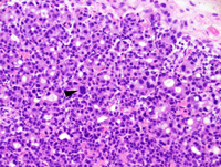
|
|
Image ID:1502 |
|
Source of Image:Ward JM |
|
Pathologist:Mikaelian I |
|
Method / Stain:H&E |
|
|
Image Caption:Glandular adenoma of the mammary gland: the neoplasm is composed of closely-packed glands lined by a one cell-thick cuboidal epithelium supported by a moderate amount of fibrovascular stroma. Most of these glands show mild to moderate ectasia and are filled by a proteinaceous fluid. Neoplastic cells have indistinct cell borders and a moderate amount of acidophilic cytoplasm that often contains numerous lipid vacuoles and proteinaceous droplets of various sizes.
|
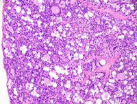
|
|
Image ID:1496 |
|
Source of Image:Ward JM |
|
Pathologist:Mikaelian I |
|
Method / Stain:H&E |
|
|
Image Caption:Glandular adenoma of the mammary gland: this photomicrograph illustrates a portion of the neoplasm that is surrounded by a capsule infiltrated by a small number of lymphocytes and plasma cells. Inflammation extends into the neoplasm which comprises a moderate amount of stroma. There is also moderate anisokaryosis.
|
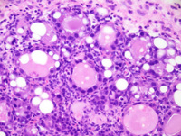
|
|
Image ID:1499 |
|
Source of Image:Ward JM |
|
Pathologist:Mikaelian I |
|
Method / Stain:H&E |
|
|
Image Caption:Mammary gland: the mammary gland is replaced by a large, nodular, well-delineated, encapsulated, expensile, densely cellular neoplasm.
|
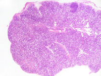
|
|
Image ID:1493 |
|
Source of Image:Ward JM |
|
Pathologist:Mikaelian I |
|
Method / Stain:H&E |
|
|
Image Caption:Glandular adenoma of the mammary gland: the neoplasm is composed of closely-packed glands lined by a one cell-thick cuboidal epithelium supported by a moderate amount of fibrovascular stroma. Most of these glands show mild to moderate ectasia and are filled by a proteinaceous fluid. Neoplastic cells have indistinct cell borders and a moderate amount of acidophilic cytoplasm that often contains numerous lipid vacuoles and proteinaceous droplets of various sizes. The nucleus is often basal, round, and hyperchromatic. Anisokaryosis and anisocytosis are moderate. Mitoses are not found in this field.
|
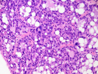
|
|
Image ID:1497 |
|
Source of Image:Ward JM |
|
Pathologist:Mikaelian I |
|
Method / Stain:H&E |
|
|
Image Caption:Glandular adenoma of the mammary gland: this photomicrograph illustrates a portion of the neoplasm that is more cellular and shows more atypia than the rest of the neoplasm.
|
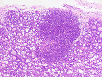
|
|
Image ID:1500 |
|
Source of Image:Ward JM |
|
Pathologist:Mikaelian I |
|
Method / Stain:H&E |
|
|
Image Caption:Mammary gland: the mammary gland is replaced by a large, nodular, well-delineated, encapsulated, expensile, densely cellular neoplasm. The neoplasm is composed of closely-packed glands lined by a one cell-thick cuboidal epithelium supported by a moderate amount of fibrovascular stroma. Most of these glands show mild to moderate ectasia and are filled by a proteinaceous fluid. Neoplastic cells have indistinct cell borders and a moderate amount of acidophilic cytoplasm that often contains numerous lipid vacuoles and proteinaceous droplets of various sizes.
|
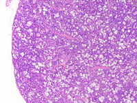
|
|
Image ID:1495 |
|
Source of Image:Ward JM |
|
Pathologist:Mikaelian I |
|
Method / Stain:H&E |
|
|
Image Caption:Mammary gland: the mammary gland is replaced by a large, nodular, well-delineated, encapsulated, expensile, densely cellular neoplasm. The neoplasm is composed of closely-packed glands supported by a moderate amount of fibrovascular stroma. Most of these glands show mild to moderate ectasia and are filled by a proteinaceous fluid.
|
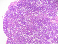
|
|
Image ID:1494 |
|
Source of Image:Ward JM |
|
Pathologist:Mikaelian I |
|
Method / Stain:H&E |
|
|
Image Caption:Mammary gland: the mammary gland is replaced by a large, nodular, well-delineated, encapsulated, expensile, densely cellular neoplasm.
|
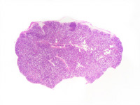
|
|
Image ID:1492 |
|
Source of Image:Ward JM |
|
Pathologist:Mikaelian I |
|
Method / Stain:H&E |
|
|
|
| MTB ID |
Tumor Name |
Organ(s) Affected |
Treatment Type |
Agents |
Strain Name |
Strain Sex |
Reproductive Status |
Tumor Frequency |
Age at Necropsy |
Description |
Reference |
| MTB:29184 |
Mammary gland cyst - macrocyst |
Mammary gland |
None (spontaneous) |
|
|
Female |
reproductive status not specified |
observed |
unknown |
Macrocyst, mammary gland |
J:94320 |
|
Image Caption:Macrocyst: this photomicrograph represents the periphery of a macrocyst. The cystic structures are interpreted as dilated mammary acini. The epithelium lining these acini is hyperplastic and some of its' cells contain very large coalescing proteinaceous droplets.
|
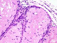
|
|
Image ID:1507 |
|
Source of Image:Ward JM |
|
Pathologist:Mikaelian I |
|
Method / Stain:H&E |
|
|
Image Caption:Macrocyst: this photomicrograph represents a more cellular portion of a macrocyst. This area is composed of a few closely-packed glands supported by a small amount of fibrovascular stroma with a mild interstitial lymphocytic and plasmacytic inflammation. This cellular area may represent an evidence of early neoplastic transformation since neoplasms may arise within macrocysts.
|
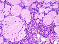
|
|
Image ID:1510 |
|
Source of Image:Ward JM |
|
Pathologist:Mikaelian I |
|
Method / Stain:H&E |
|
|
Image Caption:Mammary gland: the mammary gland is markedly expanded by a large nodule that resembles a mammary lobule that has been expanded by a considerable amount of proteinaceous fluid. The large central cysts are interpreted as intralobular ducts. The club-shaped glands that radiate from the central cysts are interpreted as mammary acini.
|
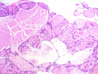
|
|
Image ID:1503 |
|
Source of Image:Ward JM |
|
Pathologist:Mikaelian I |
|
Method / Stain:H&E |
|
|
Image Caption:Macrocyst: this photomicrograph represents a more cellular portion of a macrocyst. Neoplasms may arise within macrocysts.
|
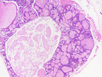
|
|
Image ID:1508 |
|
Source of Image:Ward JM |
|
Pathologist:Mikaelian I |
|
Method / Stain:H&E |
|
|
Image Caption:Macrocyst: this photomicrograph represents a more cellular portion of a macrocyst. This area is composed of a few closely-packed glands supported by a small amount of fibrovascular stroma with a mild interstitial lymphocytic and plasmacytic inflammation. This cellular area may represent an evidence of early neoplastic transformation since neoplasms may arise within macrocysts.
|
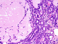
|
|
Image ID:1511 |
|
Source of Image:Ward JM |
|
Pathologist:Mikaelian I |
|
Method / Stain:H&E |
|
|
Image Caption:Macrocyst: this photomicrograph represents the periphery of a macrocyst. The cystic structures are interpreted as dilated mammary acini. The epithelium lining these acini is hyperplastic and some of its' cells contain very large coalescing proteinaceous droplets.
|
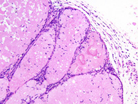
|
|
Image ID:1506 |
|
Source of Image:Ward JM |
|
Pathologist:Mikaelian I |
|
Method / Stain:H&E |
|
|
Image Caption:Macrocyst: this photomicrograph represents a more cellular portion of a macrocyst. This area is composed of a few closely-packed glands supported by a small amount of fibrovascular stroma with a mild interstitial lymphocytic and plasmacytic inflammation. This cellular area may represent an evidence of early neoplastic transformation since neoplasms may arise within macrocysts.
|
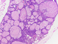
|
|
Image ID:1509 |
|
Source of Image:Ward JM |
|
Pathologist:Mikaelian I |
|
Method / Stain:H&E |
|
|
Image Caption:Macrocyst: this photomicrograph represents the periphery of a macrocyst. The cystic structures are interpreted as dilated mammary acini. The epithelium lining these acini is hyperplastic and some of its' cells contain very large coalescing proteinaceous droplets.
|
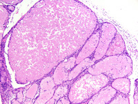
|
|
Image ID:1505 |
|
Source of Image:Ward JM |
|
Pathologist:Mikaelian I |
|
Method / Stain:H&E |
|
|
Image Caption:Mammary gland: the mammary gland is markedly expanded by a large nodule that resembles a mammary lobule that has been expanded by a considerable amount of proteinaceous fluid. The large central cysts are interpreted as intralobular ducts. The club-shaped glands that radiate from the central cysts are interpreted as mammary acini.
|
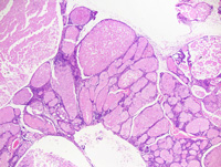
|
|
Image ID:1504 |
|
Source of Image:Ward JM |
|
Pathologist:Mikaelian I |
|
Method / Stain:H&E |
|
|
|
| MTB ID |
Tumor Name |
Organ(s) Affected |
Treatment Type |
Agents |
Strain Name |
Strain Sex |
Reproductive Status |
Tumor Frequency |
Age at Necropsy |
Description |
Reference |
| MTB:29185 |
Mammary gland lesion |
Mammary gland |
None (spontaneous) |
|
|
Female |
reproductive status not specified |
observed |
unknown |
Lactation responsive plaque, mammary gland |
J:94320 |
|
Image Caption:Lactation responsive plaque: the periphery of the mass is composed of closely-packed glands supported by a small amount of fibrovascular stroma. There is no evidence of atypia. A few mitoses are present.
|
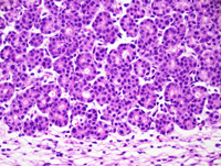
|
|
Image ID:1516 |
|
Source of Image:Ward JM |
|
Pathologist:Mikaelian I |
|
Method / Stain:H&E |
|
|
Image Caption:Mammary gland: the mammary gland is expanded by a nodular, well-defined, unencapsulated, densely cellular mass. The periphery of this mass is composed of closely-packed glands resembling mammary acini. The center of this mass is composed of duct-like structures embedded in a large amount of loose fibrovascular stroma. These features are consistent with a diagnosis of a "lactation responsive plaque", although this plaque is unusually large.
|
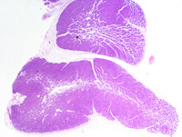
|
|
Image ID:1512 |
|
Source of Image:Ward JM |
|
Pathologist:Mikaelian I |
|
Method / Stain:H&E |
|
|
Image Caption:Lactation responsive plaque: the periphery of the mass is composed of closely-packed glands supported by a small amount of fibrovascular stroma. There is no evidence of atypia.
|
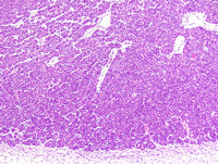
|
|
Image ID:1514 |
|
Source of Image:Ward JM |
|
Pathologist:Mikaelian I |
|
Method / Stain:H&E |
|
|
Image Caption:Lactation responsive plaque: the periphery of the mass is composed of closely-packed glands supported by a small amount of fibrovascular stroma. There is no evidence of atypia.
|
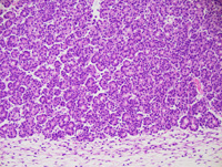
|
|
Image ID:1515 |
|
Source of Image:Ward JM |
|
Pathologist:Mikaelian I |
|
|
Image Caption:Mammary gland: the mammary gland is expanded by a nodular, well-defined, unencapsulated, densely cellular mass. The periphery of this mass is composed of closely-packed glands resembling mammary acini. The center of this mass is composed of duct-like structures embedded in a large amount of loose fibrovascular stroma. These features are consistent with a diagnosis of a "lactation responsive plaque", although this plaque is unusually large.
|
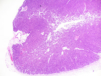
|
|
Image ID:1513 |
|
Source of Image:Ward JM |
|
Pathologist:Mikaelian I |
|
Method / Stain:H&E |
|
|
|
| MTB ID |
Tumor Name |
Organ(s) Affected |
Treatment Type |
Agents |
Strain Name |
Strain Sex |
Reproductive Status |
Tumor Frequency |
Age at Necropsy |
Description |
Reference |
| MTB:29186 |
Mammary gland lesion |
Mammary gland |
None (spontaneous) |
|
|
Female |
reproductive status not specified |
observed |
unknown |
Lactation-responsive plaque, mammary gland |
J:94320 |
|
Image Caption:Lactation-responsive plaque: there is no evidence of atypia. The interstitium shows mild to moderate diffuse edema, and is infiltrated by a small number of granulocytes, lymphocytes, plasma cells and macrophages.
|
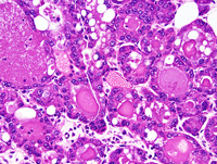
|
|
Image ID:1520 |
|
Source of Image:Ward JM |
|
Pathologist:Mikaelian I |
|
Method / Stain:H&E |
|
|
Image Caption:Mammary gland: the mammary gland is expanded by numerous ill-defined to nodular masses characterized by a radiating arrangement of intralobular ducts and acini.
|
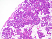
|
|
Image ID:1517 |
|
Source of Image:Ward JM |
|
Pathologist:Mikaelian I |
|
Method / Stain:H&E |
|
|
Image Caption:Lactation-responsive plaque: lactation responsive plaques are ill-defined to discoid unencapsulated masses composed of acini at the periphery, and ducts at the center. The stroma is more abundant and more edematous at the center at the plaque than at its' periphery. There is no evidence of atypia of the epithelium. Secretory activity is abundant in this case.
|
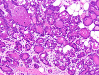
|
|
Image ID:1519 |
|
Source of Image:Ward JM |
|
Pathologist:Mikaelian I |
|
Method / Stain:H&E |
|
|
Image Caption:Lactation-responsive plaque: lactation responsive plaques are ill-defined to discoid unencapsulated masses composed of acini at the periphery, and ducts at the center. The stroma is more abundant and more edematous at the center at the plaque than at its' periphery. There is no evidence of atypia of the epithelium. Secretory activity is abundant in this case.
|
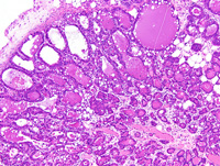
|
|
Image ID:1518 |
|
Source of Image:Ward JM |
|
Pathologist:Mikaelian I |
|
Method / Stain:H&E |
|
|
|
| MTB ID |
Tumor Name |
Organ(s) Affected |
Treatment Type |
Agents |
Strain Name |
Strain Sex |
Reproductive Status |
Tumor Frequency |
Age at Necropsy |
Description |
Reference |
| MTB:29187 |
Mammary gland fibroadenoma |
Mammary gland |
None (spontaneous) |
|
|
Female |
reproductive status not specified |
observed |
unknown |
Fibroadenoma, mammary gland |
J:94320 |
|
Image Caption:Mammary gland: the mammary gland is effaced by a moderately cellular, encapsulated, well-delineated, expensile, multilobular neoplastic mass. This neoplasm is composed of branching ducts supported by a large amount of cellular stroma. Neoplastic ducts are lined by a one cell-thick cuboidal epithelium. Neoplastic ducts are often distended by a proteinaceous fluid. Neoplastic cells have ill-defined cell borders and a moderate amount of a strongly amphophilic cytoplasm that often contains lipid vacuoles. The nucleus is central, round, medium-sized and normochromatic. Anisokaryosis and anisocytosis are mild. A few mitoses (arrowhead) are detected. The stroma is abundant, contains numerous myofibroblasts, a small number of inflammatory cells, and loosely arranged wavy and thin collagen fibers.
|
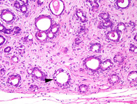
|
|
Image ID:1524 |
|
Source of Image:Ward JM |
|
Pathologist:Mikaelian I |
|
Method / Stain:H&E |
|
|
Image Caption:Fibroadenoma: this photomicrograph illustrates the branching pattern of neoplastic ducts.
|
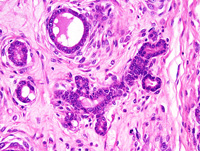
|
|
Image ID:1526 |
|
Source of Image:Ward JM |
|
Pathologist:Mikaelian I |
|
Method / Stain:H&E |
|
|
Image Caption:Mammary gland: the mammary gland is effaced by a moderately cellular, encapsulated, well-delineated, expensile, multilobular neoplastic mass. This neoplasm is composed of branching ducts supported by a large amount of cellular stroma.
|
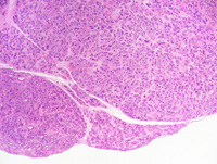
|
|
Image ID:1521 |
|
Source of Image:Ward JM |
|
Pathologist:Mikaelian I |
|
Method / Stain:H&E |
|
|
Image Caption:Mammary gland: neoplastic ducts are lined by a one cell-thick cuboidal epithelium. Neoplastic ducts are often distended by a proteinaceous fluid. Neoplastic cells have ill-defined cell borders and a moderate amount of a strongly amphophilic cytoplasm that often contains lipid vacuoles. The nucleus is central, round, medium-sized and normochromatic. Anisokaryosis and anisocytosis are mild. A few mitoses (arrowhead) are detected. Myoepithelial differentiation (arrows) is present. The stroma is abundant, contains numerous myofibroblasts, a small number of inflammatory cells, and loosely arranged wavy and thin collagen fibers.
|
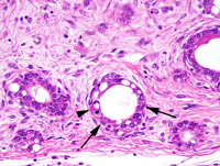
|
|
Image ID:1525 |
|
Source of Image:Ward JM |
|
Pathologist:Mikaelian I |
|
Method / Stain:H&E |
|
|
Image Caption:Mammary gland: the mammary gland is effaced by a moderately cellular, encapsulated, well-delineated, expensile, multilobular neoplastic mass. This neoplasm is composed of branching ducts supported by a large amount of cellular stroma. Neoplastic ducts are lined by a one cell-thick cuboidal epithelium. Neoplastic ducts are often distended by a proteinaceous fluid. Neoplastic cells have ill-defined cell borders and a moderate amount of a strongly amphophilic cytoplasm that often contains lipid vacuoles.
|
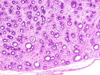
|
|
Image ID:1523 |
|
Source of Image:Ward JM |
|
Pathologist:Mikaelian I |
|
Method / Stain:H&E |
|
|
Image Caption:Fibroadenoma: this photomicrograph illustrates a portion of the neoplasm where myoepithelial differentiation (arrowheads) is prominent.
|
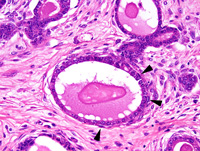
|
|
Image ID:1527 |
|
Source of Image:Ward JM |
|
Pathologist:Mikaelian I |
|
Method / Stain:H&E |
|
|
Image Caption:Mammary gland: the mammary gland is effaced by a moderately cellular, encapsulated, well-delineated, expensile, multilobular neoplastic mass. This neoplasm is composed of branching ducts supported by a large amount of cellular stroma. Neoplastic ducts are lined by a one cell-thick cuboidal epithelium.
|
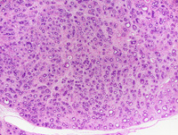
|
|
Image ID:1522 |
|
Source of Image:Ward JM |
|
Pathologist:Mikaelian I |
|
Method / Stain:H&E |
|
|
|
| MTB ID |
Tumor Name |
Organ(s) Affected |
Treatment Type |
Agents |
Strain Name |
Strain Sex |
Reproductive Status |
Tumor Frequency |
Age at Necropsy |
Description |
Reference |
| MTB:29188 |
Mammary gland hyperplasia - alveolar |
Mammary gland |
None (spontaneous) |
|
|
Female |
reproductive status not specified |
observed |
unknown |
Hyperlastic alveolar nodule, mammary gland |
J:94320 |
|
Image Caption:Mammary gland: a mammary lobule is very prominent. There is marked ectasia of some but not all acini. All acini and intralobular ducts are lined by a cuboidal epithelium. There is mild interstitial lymphocytic and plasmacytic inflammation. A few pigment-ladden macrophages are scattered in the interstitium.
|
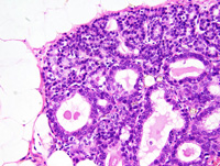
|
|
Image ID:1530 |
|
Source of Image:Ward JM |
|
Pathologist:Mikaelian I |
|
Method / Stain:H&E |
|
|
Image Caption:Mammary gland: mammary lobules are prominent and, although they do not form discrete masses, they contain a proteinaceous fluid and are composed of cuboidal cells with prominent lipid vacuoles. There are least two possible interpretations to this photomicrograph: these alveoli may represent early stages of hyperplastic alveolar nodules, or they may indicate pregnancy.
|
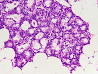
|
|
Image ID:1533 |
|
Source of Image:Ward JM |
|
Pathologist:Mikaelian I |
|
Method / Stain:H&E |
|
|
Image Caption:Mammary gland: a mammary lobule is very prominent. There is marked ectasia of some but not all acini. All acini and intralobular ducts are lined by a cuboidal epithelium. There is mild interstitial lymphocytic and plasmacytic inflammation. Interlobular ducts located in the nearby panniculus are normal.
|
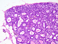
|
|
Image ID:1529 |
|
Source of Image:Ward JM |
|
Pathologist:Mikaelian I |
|
Method / Stain:H&E |
|
|
Image Caption:Mammary gland: a large, encapsulated, well-delineated expensile nodules is composed of closely packed mammary glands with prominent secretion of lipids and proteins. The epithelium lining of these glands do not show atypia. There is a mild to moderate interstitial infiltration by lymphocytes, plasma cells, and pigment-ladden macrophages.
|
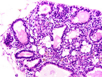
|
|
Image ID:1536 |
|
Source of Image:Ward JM |
|
Pathologist:Mikaelian I |
|
Method / Stain:H&E |
|
|
Image Caption:Mammary gland: a large, encapsulated, well-delineated expensile nodules is composed of closely packed mammary glands with prominent secretion of lipids and proteins.
|
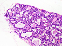
|
|
Image ID:1535 |
|
Source of Image:Ward JM |
|
Pathologist:Mikaelian I |
|
Method / Stain:H&E |
|
|
Image Caption:Mammary gland: mammary lobules are prominent and, although they do not form discrete masses, they contain a proteinaceous fluid and are composed of cuboidal cells with prominent lipid vacuoles. There are least two possible interpretations to this photomicrograph: these alveoli may represent early stages of hyperplastic alveolar nodules, or they may indicate pregnancy.
|
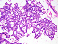
|
|
Image ID:1532 |
|
Source of Image:Ward JM |
|
Pathologist:Mikaelian I |
|
Method / Stain:H&E |
|
|
Image Caption:Mammary gland: a large, encapsulated, well-delineated expensile nodules is composed of closely packed mammary glands with prominent secretion of lipids and proteins.
|
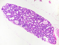
|
|
Image ID:1534 |
|
Source of Image:Ward JM |
|
Pathologist:Mikaelian I |
|
Method / Stain:H&E |
|
|
Image Caption:Mammary gland: mammary lobules are prominent and, although they do not form discrete masses, they contain a proteinaceous fluid and are composed of cuboidal cells with prominent lipid vacuoles. There are least two possible interpretations to this photomicrograph: these alveoli may represent early stages of hyperplastic alveolar nodules, or they may indicate pregnancy.
|
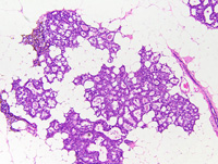
|
|
Image ID:1531 |
|
Source of Image:Ward JM |
|
Pathologist:Mikaelian I |
|
Method / Stain:H&E |
|
|
Image Caption:Mammary gland: a mammary lobule is very prominent. There is marked ectasia of some but not all acini. All acini and intralobular ducts are lined by a cuboidal epithelium. There is mild interstitial lymphocytic and plasmacytic inflammation. Interlobular ducts located in the nearby panniculus are normal.
|
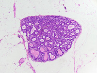
|
|
Image ID:1528 |
|
Source of Image:Ward JM |
|
Pathologist:Mikaelian I |
|
Method / Stain:H&E |
|
|
|
| MTB ID |
Tumor Name |
Organ(s) Affected |
Treatment Type |
Agents |
Strain Name |
Strain Sex |
Reproductive Status |
Tumor Frequency |
Age at Necropsy |
Description |
Reference |
| MTB:29189 |
Mammary gland adenoma - mixed |
Mammary gland |
None (spontaneous) |
|
|
Female |
reproductive status not specified |
observed |
unknown |
Adenoma, tubular and glandular, mammary gland |
J:94320 |
|
Image Caption:Mammary adenoma: neoplastic cells form glands with morphologic features consistent with the gestational mammary gland.
|
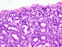
|
|
Image ID:1541 |
|
Source of Image:Ward JM |
|
Pathologist:Mikaelian I |
|
Method / Stain:H&E |
|
|
Image Caption:Mammary gland: the mammary gland is focally effaced by a small, well-delineated, irregularly-shaped, densely cellular mass. This mass is composed of closely-packed contoured and anastomosed one cell-thick tubules and glands supported by a scant amount of fibrovascular stroma.
|
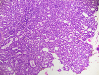
|
|
Image ID:1538 |
|
Source of Image:Ward JM |
|
Pathologist:Mikaelian I |
|
Method / Stain:H&E |
|
|
Image Caption:Mammary gland: the neoplasm is composed of closely packed contoured and anastomosed one cell-thick tubules and glands supported by a scant amount of fibrovascular stroma. Neoplastic cells are cuboidal, with indistinct cell borders and a moderate amount of amphophilic cytoplasm that often contains lipid vacuoles. The nucleus is central, round, and medium-sized. Anisokaryosis and anisocytosis are mild. Tubules (*) have a "crowded" appearance and resemble intralobular ducts. The glands have morphologic features consistent with the epithelium of mammary acini. There is a mild interstitial infiltration by lymphocytes, plasma cells and macrophages.
|
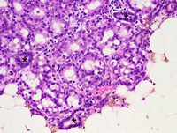
|
|
Image ID:1540 |
|
Source of Image:Ward JM |
|
Pathologist:Mikaelian I |
|
Method / Stain:H&E |
|
|
Image Caption:Mammary gland: the mammary gland is focally effaced by a small, well-delineated, irregularly-shaped, densely cellular mass. This mass is composed of closely-packed contoured and anastomosed one cell-thick tubules and glands supported by a scant amount of fibrovascular stroma.
|
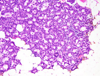
|
|
Image ID:1539 |
|
Source of Image:Ward JM |
|
Pathologist:Mikaelian I |
|
Method / Stain:H&E |
|
|
Image Caption:Mammary gland: the mammary gland is focally effaced by a small, well-delineated, irregularly-shaped, densely cellular mass. This mass is composed of closely-packed contoured and anastomosed tubules and glands supported by a scant amount of fibrovascular stroma.
|
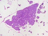
|
|
Image ID:1537 |
|
Source of Image:Ward JM |
|
Pathologist:Mikaelian I |
|
Method / Stain:H&E |
|
|
|
| MTB ID |
Tumor Name |
Organ(s) Affected |
Treatment Type |
Agents |
Strain Name |
Strain Sex |
Reproductive Status |
Tumor Frequency |
Age at Necropsy |
Description |
Reference |
| MTB:29190 |
Mammary gland adenocarcinoma - solid |
Mammary gland |
None (spontaneous) |
|
|
Female |
reproductive status not specified |
observed |
unknown |
Adenocarcinoma, solid, mammary gland |
J:94320 |
|
Image Caption:Mammary gland: this photomicrograph represents invasion of a mammary adenocarcinoma into an interlobular mammary duct. In the duct, the neoplastic epithelium forms a 2-25 cells-thick layer of cells that occasionally form inconspicuous lumens.
|
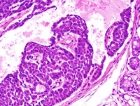
|
|
Image ID:1548 |
|
Source of Image:Ward JM |
|
Pathologist:Mikaelian I |
|
Method / Stain:H&E |
|
|
Image Caption:Mammary gland: the mammary gland is effaced by a nodular, unencapsulated, expensile, densely cellular neoplasm. This neoplasm is composed of solid areas with a few minute areas of liquefactive necrosis and cavitation.
|
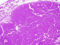
|
|
Image ID:1542 |
|
Source of Image:Ward JM |
|
Pathologist:Mikaelian I |
|
Method / Stain:H&E |
|
|
Image Caption:Mammary gland: this photomicrograph represents invasion of a mammary adenocarcinoma into an interlobular mammary duct. In the duct, the neoplastic epithelium forms a 2-25 cells-thick layer of cells that occasionally form inconspicuous lumens.
|
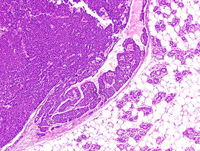
|
|
Image ID:1546 |
|
Source of Image:Ward JM |
|
Pathologist:Mikaelian I |
|
Method / Stain:H&E |
|
|
Image Caption:Mammary gland: the mammary gland is effaced by a nodular, unencapsulated, expensile, densely cellular neoplasm. This neoplasm is composed of solid areas of epithelial neoplastic cells supported by a scant amount of fibrovascular stroma.
|
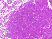
|
|
Image ID:1543 |
|
Source of Image:Ward JM |
|
Pathologist:Mikaelian I |
|
Method / Stain:H&E |
|
|
Image Caption:Mammary gland: the mammary gland is effaced by a nodular, unencapsulated, expensile, densely cellular neoplasm. This neoplasm is composed of solid areas of epithelial neoplastic cells supported by a scant amount of fibrovascular stroma. Neoplastic cells are medium-sized, polygonal, often with distinct cell borders, and a moderate amount of strongly amphophilic cytoplasm. The nucleus is central, medium-sized and slightly hyperchromatic. Anisokaryosis and anisocytosis are mild.
|
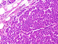
|
|
Image ID:1545 |
|
Source of Image:Ward JM |
|
Pathologist:Mikaelian I |
|
Method / Stain:H&E |
|
|
Image Caption:Mammary gland: the mammary gland is effaced by a nodular, unencapsulated, expensile, densely cellular neoplasm. This neoplasm is composed of solid areas of epithelial neoplastic cells supported by a scant amount of fibrovascular stroma.
|
|
|
Image ID:1544 |
|
Source of Image:Ward JM |
|
Pathologist:Mikaelian I |
|
Method / Stain:H&E |
|
|
Image Caption:Mammary gland: this photomicrograph represents invasion of a mammary adenocarcinoma into an interlobular mammary duct. In the duct, the neoplastic epithelium forms a 2-25 cells-thick layer of cells that occasionally form inconspicuous lumens.
|
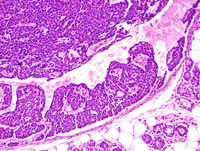
|
|
Image ID:1547 |
|
Source of Image:Ward JM |
|
Pathologist:Mikaelian I |
|
Method / Stain:H&E |
|
|
|
| MTB ID |
Tumor Name |
Organ(s) Affected |
Treatment Type |
Agents |
Strain Name |
Strain Sex |
Reproductive Status |
Tumor Frequency |
Age at Necropsy |
Description |
Reference |
| MTB:29191 |
Mammary gland carcinoma in situ |
Mammary gland |
None (spontaneous) |
|
|
Female |
reproductive status not specified |
observed |
unknown |
Carcinoma in situ, mammary gland |
J:94320 |
|
Image Caption:Mammary gland: there was, at the periphery of the solid mammary carcinoma, a dilated interlobular duct that was lined by a 5-10 cells-thick epithelium that often formed small secondary lumens. Myoepithelial differentiation was not identified in this duct. This lesion may represent invasion of an interlobular duct by the solid carcinoma, or may indicate in situ carcinoma in an interlobular duct adjancent to a mammary carcinoma. The formation of a relatively large number of secondary lumens, a feature that was not detected in the solid carcinoma, supports a diagnosis of in situ carcinoma.
|
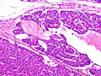
|
|
Image ID:1550 |
|
Source of Image:Ward JM |
|
Pathologist:Mikaelian I |
|
Method / Stain:H&E |
|
|
Image Caption:Mammary gland: there was, at the periphery of the solid mammary carcinoma, a dilated interlobular duct that was lined by a 5-10 cells-thick epithelium that often formed small secondary lumens. Myoepithelial differentiation was not identified in this duct. This lesion may represent invasion of an interlobular duct by the solid carcinoma, or may indicate in situ carcinoma in an interlobular duct adjancent to a mammary carcinoma. The formation of a relatively large number of secondary lumens, a feature that was not detected in the solid carcinoma, supports a diagnosis of in situ carcinoma.
|
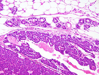
|
|
Image ID:1549 |
|
Source of Image:Ward JM |
|
Pathologist:Mikaelian I |
|
Method / Stain:H&E |
|
|
|
| MTB ID |
Tumor Name |
Organ(s) Affected |
Treatment Type |
Agents |
Strain Name |
Strain Sex |
Reproductive Status |
Tumor Frequency |
Age at Necropsy |
Description |
Reference |
| MTB:29192 |
Mammary gland adenocarcinoma |
Mammary gland |
None (spontaneous) |
|
|
Female |
reproductive status not specified |
observed |
unknown |
Adenocarcinoma, glandular, secretory, with prominent interstitial fibrosis, mammary gland |
J:94320 |
|
Image Caption:Mammary gland: the mammary gland is effaced by a large, nodular, encapsulated, and moderately cellular neoplasm. The neoplasm is composed of interconnected branching ducts embedded is a large amount of a fibrovascular stroma characterized with interstitial edema, deposition of numerous and thick wavy collagen fibers, and the presence of numerous spindloid cells consistent with myofibroblasts.
|
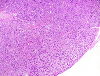
|
|
Image ID:1552 |
|
Source of Image:Ward JM |
|
Pathologist:Mikaelian I |
|
Method / Stain:H&E |
|
|
Image Caption:Glandular and secretory adenocarcinoma: myoepithelial differentiation (arrowheads) is often prominent.
|
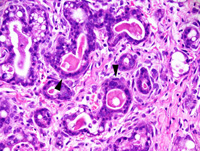
|
|
Image ID:1557 |
|
Source of Image:Ward JM |
|
Pathologist:Mikaelian I |
|
Method / Stain:H&E |
|
|
Image Caption:Mammary gland: the mammary gland is effaced by a large, nodular, encapsulated, and moderately cellular neoplasm. The neoplasm is composed of interconnected branching ducts embedded is a large amount of a fibrovascular stroma characterized with interstitial edema, deposition of numerous and thick wavy collagen fibers, and the presence of numerous spindloid cells consistent with myofibroblasts. Neoplastic glands are lined by a two cells-thick epithelium: cells of the basal layer resemble myoepithelial cells of the mammary gland while cells of the luminal layer resemble luminal cells of the mammary gland. Neoplastic luminal cells often contain lipid vacuoles of various sizes.
|
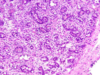
|
|
Image ID:1554 |
|
Source of Image:Ward JM |
|
Pathologist:Mikaelian I |
|
Method / Stain:H&E |
|
|
Image Caption:Mammary gland: the mammary gland is effaced by a large, nodular, encapsulated, moderately cellular neoplasm (only a portion of the neoplasm is present on the slide).
|
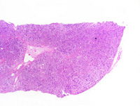
|
|
Image ID:1551 |
|
Source of Image:Ward JM |
|
Pathologist:Mikaelian I |
|
Method / Stain:H&E |
|
|
Image Caption:Glandular and secretory adenocarcinoma: this photomicrograph illustrates the ratio between the stroma (approximately 60% of the surface of the section) and the neoplastic epithelial cells (approximately 40% if the surface of the section). Cells in the stroma are oat-shaped to spindloid and resemble myofibroblasts.
|
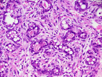
|
|
Image ID:1555 |
|
Source of Image:Ward JM |
|
Pathologist:Mikaelian I |
|
Method / Stain:H&E |
|
|
Image Caption:Mammary gland: the mammary gland is effaced by a large, nodular, encapsulated, and moderately cellular neoplasm. The neoplasm is composed of interconnected branching ducts embedded is a large amount of a fibrovascular stroma characterized with interstitial edema, deposition of numerous and thick wavy collagen fibers, and the presence of numerous spindloid cells consistent with myofibroblasts. Neoplastic glands are lined by a two cells-thick epithelium: cells of the basal layer resemble myoepithelial cells of the mammary gland while cells of the luminal layer resemble luminal cells of the mammary gland. Neoplastic luminal cells often contain lipid vacuoles of various sizes.
|
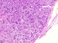
|
|
Image ID:1553 |
|
Source of Image:Ward JM |
|
Pathologist:Mikaelian I |
|
Method / Stain:H&E |
|
|
Image Caption:Glandular and secretory adenocarcinoma: mitoses (arrowheads) are detected in myofibroblasts and neoplastic epithelial cells.
|

|
|
Image ID:1556 |
|
Source of Image:Ward JM |
|
Pathologist:Mikaelian I |
|
Method / Stain:H&E |
|
|
|
| MTB ID |
Tumor Name |
Organ(s) Affected |
Treatment Type |
Agents |
Strain Name |
Strain Sex |
Reproductive Status |
Tumor Frequency |
Age at Necropsy |
Description |
Reference |
| MTB:29193 |
Mammary gland hyperplasia - alveolar |
Mammary gland |
None (spontaneous) |
|
|
Female |
reproductive status not specified |
observed |
unknown |
Hyperplastic alveolar nodule, mammary gland |
J:94320 |
|
Image Caption:Mammary gland: the mammary gland is expanded by small, well-delineated, unencapsulated, and densely cellular nodule. This nodule is composed of closely packed glands and intralobular ducts which show mild to moderate ectasia and contain an amorphous proteinaceous fluid. Alveoli and ducts are lined by a cuboidal one cell-thick epithelium supported by a scant amount of connective tissue. Cells lining the glands have indistinct cell borders and a moderate amount of acidophilic cytoplasm that often contains lipid vacuoles and proteinaceous droplets. There is no evidence of atypia and cells do not pile-up.
|
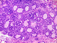
|
|
Image ID:1560 |
|
Source of Image:Ward JM |
|
Pathologist:Mikaelian I |
|
Method / Stain:H&E |
|
|
Image Caption:Mammary gland: the mammary gland is moderately expanded by small nodules of various sizes. Most of these nodules are well-delineated, unencapsulated, and densely cellular. Some are encapsulated and slightly compress the adjacent tissues. There is mild to moderate interstitial fibrosis and moderate ductal and alveolar ectasia in the remaining portions of the mammary gland.
|
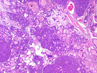
|
|
Image ID:1558 |
|
Source of Image:Ward JM |
|
Pathologist:Mikaelian I |
|
Method / Stain:H&E |
|
|
Image Caption:Mammary gland: the mammary gland is expanded by small, well-delineated, unencapsulated, and densely cellular nodule. This nodule is composed of closely packed glands and intralobular ducts which show mild to moderate ectasia and contain an amorphous proteinaceous fluid. Alveoli and ducts are lined by a cuboidal epithelium.
|
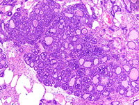
|
|
Image ID:1559 |
|
Source of Image:Ward JM |
|
Pathologist:Mikaelian I |
|
Method / Stain:H&E |
|
|
Image Caption:Mammary gland: the mammary gland is expanded by small, well-delineated, unencapsulated, and densely cellular nodule. This nodule is composed of closely packed glands and intralobular ducts which show mild to moderate ectasia and contain an amorphous proteinaceous fluid. Alveoli and ducts are lined by a cuboidal one cell-thick epithelium supported by a scant amount of connective tissue. Cells lining the glands have indistinct cell borders and a moderate amount of acidophilic cytoplasm that often contains lipid vacuoles and proteinaceous droplets. There is no evidence of atypia and cells do not pile-up. A moderate number of epithelial cells undergo apoptosis.
|
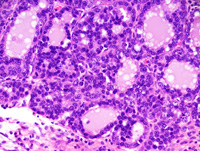
|
|
Image ID:1561 |
|
Source of Image:Ward JM |
|
Pathologist:Mikaelian I |
|
Method / Stain:H&E |
|
|
|
| MTB ID |
Tumor Name |
Organ(s) Affected |
Treatment Type |
Agents |
Strain Name |
Strain Sex |
Reproductive Status |
Tumor Frequency |
Age at Necropsy |
Description |
Reference |
| MTB:29194 |
Mammary gland cyst - macrocyst |
Mammary gland |
None (spontaneous) |
|
|
Female |
reproductive status not specified |
observed |
unknown |
Macrocyst, mammary gland |
J:94320 |
|
Image Caption:Mammary gland: the mammary gland is effaced by a large, multilocular mass. The cysts that make up most of this mass contain an inspissated proteinaceous material and are lined by a 2 cells-thick cuboidal epithelium that, in a few areas, forms secondary glands.
|
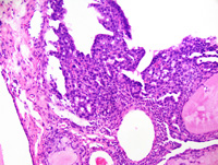
|
|
Image ID:1564 |
|
Source of Image:Ward JM |
|
Pathologist:Mikaelian I |
|
Method / Stain:H&E |
|
|
Image Caption:Macrocyst: the cystic spaces are filled with an amorphous acidophilic material. Numerous small glands radiate from the smaller cysts.
|
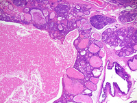
|
|
Image ID:1567 |
|
Source of Image:Ward JM |
|
Pathologist:Mikaelian I |
|
Method / Stain:H&E |
|
|
Image Caption:Macrocysts: the epithelium surrounding the major cyst is once cell-thick and is composed of attenuated cells. The epithelium lining the glands is cuboidal. There is no evidence of atypia. The proteinaceous material in the lumen of the glands often is inspissated. There is no evidence of atypia.
|
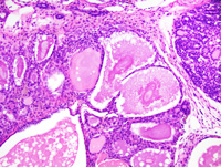
|
|
Image ID:1569 |
|
Source of Image:Ward JM |
|
Pathologist:Mikaelian I |
|
Method / Stain:H&E |
|
|
Image Caption:Mammary gland: the mammary gland is effaced by a large, multilocular mass. The cysts that make up most of this mass contain an inspissated proteinaceous material and are lined by a 2 cells-thick cuboidal epithelium.
|
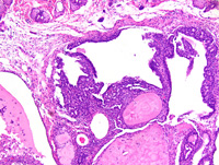
|
|
Image ID:1563 |
|
Source of Image:Ward JM |
|
Pathologist:Mikaelian I |
|
Method / Stain:H&E |
|
|
Image Caption:Macrocyst: this low power magnification illustrates the cystic appearance of most macrocyts, and also the connection between the larger cyst and smaller cysts that radiate from the largest cyst. Small adenomas are present at the periphery of the macrocyst.
|
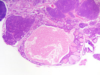
|
|
Image ID:1566 |
|
Source of Image:Ward JM |
|
Pathologist:Mikaelian I |
|
Method / Stain:H&E |
|
|
Image Caption:Mammary gland: the mammary gland is effaced by a large, multilocular mass.
|
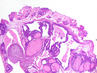
|
|
Image ID:1562 |
|
Source of Image:Ward JM |
|
Pathologist:Mikaelian I |
|
Method / Stain:H&E |
|
|
Image Caption:Macrocyst: as exemplified on this photomicrograph, mitotic activity (arrowheads) is multifocally prominent. The cells lining the ducts and the cysts are cuboidal to low columnar, have indistinct cell borders and a moderate amount of strongly amphophilic cytoplasm that often contains lipid vacuoles. The nucleus tends to be basal, is medium-sized, round to oval and normochromatic. Anisokaryosis and anisocytosis are mild. There is a moderate interstitial granulocytic (eosinophilic?) infiltration.
|
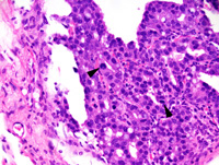
|
|
Image ID:1565 |
|
Source of Image:Ward JM |
|
Pathologist:Mikaelian I |
|
Method / Stain:H&E |
|
|
Image Caption:Macrocyst: the cystic spaces are filled with an amorphous acidophilic material. Numerous small glands radiate from the smaller cysts. The cysts and the glands are lined by a 1-2 cells-thick epithelium supported by a small to moderate amount of fibrovascular stroma.
|
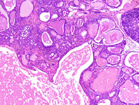
|
|
Image ID:1568 |
|
Source of Image:Ward JM |
|
Pathologist:Mikaelian I |
|
Method / Stain:H&E |
|
|
|
| MTB ID |
Tumor Name |
Organ(s) Affected |
Treatment Type |
Agents |
Strain Name |
Strain Sex |
Reproductive Status |
Tumor Frequency |
Age at Necropsy |
Description |
Reference |
| MTB:29195 |
Mammary gland adenoma |
Mammary gland |
None (spontaneous) |
|
|
Female |
reproductive status not specified |
observed |
unknown |
Adenoma, glandular, secretory, mammary gland |
J:94320 |
|
Image Caption:Mammary gland, small glandular adenoma: the mammary gland is effaced by a large, nodular, encapsulated, densely cellular mass. This mass is composed of small glands lined by a one cell-thick cuboidal epithelium supported by a scant amount of fibrovascular stroma. Some glands are ectatic and are filled with an amorphous proteinaceous fluid.
|
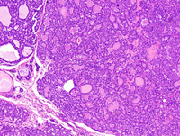
|
|
Image ID:1571 |
|
Source of Image:Ward JM |
|
Pathologist:Mikaelian I |
|
Method / Stain:H&E |
|
|
Image Caption:Glandular adenoma, small glandular adenoma: cells lining neoplastic glands are cuboidal, with indistinct cell borders and a moderate amount of strongly amphophilic cytoplasm that often contains small lipid vacuoles.
|
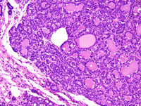
|
|
Image ID:1572 |
|
Source of Image:Ward JM |
|
Pathologist:Mikaelian I |
|
Method / Stain:H&E |
|
|
Image Caption:Mammary glandular adenoma (large): the mammary gland is effaced by a large, nodular, encapsulated, densely cellular mass. The neoplasm is composed of closely-packed glands lined by a one cell-thick cuboidal epithelium supported by a scant amount of fibrovascular stroma.
|
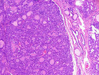
|
|
Image ID:1577 |
|
Source of Image:Ward JM |
|
Pathologist:Mikaelian I |
|
Method / Stain:H&E |
|
|
Image Caption:Mammary glandular adenoma (large): the mammary gland is effaced by a large, nodular, encapsulated, densely cellular mass. The neoplasm is composed of closely-packed glands lined by a one cell-thick cuboidal epithelium supported by a scant amount of fibrovascular stroma.
|
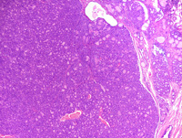
|
|
Image ID:1576 |
|
Source of Image:Ward JM |
|
Pathologist:Mikaelian I |
|
Method / Stain:H&E |
|
|
Image Caption:Mammary glandular adenoma (large): the mammary gland is effaced by a large, nodular, encapsulated, densely cellular mass. Similar smaller masses and hyperplastic alveolar nodules distort the remaining portions of the mammary gland.
|
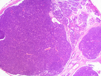
|
|
Image ID:1575 |
|
Source of Image:Ward JM |
|
Pathologist:Mikaelian I |
|
Method / Stain:H&E |
|
|
Image Caption:Mammary glandular adenoma (large): mitoses (arrowheads) are numerous in some fields.
|
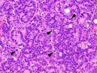
|
|
Image ID:1579 |
|
Source of Image:Ward JM |
|
Pathologist:Mikaelian I |
|
Method / Stain:H&E |
|
|
Image Caption:Mammary glandular adenoma (large): the mammary gland is effaced by a large, nodular, encapsulated, densely cellular mass. Similar smaller masses and hyperplastic alveolar nodules distort the remaining portions of the mammary gland.
|
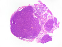
|
|
Image ID:1574 |
|
Source of Image:Ward JM |
|
Pathologist:Mikaelian I |
|
Method / Stain:H&E |
|
|
Image Caption:Mammary glandular adenoma (large): this photomicrograph illustrates the glandular pattern of this adenoma.
|
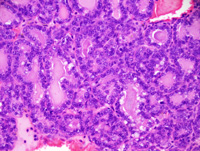
|
|
Image ID:1580 |
|
Source of Image:Ward JM |
|
Pathologist:Mikaelian I |
|
Method / Stain:H&E |
|
|
Image Caption:Glandular adenoma (small): cells lining neoplastic glands are cuboidal, with indistinct cell borders and a moderate amount of strongly amphophilic cytoplasm that often contains proteinaceous droplets (arrow) or small lipid vacuoles. The nucleus is central, round, medium-sized and normochromatic. Anisocytosis and anisokaryosis are mild. A moderate number of mitoses (arrowheads) is present.
|
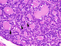
|
|
Image ID:1573 |
|
Source of Image:Ward JM |
|
Pathologist:Mikaelian I |
|
Method / Stain:H&E |
|
|
Image Caption:Mammary glandular adenoma (large): the mammary gland is effaced by a large, nodular, encapsulated, densely cellular mass. The neoplasm is composed of closely-packed glands lined by a one cell-thick cuboidal epithelium supported by a scant amount of fibrovascular stroma. Most glands are filled with an occasionally inspissated proteinaceous fluid. Neoplastic cells are cuboidal, with indistinct cell borders and a moderate amount of strongly amphophilic cytoplasm that often contains a few small lipid droplets. The nucleus generally is basal, round to oval, medium-sized, and normochromatic. Anisokaryosis and anisocytosis are mild.
|
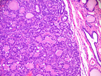
|
|
Image ID:1578 |
|
Source of Image:Ward JM |
|
Pathologist:Mikaelian I |
|
Method / Stain:H&E |
|
|
Image Caption:Mammary gland, small glandular adenoma: the mammary gland is effaced by a large, nodular, encapsulated, densely cellular mass. This mass is composed of small glands supported by a scant amount of fibrovascular stroma.
|
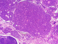
|
|
Image ID:1570 |
|
Source of Image:Ward JM |
|
Pathologist:Mikaelian I |
|
Method / Stain:H&E |
|
|
|
| MTB ID |
Tumor Name |
Organ(s) Affected |
Treatment Type |
Agents |
Strain Name |
Strain Sex |
Reproductive Status |
Tumor Frequency |
Age at Necropsy |
Description |
Reference |
| MTB:29196 |
Mammary gland hyperplasia - alveolar |
Mammary gland |
None (spontaneous) |
|
|
Female |
reproductive status not specified |
observed |
unknown |
Hyperplastic alveolar nodule, mammary gland |
J:94320 |
|
Image Caption:Hyperplastic alveolar nodule (early stage): the lobular architecture of the mammary gland is enhanced. Alveoli and intra-lobular ducts are lined by a one cell-thick cuboidal epithelium and are often filled with a moderate amount of proteinaceous material. There is mild interstitial fibrosis and lymphoplasmacytic inflammation. There is no evidence of atypia.
|
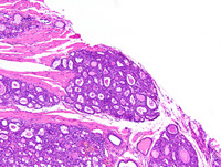
|
|
Image ID:1581 |
|
Source of Image:Ward JM |
|
Pathologist:Mikaelian I |
|
Method / Stain:H&E |
|
|
Image Caption:Hyperplastic alveolar nodule (early stage): the lobular architecture of the mammary gland is enhanced. Alveoli and intra-lobular ducts are lined by a one cell-thick cuboidal epithelium and are often filled with a moderate amount of proteinaceous material. There is mild interstitial fibrosis and lymphoplasmacytic inflammation. There is no evidence of atypia.
|
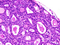
|
|
Image ID:1583 |
|
Source of Image:Ward JM |
|
Pathologist:Mikaelian I |
|
Method / Stain:H&E |
|
|
Image Caption:Hyperplastic alveolar nodule (early stage): the lobular architecture of the mammary gland is enhanced. Alveoli and intra-lobular ducts are lined by a one cell-thick cuboidal epithelium and are often filled with a moderate amount of proteinaceous material. There is mild interstitial fibrosis and lymphoplasmacytic inflammation. There is no evidence of atypia.
|
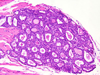
|
|
Image ID:1582 |
|
Source of Image:Ward JM |
|
Pathologist:Mikaelian I |
|
Method / Stain:H&E |
|
|
|
| MTB ID |
Tumor Name |
Organ(s) Affected |
Treatment Type |
Agents |
Strain Name |
Strain Sex |
Reproductive Status |
Tumor Frequency |
Age at Necropsy |
Description |
Reference |
| MTB:29197 |
Mammary gland fibroadenoma |
Mammary gland |
None (spontaneous) |
|
|
Female |
reproductive status not specified |
observed |
300 days |
Fibroadenoma, secretory, with atypia, mammary gland |
J:94320 |
|
Image Caption:Mammary gland: the mammary gland is effaced by a large, nodular, encapsulated, moderately cellular, expensile neoplasm. There is moderate diffuse lobular hyperplasia and secretory activity in the adjacent mammary parenchyma. The neoplasm is composed of contoured glands lined by a two cells-thick epithelium supported by a large amount of fibrovascular stroma. Many glands are ectatic and are filled with an amorphous proteinaceous fluid.
|
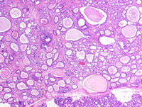
|
|
Image ID:1586 |
|
Source of Image:Ward JM |
|
Pathologist:Mikaelian I |
|
Method / Stain:H&E |
|
|
Image Caption:Fibroadenoma with atypia: this photomicrograph illustrates a portion of the neoplasm composed of small glands with atypia. Neoplastic cells are large and most of them contain a large lipid vacuole. The nuclei are large, too, and there is moderate anisokaryosis. There is a moderate granulocytic (eosinophilic?; predominantly), lymphocytic and plasmacytic inflammation of the stroma. A few neoplastic cells are multinucleated (arrowhead).
|
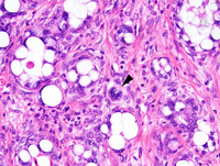
|
|
Image ID:1593 |
|
Source of Image:Ward JM |
|
Pathologist:Mikaelian I |
|
Method / Stain:H&E |
|
|
Image Caption:Mammary gland: the mammary gland is effaced by a large, nodular, encapsulated, moderately cellular, expensile neoplasm. There is moderate diffuse lobular hyperplasia and secretory activity in the adjacent mammary parenchyma. The neoplasm is composed of contoured glands lined by a two cells-thick epithelium supported by a large amount of fibrovascular stroma. Many glands are ectatic and are filled with an amorphous proteinaceous fluid.
|
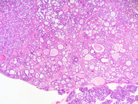
|
|
Image ID:1585 |
|
Source of Image:Ward JM |
|
Pathologist:Mikaelian I |
|
Method / Stain:H&E |
|
|
Image Caption:Fibroadenoma with atypia: this photomicrograph illustrates the bi-stratified feature of the neoplasm. Arrowheads point at cells with myoepithelial differentiation.
|
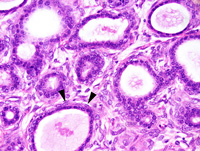
|
|
Image ID:1597 |
|
Source of Image:Ward JM |
|
Pathologist:Mikaelian I |
|
Method / Stain:H&E |
|
|
Image Caption:Fibroadenoma with atypia: this photomicrograph illustrates a portion of the neoplasm composed of small glands with atypia. Neoplastic cells are large and most of them contain a large lipid vacuole. The nuclei are large, too, and there is moderate anisokaryosis. There is a moderate granulocytic (eosinophilic?; predominantly), lymphocytic and plasmacytic inflammation of the stroma. Small clusters of cells seem to be engaged in the early stages of cornificatin (arrowhead).
|
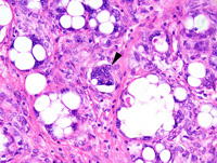
|
|
Image ID:1594 |
|
Source of Image:Ward JM |
|
Pathologist:Mikaelian I |
|
Method / Stain:H&E |
|
|
Image Caption:Fibroadenoma with atypia: this photomicrograph illustrates portion of the neoplasm with a fibrous stroma (to the left of the photomicrograph) and other portions of the neoplasm with a cellular stroma.
|
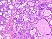
|
|
Image ID:1595 |
|
Source of Image:Ward JM |
|
Pathologist:Mikaelian I |
|
Method / Stain:H&E |
|
|
Image Caption:Fibroadenoma with atypia: this photomicrograph illustrates the bi-stratified feature of the neoplasm. Cells with myoepithelial differentiation are difficult to differentiate from stromal cells, and are best identified with immunolabeling for myoepithelial cell markers (keratins 5, 14 and 17, alpha smooth muscle actin, p63, caldesmon, ...).
|
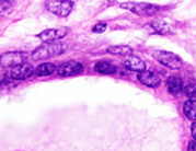
|
|
Image ID:1598 |
|
Source of Image:Ward JM |
|
Pathologist:Mikaelian I |
|
Method / Stain:H&E |
|
|
Image Caption:Mammary gland: the mammary gland is effaced by a large, nodular, encapsulated, moderately cellular, expensile neoplasm. There is moderate diffuse lobular hyperplasia and secretory activity in the adjacent mammary parenchyma. The neoplasm is composed of contoured glands lined by a two cells-thick epithelium supported by a large amount of fibrovascular stroma. Many glands are ectatic and are filled with an amorphous proteinaceous fluid oaccasionally admixed with a coarse granular yellow-brown pigment.
|

|
|
Image ID:1587 |
|
Source of Image:Ward JM |
|
Pathologist:Mikaelian I |
|
Method / Stain:H&E |
|
|
Image Caption:Fibroadenoma with atypia: this photomicrograph illustrates portion of the neoplasm with a fibrous srtoma (to the left of the photomicrograph) and other portions of the neoplasm with a cellular stroma. Mitoses are not rare, even in the most immature portions of the neoplasm.
|
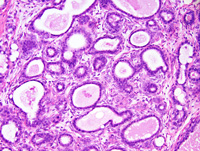
|
|
Image ID:1596 |
|
Source of Image:Ward JM |
|
Pathologist:Mikaelian I |
|
Method / Stain:H&E |
|
|
Image Caption:Mammary gland: the mammary gland is effaced by a large, nodular, encapsulated, moderately cellular, expensile neoplasm. There is moderate diffuse lobular hyperplasia and secretory activity in the adjacent mammary parenchyma.
|
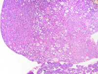
|
|
Image ID:1584 |
|
Source of Image:Ward JM |
|
Pathologist:Mikaelian I |
|
Method / Stain:H&E |
|
|
Image Caption:Fibroadenoma with atypia: the neoplasm is composed of dilated glands filled with a proteinaceous material and embedded in a large amount of dense fibrovascular stroma. There are two layers to the neoplastic epithelium: (1) a luminal layer composed of cuboidal cells that often contain lipid vacuoles and occasionally proteinaceous droplets; (2) an inconspicuous basal (=myoepithelial) layer. There is no evidence of atypia in this microscopic field.
|
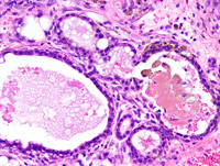
|
|
Image ID:1589 |
|
Source of Image:Ward JM |
|
Pathologist:Mikaelian I |
|
Method / Stain:H&E |
|
|
Image Caption:Fibroadenoma with atypia: this photomicrograph illustrates a portion of the neoplasm composed of small glands with atypia.
|
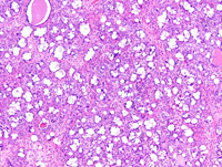
|
|
Image ID:1590 |
|
Source of Image:Ward JM |
|
Pathologist:Mikaelian I |
|
Method / Stain:H&E |
|
|
Image Caption:Mammary gland: the mammary gland is effaced by a large, nodular, encapsulated, moderately cellular, expensile neoplasm. There is moderate diffuse lobular hyperplasia and secretory activity in the adjacent mammary parenchyma. The neoplasm is composed of contoured glands lined by a two cells-thick epithelium supported by a large amount of fibrovascular stroma. Many glands are ectatic and are filled with an amorphous proteinaceous fluid oaccasionally admixed with a coarse granular yellow-brown pigment.
|
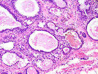
|
|
Image ID:1588 |
|
Source of Image:Ward JM |
|
Pathologist:Mikaelian I |
|
Method / Stain:H&E |
|
|
Image Caption:Fibroadenoma with atypia: this photomicrograph illustrates a portion of the neoplasm composed of small glands with atypia. Neoplastic cells are large and most of them contain a large lipid vacuole. The nuclei are large, too.
|
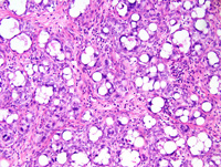
|
|
Image ID:1591 |
|
Source of Image:Ward JM |
|
Pathologist:Mikaelian I |
|
Method / Stain:H&E |
|
|
Image Caption:Fibroadenoma with atypia: this photomicrograph illustrates a portion of the neoplasm composed of small glands with atypia. Neoplastic cells are large and most of them contain a large lipid vacuole. The nuclei are large, too, and there is moderate anisokaryosis. There is a moderate granulocytic (eosinophilic?; predominantly), lymphocytic and plasmacytic inflammation of the stroma.
|
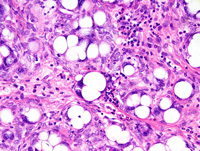
|
|
Image ID:1592 |
|
Source of Image:Ward JM |
|
Pathologist:Mikaelian I |
|
Method / Stain:H&E |
|
|
|
| MTB ID |
Tumor Name |
Organ(s) Affected |
Treatment Type |
Agents |
Strain Name |
Strain Sex |
Reproductive Status |
Tumor Frequency |
Age at Necropsy |
Description |
Reference |
| MTB:29198 |
Mammary gland adenocarcinoma - mixed |
Mammary gland |
None (spontaneous) |
|
|
Female |
reproductive status not specified |
observed |
120 days |
Adenocarcinoma, solid, comedo, mammary gland |
J:94320 |
|
Image Caption:Comedo mammary adenocarcinoma: neoplastic cells are medium-size, polygonal, with indistinct cell borders and a moderate amount of strongly amphophilic cytoplasm. The nucleus is medium-size, oval, hypochromatic, with a finely stippled chromatin and 1-3 large basophilic nucleoli. Numerous neoplastic cells undergo single cell necrosis. Anisokaryosis and anisocytosis are mild. Mitoses are numerous.
|
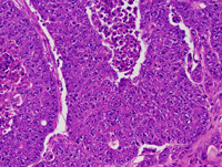
|
|
Image ID:1603 |
|
Source of Image:Ward JM |
|
Pathologist:Mikaelian I |
|
Method / Stain:H&E |
|
|
Image Caption:Mammary gland: the mammary gland is effaced by a nodular, invasive, densely cellular neoplasm. The neoplasm is composed of thick interconnected cords with prominent central liquefactive necrosis and supported by a small to moderate amount of fibrovascular stroma.
|
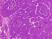
|
|
Image ID:1599 |
|
Source of Image:Ward JM |
|
Pathologist:Mikaelian I |
|
Method / Stain:H&E |
|
|
Image Caption:Comedo mammary adenocarcinoma: this photomicrograph illustrates a portion of the neoplasm where cells form 1-2 cells thick cords. It is often at the vicinity of these areas that epithelial to mesenchymal transition occurs.
|
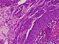
|
|
Image ID:1601 |
|
Source of Image:Ward JM |
|
Pathologist:Mikaelian I |
|
Method / Stain:H&E |
|
|
Image Caption:Mammary gland: the mammary gland is effaced by a nodular, invasive, densely cellular neoplasm. The neoplasm is composed of thick interconnected cords with prominent central liquefactive necrosis and supported by a small to moderate amount of fibrovascular stroma. Small secondary lumens are occasionally detected (arrowheads). Neoplastic cells are medium-sized, polygonal to spindloid, with indistinct cell borders and they have a moderate amount of strongly amphophilic cytoplasm.
|
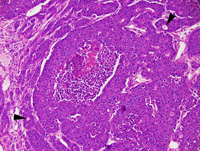
|
|
Image ID:1600 |
|
Source of Image:Ward JM |
|
Pathologist:Mikaelian I |
|
Method / Stain:H&E |
|
|
Image Caption:Comedo mammary adenocarcinoma: this photomicrograph illustrates areas of the neoplasm that is predominantly solid with large central areas of liquefactive necrosis. In addition, neoplastic cells form a few lumens.
|
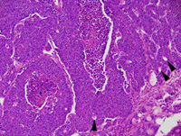
|
|
Image ID:1602 |
|
Source of Image:Ward JM |
|
Pathologist:Mikaelian I |
|
Method / Stain:H&E |
|
|
|
| MTB ID |
Tumor Name |
Organ(s) Affected |
Treatment Type |
Agents |
Strain Name |
Strain Sex |
Reproductive Status |
Tumor Frequency |
Age at Necropsy |
Description |
Reference |
| MTB:29199 |
Mammary gland adenocarcinoma - mixed |
Mammary gland |
None (spontaneous) |
|
|
Female |
reproductive status not specified |
observed |
120 days |
Adenocarcinoma, circumscribed, solid, comedo, glandular, mammary gland |
J:94320 |
|
Image Caption:Comedo mammary adenocarcinoma: the neoplasm is composed of thick trabeculae and solid areas supported by a small amount of fibrovascular stroma. Neoplastic cells occasionally form inconspicuous lumens (arrowhead). Neoplastic cells are medium-sized, polygonal, with indistinct cell borders and a moderate amount of strongly amphophilic cytoplasm. The nucleus is central and oval.
|
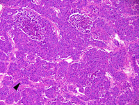
|
|
Image ID:1607 |
|
Source of Image:Ward JM |
|
Pathologist:Mikaelian I |
|
Method / Stain:H&E |
|
|
Image Caption:Comedo mammary adenocarcinoma: the neoplasm is composed of thick trabeculae and solid areas supported by a small amount of fibrovascular stroma. Neoplastic cells are medium-sized, polygonal, with indistinct cell borders and a moderate amount of strongly amphophilic cytoplasm. The nucleus is central, oval, medium-sized, hypochromatic, with 1-2 large basophilic nucleoli. Anisokaryosis and anisocytosis are mild. Numerous neoplastic cells undergo single cell necrosis.
|
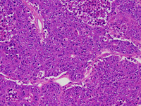
|
|
Image ID:1608 |
|
Source of Image:Ward JM |
|
Pathologist:Mikaelian I |
|
Method / Stain:H&E |
|
|
Image Caption:Mammary gland: the mammary gland is expanded by a nodular, unencapsulated, expensile, densely cellular neoplasm. This neoplasm is composed of closely packed glands and thick trabeculae with prominent central liquefactive necrosis.
|
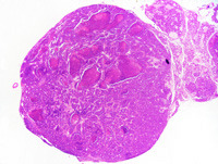
|
|
Image ID:1604 |
|
Source of Image:Ward JM |
|
Pathologist:Mikaelian I |
|
Method / Stain:H&E |
|
|
Image Caption:Mammary gland: the mammary gland is expanded by a nodular, unencapsulated, expensile, densely cellular neoplasm. This neoplasm is composed of closely packed glands and thick trabeculae with prominent central liquefactive necrosis.
|
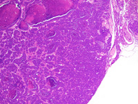
|
|
Image ID:1605 |
|
Source of Image:Ward JM |
|
Pathologist:Mikaelian I |
|
Method / Stain:H&E |
|
|
Image Caption:Mammary gland: the mammary gland is expanded by a nodular, unencapsulated, expensile, densely cellular neoplasm. This neoplasm is composed of closely packed glands and thick trabeculae with prominent central liquefactive necrosis.
|
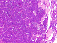
|
|
Image ID:1606 |
|
Source of Image:Ward JM |
|
Pathologist:Mikaelian I |
|
Method / Stain:H&E |
|
|
|
| MTB ID |
Tumor Name |
Organ(s) Affected |
Treatment Type |
Agents |
Strain Name |
Strain Sex |
Reproductive Status |
Tumor Frequency |
Age at Necropsy |
Description |
Reference |
| MTB:29200 |
Mammary gland adenocarcinoma - papillary |
Mammary gland |
None (spontaneous) |
|
|
Female |
reproductive status not specified |
observed |
120 days |
Adenocarcinoma, papillary, circumscribed, mammary gland |
J:94320 |
|
Image Caption:Papillary adenocarcinoma: neoplastic cells are columnar to polygonal, with indistinct cell borders and a moderate amount of strongly amphophilic cytoplasm. The nucleus is central, oval, medium-sized, hypochromatic, and with 1-3 small to medium-sized strongly basophilic nucleoli. Anisokaryosis and anisocytosis are mild. Numerous degenerated neoplastic cells have desquamated.
|
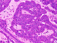
|
|
Image ID:1611 |
|
Source of Image:Ward JM |
|
Pathologist:Mikaelian I |
|
Method / Stain:H&E |
|
|
Image Caption:Mammary gland: the mammary gland is effaced by a small, nodular, unencapsulated, densely cellular neoplasm. This neoplasm is composed of coalescing papillae supported by a small amount of fibrovascular stroma and lined by a (predominantly) one cell-thick columnar epithelium. Neoplastic cells often pile-up disorderly.
|
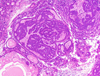
|
|
Image ID:1609 |
|
Source of Image:Ward JM |
|
Pathologist:Mikaelian I |
|
Method / Stain:H&E |
|
|
Image Caption:Mammary gland: the mammary gland is effaced by a small, nodular, unencapsulated, densely cellular neoplasm. This neoplasm is composed of coalescing papillae supported by a small amount of fibrovascular stroma and lined by a (predominantly) one cell-thick columnar epithelium. Neoplastic cells often pile-up disorderly or form small secondary lumens.
|
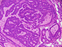
|
|
Image ID:1610 |
|
Source of Image:Ward JM |
|
Pathologist:Mikaelian I |
|
Method / Stain:H&E |
|
|
|
| MTB ID |
Tumor Name |
Organ(s) Affected |
Treatment Type |
Agents |
Strain Name |
Strain Sex |
Reproductive Status |
Tumor Frequency |
Age at Necropsy |
Description |
Reference |
| MTB:29201 |
Mammary gland adenocarcinoma - solid |
Mammary gland |
None (spontaneous) |
|
|
Female |
reproductive status not specified |
observed |
120 days |
Adenocarcinoma, solid, with secondary lumens and epithelial to mesenchymal transition |
J:94320 |
|
Image Caption:Mammary gland: the mammary gland is effaced by a large, multilobular, unencapsulated, densely cellular neoplasm. This neoplasm is composed of lobules that are made up of solid areas with secondary lumens.
|
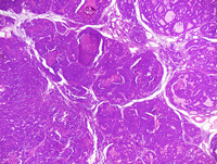
|
|
Image ID:1614 |
|
Source of Image:Ward JM |
|
Pathologist:Mikaelian I |
|
Method / Stain:H&E |
|
|
Image Caption:Solid mammary adenocarcinoma: this photomicrograph illustrates a portion of the neoplasm with epithelial to mesenchymal transition. Neoplastic cells form one cell-thick trabeculae and become spindloid. Neoplastic spindloid cells are difficult to differentiate from stromal myofibroblasts. Individual spindloid neoplastic cells are present in the stroma.
|
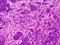
|
|
Image ID:1624 |
|
Source of Image:Ward JM |
|
Pathologist:Mikaelian I |
|
Method / Stain:H&E |
|
|
Image Caption:Mammary gland: the mammary gland is effaced by a large, multilobular, unencapsulated, densely cellular neoplasm. This neoplasm is composed of lobules of various size that are made up of glands (predominantly) and solid areas. These lobules are separated by a small amount of fibrovascular stroma.
|
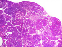
|
|
Image ID:1612 |
|
Source of Image:Ward JM |
|
Pathologist:Mikaelian I |
|
Method / Stain:H&E |
|
|
Image Caption:Solid mammary adenocarcinoma: a neoplastic lobule with prominent secondary lumens has prominent central coagulation necrosis.
|
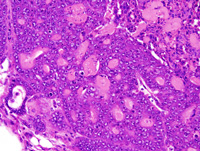
|
|
Image ID:1621 |
|
Source of Image:Ward JM |
|
Pathologist:Mikaelian I |
|
Method / Stain:H&E |
|
|
Image Caption:Solid mammary adenocarcinoma: the largest solid areas have central liquefactive necrosis and have a comedo-type pattern. All solid areas contain secondary lumens filled by a proteinaceous fluid.
|
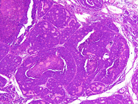
|
|
Image ID:1615 |
|
Source of Image:Ward JM |
|
Pathologist:Mikaelian I |
|
Method / Stain:H&E |
|
|
Image Caption:Solid mammary adenocarcinoma: this photomicrograph illustrates a portion of the neoplasm with epithelial to mesenchymal transition. Neoplastic cells form one cell-thick trabeculae and become spindloid.
|
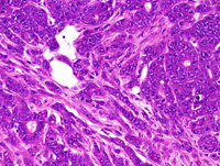
|
|
Image ID:1622 |
|
Source of Image:Ward JM |
|
Pathologist:Mikaelian I |
|
Method / Stain:H&E |
|
|
Image Caption:Solid mammary adenocarcinoma: this photomicrograph illustrates a portion of the neoplasm with epithelial to mesenchymal transition. Neoplastic cells form one cell-thick trabeculae and become spindloid. Neoplastic spindloid cells are difficult to differentiate from stromal myofibroblasts.
|
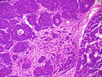
|
|
Image ID:1623 |
|
Source of Image:Ward JM |
|
Pathologist:Mikaelian I |
|
Method / Stain:H&E |
|
|
Image Caption:Solid mammary adenocarcinoma: the largest solid areas have central liquefactive necrosis and have a comedo-type pattern (*). All solid areas contain secondary lumens filled by a proteinaceous fluid.
|
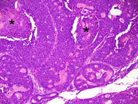
|
|
Image ID:1616 |
|
Source of Image:Ward JM |
|
Pathologist:Mikaelian I |
|
Method / Stain:H&E |
|
|
Image Caption:Solid mammary adenocarcinoma: this photomicrograph illustrates a portion of the neoplasm with a a solid pattern and with numerous secondary lumens (*). Nearby neoplastic lobules have a more classical solid pattern with large central areas of liquefactive necrosis ("comedo" pattern).
|
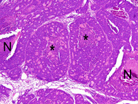
|
|
Image ID:1619 |
|
Source of Image:Ward JM |
|
Pathologist:Mikaelian I |
|
Method / Stain:H&E |
|
|
Image Caption:Solid mammary adenocarcinoma: this photomicrograph illustrates a portion of the neoplasm with a pattern intermediate between a glandular pattern and a solid pattern. Numerous secondary lumens (*) are present.
|
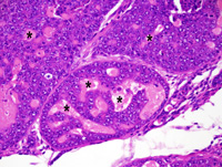
|
|
Image ID:1618 |
|
Source of Image:Ward JM |
|
Pathologist:Mikaelian I |
|
Method / Stain:H&E |
|
|
Image Caption:Solid mammary adenocarcinoma: a neoplastic lobule with prominent secondary lumens has prominent central coagulation necrosis.
|
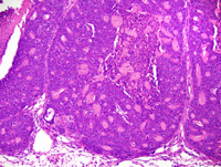
|
|
Image ID:1620 |
|
Source of Image:Ward JM |
|
Pathologist:Mikaelian I |
|
Method / Stain:H&E |
|
|
Image Caption:Mammary gland: the mammary gland is effaced by a large, multilobular, unencapsulated, densely cellular neoplasm. This neoplasm is composed of lobules that are made up of solid areas with secondary lumens.
|
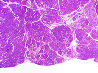
|
|
Image ID:1613 |
|
Source of Image:Ward JM |
|
Pathologist:Mikaelian I |
|
Method / Stain:H&E |
|
|
Image Caption:Solid mammary adenocarcinoma: the secondary lumens are filled by a proteinaceous fluid. Neoplastic cells are cuboidal to polygonal, with indistinct cell borders and a moderate amount of strongly amphophilic cytoplasm. The nucleus is central, round to oval, slightly hypochromatic, with a stippled chromatin and 1-2 medium-sized and strongly basophilic nucleoli. Anisokaryosis and anisocytosis are mild. Mitoses are numerous.
|
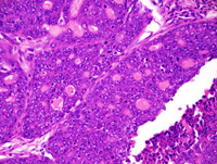
|
|
Image ID:1617 |
|
Source of Image:Ward JM |
|
Pathologist:Mikaelian I |
|
Method / Stain:H&E |
|
|
|
| MTB ID |
Tumor Name |
Organ(s) Affected |
Treatment Type |
Agents |
Strain Name |
Strain Sex |
Reproductive Status |
Tumor Frequency |
Age at Necropsy |
Description |
Reference |
| MTB:29202 |
Mammary gland adenocarcinoma - mixed |
Mammary gland |
None (spontaneous) |
|
|
Female |
reproductive status not specified |
observed |
unknown |
Adenocarcinoma, solid, comedo, mammary gland |
J:94320 |
|
Image Caption:Mammary gland: the mammary gland is expanded by a nodular, unencapsulated, expensile, densely cellular neoplasm. This neoplasm is composed of solid areas with central necrosis supported by a small amount of fibrovascular stroma. Neoplastic cells are medium-sized to large, polygonal, with ill-defined cell borders and a moderate amount of strongly amphophilic cytoplasm. The nucleus is central, round to oval, medium-size, occasionally with a slightly contoured nuclear membrane, slightly hypochromatic and with 1-3 medium size basophilic nucleoli. Anisokaryosis and anisocytosis are mild. Mitotic rate is elevated. A moderate number of neoplastic cells undergoes apoptosis.
|
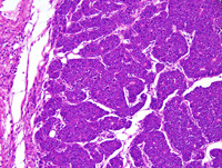
|
|
Image ID:1627 |
|
Source of Image:Ward JM |
|
Pathologist:Mikaelian I |
|
Method / Stain:H&E |
|
|
Image Caption:Mammary gland: the mammary gland is expanded by a nodular, unencapsulated, expensile, densely cellular neoplasm. This neoplasm is composed of solid areas with central necrosis, which gives a pseudo-glandular pattern to the neoplasm when examined at low magnification.
|
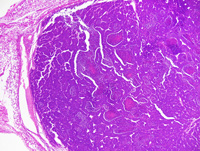
|
|
Image ID:1625 |
|
Source of Image:Ward JM |
|
Pathologist:Mikaelian I |
|
Method / Stain:H&E |
|
|
Image Caption:Mammary gland: the mammary gland is expanded by a nodular, unencapsulated, expensile, densely cellular neoplasm. This neoplasm is composed of solid areas supported by a small amount of fibrovascular stroma. Neoplastic cells are medium-sized to large, polygonal, with ill-defined cell borders and a moderate amount of strongly amphophilic cytoplasm. The nucleus is central, round to oval, medium-size, occasionally with a slightly contoured nuclear membrane, slightly hypochromatic and with 1-3 medium size basophilic nucleoli. Anisokaryosis and anisocytosis are mild. Mitotic rate is elevated. A moderate number of neoplastic cells undergoes apoptosis. Necrosis and cavitation are present at the center of the largest trabeculae.
|
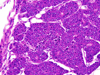
|
|
Image ID:1628 |
|
Source of Image:Ward JM |
|
Pathologist:Mikaelian I |
|
Method / Stain:H&E |
|
|
Image Caption:Solid mammary adenocarcinoma with comedo pattern: this photomicrograph illustrates a portion of the neoplasm with prominent necrosis and early cavitation at the center of the solid areas. The cavitated area contains degenerated neoplastic cells.
|
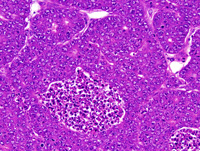
|
|
Image ID:1630 |
|
Source of Image:Ward JM |
|
Pathologist:Mikaelian I |
|
Method / Stain:H&E |
|
|
Image Caption:Solid mammary adenocarcinoma with comedo pattern: this photomicrograph illustrates a portion of the neoplasm with prominent necrosis and early cavitation at the center of the solid areas.
|
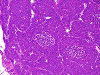
|
|
Image ID:1629 |
|
Source of Image:Ward JM |
|
Pathologist:Mikaelian I |
|
Method / Stain:H&E |
|
|
Image Caption:Mammary gland: the mammary gland is expanded by a nodular, unencapsulated, expensile, densely cellular neoplasm. This neoplasm is composed of solid areas with central necrosis supported by a small amount of fibrovascular stroma.
|
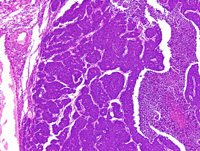
|
|
Image ID:1626 |
|
Source of Image:Ward JM |
|
Pathologist:Mikaelian I |
|
Method / Stain:H&E |
|
|
|
| MTB ID |
Tumor Name |
Organ(s) Affected |
Treatment Type |
Agents |
Strain Name |
Strain Sex |
Reproductive Status |
Tumor Frequency |
Age at Necropsy |
Description |
Reference |
| MTB:29203 |
Mammary gland adenocarcinoma |
Mammary gland |
None (spontaneous) |
|
|
Female |
reproductive status not specified |
observed |
unknown |
Adenocarcinoma, glandular, mammary gland |
J:94320 |
|
Image Caption:Mammary gland: the mammary gland is focally expanded by a nodular, densely cellular, expensile neoplastic mass surounded by a thin fibrous capsule. This neoplasm is composed of closely-packed glands with a narrow lumen, lined by a one cell-thick cuboidal epithelium, and supported by a small amount of fibrovascular stroma.
|
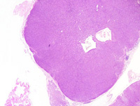
|
|
Image ID:1632 |
|
Source of Image:Ward JM |
|
Pathologist:Mikaelian I |
|
Method / Stain:H&E |
|
|
Image Caption:Mammary gland: the mammary gland is focally expanded by a nodular, densely cellular, expensile neoplastic mass surounded by a thin fibrous capsule. This neoplasm is composed of closely-packed glands with a narrow lumen, lined by a one cell-thick cuboidal epithelium, and supported by a small amount of fibrovascular stroma.
|
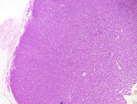
|
|
Image ID:1633 |
|
Source of Image:Ward JM |
|
Pathologist:Mikaelian I |
|
Method / Stain:H&E |
|
|
Image Caption:Mammary gland: the neoplasm is composed of closely-packed glands with a narrow lumen, lined by a one cell-thick cuboidal epithelium, and supported by a small amount of fibrovascular stroma. Neoplastic cells are cuboidal, with indistinct cell borders and a moderate amount of acidophilic cytoplasm. The nucleus is central, oval, generally basal, medium-sized, normochromatic, with a finely stippled chromatin and 1-4 medium-sized basophilic nucleoli. Anisokaryosis and anisocytosis are mild. A few mitoses are present. There is a mild lymphocytic and granulocytic infiltrate at the periphery of the neoplasm.
|
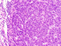
|
|
Image ID:1636 |
|
Source of Image:Ward JM |
|
Pathologist:Mikaelian I |
|
Method / Stain:H&E |
|
|
Image Caption:Mammary gland: the mammary gland is focally expanded by a nodular, densely cellular, expensile neoplastic mass surounded by a thin fibrous capsule. This neoplasm is composed of closely-packed glands with a narrow lumen, lined by a one cell-thick cuboidal epithelium, and supported by a small amount of fibrovascular stroma.
|
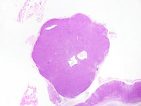
|
|
Image ID:1631 |
|
Source of Image:Ward JM |
|
Pathologist:Mikaelian I |
|
Method / Stain:H&E |
|
|
Image Caption:Mammary gland: the neoplasm is composed of closely-packed glands with a narrow lumen, lined by a one cell-thick cuboidal epithelium, and supported by a small amount of fibrovascular stroma. Neoplastic cells are cuboidal, with indistinct cell borders and a moderate amount of acidophilic cytoplasm. The nucleus is central, oval, generally basal, medium-sized, normochromatic, with a finely stippled chromatin and 1-4 medium-sized basophilic nucleoli.
|
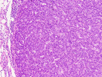
|
|
Image ID:1635 |
|
Source of Image:Ward JM |
|
Pathologist:Mikaelian I |
|
Method / Stain:H&E |
|
|
Image Caption:Mammary gland: the neoplasm is composed of closely-packed glands with a narrow lumen, lined by a one cell-thick cuboidal epithelium, and supported by a small amount of fibrovascular stroma. Neoplastic cells are cuboidal, with indistinct cell borders and a moderate amount of acidophilic cytoplasm. The nucleus is central, oval, generally basal, medium-sized, normochromatic, with a finely stippled chromatin and 1-4 medium-sized basophilic nucleoli.
|
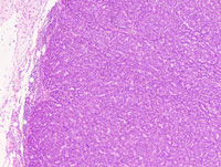
|
|
Image ID:1634 |
|
Source of Image:Ward JM |
|
Pathologist:Mikaelian I |
|
Method / Stain:H&E |
|
|
|
| MTB ID |
Tumor Name |
Organ(s) Affected |
Treatment Type |
Agents |
Strain Name |
Strain Sex |
Reproductive Status |
Tumor Frequency |
Age at Necropsy |
Description |
Reference |
| MTB:29204 |
Mammary gland carcinoma in situ |
Mammary gland |
None (spontaneous) |
|
|
Female |
reproductive status not specified |
observed |
unknown |
Carcinoma in situ, mammary gland |
J:94320 |
|
Image Caption:In situ mammary carcinoma: there is, at the vicinity of a large glandular carcinoma (*) a neoplastic lobules that shows prominent proliferation of its' cells. Cells in this lobule pile-up disorderly and distend a few alveoli (arrowhead). The architecture of the lobule is preserved. There is mild to moderate interstitial fibrosis and lymphocytic and plasmacytic inflammation.
|
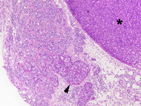
|
|
Image ID:1637 |
|
Source of Image:Ward JM |
|
Pathologist:Mikaelian I |
|
Method / Stain:H&E |
|
|
Image Caption:In situ mammary carcinoma: there is prominent proliferation of cells in all mammary alveoli and intralobular ducts. These cells occasionally form solid areas that expand mammary alveoli and acini. Proliferating cells are large, with indistinct cell borders and a moderate amount of pale acidophilic cytoplasm. The nucleus is central, oval, medium-sized and slightly hypochromatic. Anisokaryosis and anisocytosis are mild. There is no evidence of invasion. There is mild to moderate interstitial fibrosis and lymphocytic and plasmacytic inflammation.
|
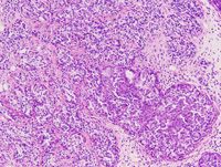
|
|
Image ID:1638 |
|
Source of Image:Ward JM |
|
Pathologist:Mikaelian I |
|
Method / Stain:H&E |
|
|
Image Caption:In situ mammary carcinoma: there is moderate proliferation of epithelial cells of all alveoli and ducts with occlusion of the ductal and alveolar lumen. Myoepithelial differentiation (arrowheads) is present in this portion of the lesion.
|
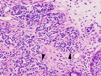
|
|
Image ID:1640 |
|
Source of Image:Ward JM |
|
Pathologist:Mikaelian I |
|
Method / Stain:H&E |
|
|
Image Caption:In situ mammary carcinoma: there is prominent proliferation of cells in a mammary alveolus. Thin fibrovascular projections subdivide the alveolus. Proliferating cells are large, with indistinct cell borders and a moderate amount of pale acidophilic cytoplasm. The nucleus is central, oval, medium-sized and slightly hypochromatic. Anisokaryosis and anisocytosis are mild. There is no evidence of invasion. There is mild to moderate interstitial fibrosis and lymphocytic and plasmacytic inflammation. Myoepithelial differentiation is not detected in this portion of the lesion.
|
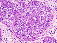
|
|
Image ID:1639 |
|
Source of Image:Ward JM |
|
Pathologist:Mikaelian I |
|
Method / Stain:H&E |
|
|
|
| MTB ID |
Tumor Name |
Organ(s) Affected |
Treatment Type |
Agents |
Strain Name |
Strain Sex |
Reproductive Status |
Tumor Frequency |
Age at Necropsy |
Description |
Reference |
| MTB:29205 |
Mammary gland carcinoma in situ |
Mammary gland |
None (spontaneous) |
|
|
Female |
reproductive status not specified |
observed |
unknown |
Carcinoma in situ, mammary gland |
J:94320 |
|
Image Caption:Mammary gland: hypercellularity of mammary lobules is the result of proliferation of luminal cells of mammary alveoli and intralobular ducts. These cells pile-up and occlude the lumen of some alveoli. There is no evidence of local invasion.
|
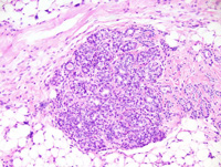
|
|
Image ID:1642 |
|
Source of Image:Ward JM |
|
Pathologist:Mikaelian I |
|
Method / Stain:H&E |
|
|
Image Caption:Mammary gland: hypercellularity of mammary lobules is the result of proliferation of luminal cells of mammary alveoli and intralobular ducts. These cells pile-up and occlude the lumen of some alveoli. The cells populating this lobule are very heterogeneous. About 30% of these cells have primitive features: they are oval, with a pale acidophilic cytoplasm and a medium-sized slightly hypochromatic nucleus. The other cells are more differentiated and have a cuboidal to low columnar shape, a more strongly acidophilic cytoplasm and a normochromatic nucleus. There is no evidence of atypia but a few mitoses (arrowheads) are detected. There is no evidence of local invasion. There is mild interstitial fibrosis and lymphocytic and plasmacytic inflammation.
|
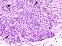
|
|
Image ID:1643 |
|
Source of Image:Ward JM |
|
Pathologist:Mikaelian I |
|
Method / Stain:H&E |
|
|
Image Caption:Mammary gland: there is marked diffuse hypercellularity of two adjacent mammary lobules.
|
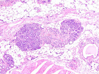
|
|
Image ID:1641 |
|
Source of Image:Ward JM |
|
Pathologist:Mikaelian I |
|
Method / Stain:H&E |
|
|
Image Caption:Mammary gland: a mammary gland is moderately increased in size. Most alveoli are slightly expanded by a proteinaceous material and are lined by an hypercellular epithelium. Some alveoli are filled with epithelial cells and resemble terminal end buds.
|
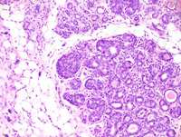
|
|
Image ID:1645 |
|
Source of Image:Ward JM |
|
Pathologist:Mikaelian I |
|
Method / Stain:H&E |
|
|
Image Caption:Mammary gland: a mammary gland is moderately increased in size. Most alveoli are slightly expanded by a proteinaceous material and are lined by an hypercellular epithelium. Some alveoli are filled with epithelial cells and resemble terminal end buds. There is mild interstitial fibrosis and lymphocytic and plasmacytic inflammation.
|
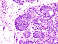
|
|
Image ID:1646 |
|
Source of Image:Ward JM |
|
Pathologist:Mikaelian I |
|
Method / Stain:H&E |
|
|
Image Caption:Mammary gland: a mammary gland is moderately increased in size. Most alveoli are slightly expanded by a proteinaceous material and are lined by an hypercellular epithelium. Some alveoli are filled with epithelial cells and resemble terminal end buds.
|
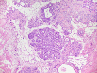
|
|
Image ID:1644 |
|
Source of Image:Ward JM |
|
Pathologist:Mikaelian I |
|
Method / Stain:H&E |
|
|
|
| MTB ID |
Tumor Name |
Organ(s) Affected |
Treatment Type |
Agents |
Strain Name |
Strain Sex |
Reproductive Status |
Tumor Frequency |
Age at Necropsy |
Description |
Reference |
| MTB:29206 |
Mammary gland adenocarcinoma |
Mammary gland |
None (spontaneous) |
|
|
Female |
reproductive status not specified |
observed |
unknown |
Adenocarcinoma, glandular, mammary gland |
J:94320 |
|
Image Caption:Mammary gland: the mammary gland is expanded by a large, multilobular, densely cellular neoplastic mass surrounded by a thin capsule. A few areas of liquefactive necrosis are scattered throughout the neoplasm.
|
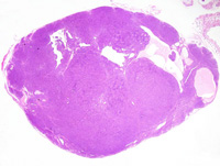
|
|
Image ID:1647 |
|
Source of Image:Ward JM |
|
Pathologist:Mikaelian I |
|
Method / Stain:H&E |
|
|
Image Caption:Mammary gland: the mammary gland is expanded by a large, multilobular, densely cellular neoplastic mass surrounded by a thin capsule. The neoplasm is composed of closely-packed branching glands with an inconspicuous lumen. These glands are lined by a one cell-thick epithelium and are supported by a small amount of fibrovascular stroma. There is mild lymphocytic and plasmacytic inflammation in the capsule of the neoplasm.
|
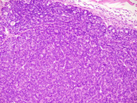
|
|
Image ID:1651 |
|
Source of Image:Ward JM |
|
Pathologist:Mikaelian I |
|
Method / Stain:H&E |
|
|
Image Caption:Mammary gland: the neoplasm is composed of closely-packed branching glands with an inconspicuous lumen. These glands are lined by a one cell-thick epithelium and are supported by a small amount of fibrovascular stroma. There is mild lymphocytic and plasmacytic inflammation in the capsule of the neoplasm. Neoplastic cells are low columnar, with indistinct cell borders and a moderate amount of acidophilic cytoplasm. The nucleus is basal, often oriented in the long axis of the cell, medium-sized,normochromatic, with a finely stippled chromatin and 1-2 small basophilic nucleoli. Anisokaryosis and anisocytosis are mild. A few mitoses are present.
|
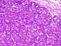
|
|
Image ID:1652 |
|
Source of Image:Ward JM |
|
Pathologist:Mikaelian I |
|
Method / Stain:H&E |
|
|
Image Caption:Mammary gland: the mammary gland is expanded by a large, multilobular, densely cellular neoplastic mass surrounded by a thin capsule. The neoplasm is composed of closely-packed branching glands with an inconspicuous lumen. These glands are lined by a one cell-thick epithelium and are supported by a small amount of fibrovascular stroma. There is mild lymphocytic and plasmacytic inflammation in the capsule of the neoplasm.
|
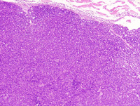
|
|
Image ID:1650 |
|
Source of Image:Ward JM |
|
Pathologist:Mikaelian I |
|
Method / Stain:H&E |
|
|
Image Caption:Mammary gland: the mammary gland is expanded by a large, multilobular, densely cellular neoplastic mass surrounded by a thin capsule. The neoplasm is composed of closely-packed branching glands with an inconspicuous lumen. These glands are lined by a one cell-thick epithelium and are supported by a small amount of fibrovascular stroma. A few areas of liquefactive necrosis are scattered throughout the neoplasm.
|
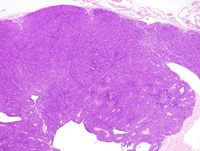
|
|
Image ID:1649 |
|
Source of Image:Ward JM |
|
Pathologist:Mikaelian I |
|
Method / Stain:H&E |
|
|
Image Caption:Mammary gland: the mammary gland is expanded by a large, multilobular, densely cellular neoplastic mass surrounded by a thin capsule. The neoplasm is composed of closely-packed branching glands with an inconspicuous lumen. These glands are lined by a one cell-thick epithelium and are supported by a small amount of fibrovascular stroma. A few areas of liquefactive necrosis are scattered throughout the neoplasm.
|
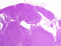
|
|
Image ID:1648 |
|
Source of Image:Ward JM |
|
Pathologist:Mikaelian I |
|
Method / Stain:H&E |
|
|
Image Caption:Mammary gland: the neoplasm is composed of closely-packed branching glands with an inconspicuous lumen. These glands are lined by a one cell-thick epithelium and are supported by a small amount of fibrovascular stroma. Neoplastic cells are low columnar, with indistinct cell borders and a moderate amount of acidophilic cytoplasm. The nucleus is basal, often oriented in the long axis of the cell, medium-sized,normochromatic, with a finely stippled chromatin and 1-2 small basophilic nucleoli. Anisokaryosis and anisocytosis are mild. A few mitoses are present.
|
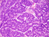
|
|
Image ID:1653 |
|
Source of Image:Ward JM |
|
Pathologist:Mikaelian I |
|
Method / Stain:H&E |
|
|
|
| MTB ID |
Tumor Name |
Organ(s) Affected |
Treatment Type |
Agents |
Strain Name |
Strain Sex |
Reproductive Status |
Tumor Frequency |
Age at Necropsy |
Description |
Reference |
| MTB:29207 |
Mammary gland carcinoma in situ |
Mammary gland |
None (spontaneous) |
|
|
Female |
reproductive status not specified |
observed |
unknown |
Carcinoma in situ, mammary gland |
J:94320 |
|
Image Caption:In situ mammary carcinoma: this photomicrograph represents the transition from the early stages of in situ mammary carcinoma to a more advanced stage of the disease (at the center of the photomicrograph). Myoepithelial differentiation is prominent in the areas of early in situ mammary carcinoma. It is still present but difficult to identify in the more advance areas. Cells in the more advanced areas are larger, with pale acidophilic cytoplasm and a medium-sized hypochromatic nucleus. Cells in the least advanced areas are smaller and have a smaller and normo- to hyperchromatic nucleus. There is no evidence of invasion. There is mild interstitial lymphocytic and plasmacytic inflammation and fibrosis.
|
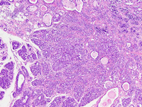
|
|
Image ID:1659 |
|
Source of Image:Ward JM |
|
Pathologist:Mikaelian I |
|
Method / Stain:H&E |
|
|
Image Caption:In situ mammary carcinoma: this photomicrograph represents the transition from the early stages of in situ mammary carcinoma to a more advanced stage of the disease (at the center of the photomicrograph). Myoepithelial differentiation is prominent in the areas of early in situ mammary carcinoma. It is still present but difficult to identify in the more advance areas. Cells in the more advanced areas are larger, with pale acidophilic cytoplasm and a medium-sized hypochromatic nucleus. Cells in the least advanced areas are smaller and have a smaller and normo- to hyperchromatic nucleus. There is no evidence of invasion. There is mild interstitial lymphocytic and plasmacytic inflammation and fibrosis.
|
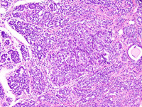
|
|
Image ID:1660 |
|
Source of Image:Ward JM |
|
Pathologist:Mikaelian I |
|
Method / Stain:H&E |
|
|
Image Caption:Mammary gland: there is moderate diffuse expansion of a mammary lobule. Within this lobule, a few acini are markedly expanded by closely packed epithelial cells that form solid areas and small glands with an inconspicuous lumen. The cells are polygonal, with distinct cell borders and a moderate amount of pale acidophilic cytoplasm. The nucleus is central, oval, medium-sized and hypochromatic. Anisokaryosis and anisocytosis are mild. There is no evidence of local invasion. Mitotic figures are numerous. A small focus of cornification (arrowhead) is present.There is moderate interstitial lymphocytic and plasmacytic inflammation and fibrosis.
|
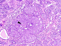
|
|
Image ID:1655 |
|
Source of Image:Ward JM |
|
Pathologist:Mikaelian I |
|
Method / Stain:H&E |
|
|
Image Caption:Mammary gland: there is moderate diffuse expansion of a mammary lobule. Within this lobule, a few acini are markedly expanded by closely packed epithelial cells that form solid areas and small glands with an inconspicuous lumen. The cells are polygonal, with distinct cell borders and a moderate amount of pale acidophilic cytoplasm. The nucleus is central, oval, medium-sized and hypochromatic. Anisokaryosis and anisocytosis are mild. There is no evidence of local invasion. Mitotic figures are numerous. There is moderate interstitial lymphocytic and plasmacytic inflammation and fibrosis. Myoepithelial differentiation is prominent and appears as a continuous layer of cells with a nucleus oriented parallel to the basement membrane and a scant amount of cytoplasm.
|
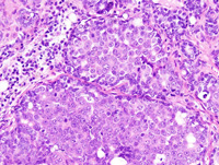
|
|
Image ID:1656 |
|
Source of Image:Ward JM |
|
Pathologist:Mikaelian I |
|
Method / Stain:H&E |
|
|
Image Caption:In situ mammary carcinoma: this represents an early stage of in situ mammary carcinoma: the architecture of the mamamry gland is preserved but mammary lobules are packed with epithelial cells and often lack a lumen. Numerous cells in the mammary gland are round, have a very pale cytoplasm with a ground glass appearance, and have a medium-sized hypochromatic nucleus. These features are consistent with the features of a primitive epithelial cell. There is mild to moderate interstitial lymphocytic and plasmacytic inflammation. Myoepithelial differentiation is still detected and myoepithelial cells appear as cells with an ellongated nucleus parallel to the basement membrane and a scant amount of cytoplasm.
|
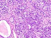
|
|
Image ID:1658 |
|
Source of Image:Ward JM |
|
Pathologist:Mikaelian I |
|
Method / Stain:H&E |
|
|
Image Caption:In situ mammary carcinoma: this low magnification photomicrograph represents portions of a mammary gland with areas of in situ mammary carcinoma (at the center of the photomicrographs) with multiple areas of atypical hyperplasia more at the periphery of the neoplasm. There is moderate intersitial fibrosis and lymphoplasmacytic inflammation that involves all mammary lobules.
|
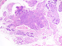
|
|
Image ID:1662 |
|
Source of Image:Ward JM |
|
Pathologist:Mikaelian I |
|
Method / Stain:H&E |
|
|
Image Caption:In situ mammary carcinoma: this represents an early stage of in situ mammary carcinoma: the architecture of the mamamry gland is preserved but mammary lobules are packed with epithelial cells and often lack a lumen. Numerous cells in the mammary gland are round, have a very pale cytoplasm with a ground glass appearance, and have a medium-sized hypochromatic nucleus. These features are consistent with the features of a primitive epithelial cell. There is mild to moderate interstitial lymphocytic and plasmacytic inflammation.
|
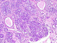
|
|
Image ID:1657 |
|
Source of Image:Ward JM |
|
Pathologist:Mikaelian I |
|
Method / Stain:H&E |
|
|
Image Caption:Mammary gland: there is moderate diffuse expansion of a mammary lobule. Within this lobule, a few acini are markedly expanded by closely-packed epithelial cells that form solid areas and small glands with an inconspicuous lumen.
|
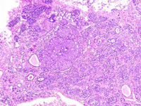
|
|
Image ID:1654 |
|
Source of Image:Ward JM |
|
Pathologist:Mikaelian I |
|
Method / Stain:H&E |
|
|
Image Caption:In situ mammary carcinoma: this photomicrograph represents the transition from the early stages of in situ mammary carcinoma to a more advanced stage of the disease (at the center of the photomicrograph). Myoepithelial differentiation is prominent in the areas of early in situ mammary carcinoma. It is still present but difficult to identify in the more advance areas. Cells in the more advanced areas are larger, with pale acidophilic cytoplasm and a medium-sized hypochromatic nucleus. Cells in the least advanced areas are smaller and have a smaller and normo- to hyperchromatic nucleus. There is no evidence of invasion. There is mild interstitial lymphocytic and plasmacytic inflammation and fibrosis.
|
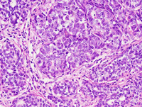
|
|
Image ID:1661 |
|
Source of Image:Ward JM |
|
Pathologist:Mikaelian I |
|
Method / Stain:H&E |
|
|
|
| MTB ID |
Tumor Name |
Organ(s) Affected |
Treatment Type |
Agents |
Strain Name |
Strain Sex |
Reproductive Status |
Tumor Frequency |
Age at Necropsy |
Description |
Reference |
| MTB:29230 |
Mammary gland adenocarcinoma - solid |
Mammary gland |
None (spontaneous) |
|
|
Female |
reproductive status not specified |
observed |
unknown |
Adenocarcinoma, solid, glandular, mammary gland |
J:94320 |
|
Image Caption:Solid mammary adenocarcinoma: the tumor is well-delineated and expensile.
|
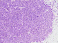
|
|
Image ID:1669 |
|
Source of Image:Ward JM |
|
Pathologist:Mikaelian I |
|
Method / Stain:H&E |
|
|
Image Caption:Solid mammary adenocarcinoma: this photomicrograph illustrates a portion of the neoplasm with glandular differentiation.
|
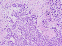
|
|
Image ID:1667 |
|
Source of Image:Ward JM |
|
Pathologist:Mikaelian I |
|
Method / Stain:H&E |
|
|
Image Caption:Solid mammary adenocarcinoma: a few areas of glandular differentiation (arrowheads) are identified in a neoplasm that otherwise has a solid phenotype.
|
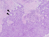
|
|
Image ID:1666 |
|
Source of Image:Ward JM |
|
Pathologist:Mikaelian I |
|
Method / Stain:H&E |
|
|
Image Caption:Mammary gland: the mammary parenchyma is entirely effaced by a densely cellular neoplasm with large areas of central liquefactive necrosis (*).
|
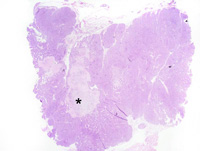
|
|
Image ID:1663 |
|
Source of Image:Ward JM |
|
Pathologist:Mikaelian I |
|
Method / Stain:H&E |
|
|
Image Caption:Mammary gland: the mammary parenchyma is entirely effaced by a densely cellular neoplasm with large areas of central liquefactive necrosis (*). This neoplasm is predominantly composed of large solid areas made-up of closely-packed neoplastic cells.
|
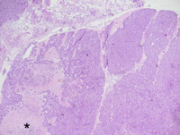
|
|
Image ID:1665 |
|
Source of Image:Ward JM |
|
Pathologist:Mikaelian I |
|
Method / Stain:H&E |
|
|
Image Caption:Solid mammary adenocarcinoma:the neoplasm is composed of thick trabeculae and solid areas with prominent nuclear palissading at the periphery of these areas.
|
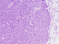
|
|
Image ID:1670 |
|
Source of Image:Ward JM |
|
Pathologist:Mikaelian I |
|
Method / Stain:H&E |
|
|
Image Caption:Mammary gland: the mammary parenchyma is entirely effaced by a densely cellular neoplasm with large areas of central liquefactive necrosis. This neoplasm is predominantly composed of large solid areas made-up of closely-packed neoplastic cells.
|
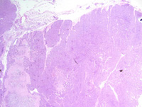
|
|
Image ID:1664 |
|
Source of Image:Ward JM |
|
Pathologist:Mikaelian I |
|
Method / Stain:H&E |
|
|
Image Caption:Solid mammary adenocarcinoma: this photomicrograph illustrates a portion of the neoplasm with glandular differentiation.
|
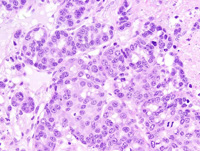
|
|
Image ID:1668 |
|
Source of Image:Ward JM |
|
Pathologist:Mikaelian I |
|
Method / Stain:H&E |
|
|
Image Caption:Solid mammary adenocarcinoma: neoplastic cells are medium-sized, polygonal, with indistinct cell borders and a moderate amount of amphophilic cytoplasm. The nucleus is central, round to oval, and slightly hypochromatic. Anisokaryosis and anisocytosis are mild.
|
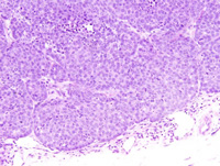
|
|
Image ID:1671 |
|
Source of Image:Ward JM |
|
Pathologist:Mikaelian I |
|
Method / Stain:H&E |
|
|
|
| MTB ID |
Tumor Name |
Organ(s) Affected |
Treatment Type |
Agents |
Strain Name |
Strain Sex |
Reproductive Status |
Tumor Frequency |
Age at Necropsy |
Description |
Reference |
| MTB:29231 |
Mammary gland adenocarcinoma - solid |
Mammary gland |
None (spontaneous) |
|
|
Female |
reproductive status not specified |
observed |
unknown |
Adenocarcinoma, solid, mammary gland |
J:94320 |
|
Image Caption:Mammary gland: the mammary gland is replaced by multiple, nodular, unencapsulated, well-delineated, densely cellular neoplasms. These neoplasm often display central liquefactive necrosis and cavitation (*).
|
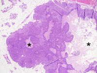
|
|
Image ID:1673 |
|
Source of Image:Ward JM |
|
Pathologist:Mikaelian I |
|
Method / Stain:H&E |
|
|
Image Caption:Solid mammary adenocarcinoma: the neoplasm is composed of solid areas of closely-packed polygonal neoplastic cells with indistinct cell borders. There are, at the periphery of the neoplasm, small clusters of neoplastic cells that have a paler cytoplasm, and a larger and more hypochromatic nucleus than neoplastic cells in the rest of the neoplasm.
|
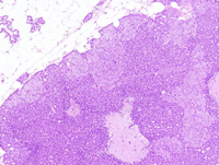
|
|
Image ID:1674 |
|
Source of Image:Ward JM |
|
Pathologist:Mikaelian I |
|
Method / Stain:H&E |
|
|
Image Caption:Solid mammary adenocarcinoma: there are three morphologic types of neoplastic cells. First, at the periphery of the neoplasms, there are small clusters of large cells with a pale cytoplasm and an hypochromatic nucleus. Second, around the areas of liquefactive necrosis, neoplastic cells are small, with a condensed nucleus and a small amount of cytoplasm. Third, acounting for the majority of neoplastic cells, there is a population of medium-sized polygonal cells with indistinct cell borders, a moderate amount of strongly acidophilic cytoplasm, and a medium-sized central normochromatic nucleolus.
|
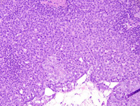
|
|
Image ID:1678 |
|
Source of Image:Ward JM |
|
Pathologist:Mikaelian I |
|
Method / Stain:H&E |
|
|
Image Caption:Solid mammary adenocarcinoma: the neoplasm is composed of solid areas of closely-packed polygonal neoplastic cells with indistinct cell borders. There are, at the periphery of the neoplasm, small clusters of neoplastic cells that have a paler cytoplasm, and a larger and more hypochromatic nucleus than neoplastic cells in the rest of the neoplasm.
|
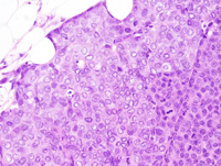
|
|
Image ID:1676 |
|
Source of Image:Ward JM |
|
Pathologist:Mikaelian I |
|
Method / Stain:H&E |
|
|
Image Caption:Solid mammary adenocarcinoma: this high magnification picture represents a peripheral portion of a neoplasm. There is a cluster of large neoplastic cells, while most neoplastic cells are medium-sized and have a more acidophilic cytoplasm. The arrowhead points to a minute lumen.
|
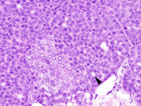
|
|
Image ID:1679 |
|
Source of Image:Ward JM |
|
Pathologist:Mikaelian I |
|
Method / Stain:H&E |
|
|
Image Caption:Solid mammary adenocarcinoma: this photomicrograph illustrates a portion of the neoplasm that comprises areas of liquefactive necrosis), a cluster of large neoplastic cells with a pale cytoplasm (*) and small areas composed of small cells with a small amount of strongly acidophilic cytoplasm and an hyperchromatic nucleus (arrowheads). The rest of the neoplasm is composed of medium-sized cells with a moderate amount of acidophilic cytoplasm and a medium-sized normochromatic nucleus.
|
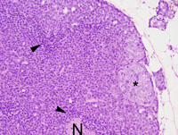
|
|
Image ID:1680 |
|
Source of Image:Ward JM |
|
Pathologist:Mikaelian I |
|
Method / Stain:H&E |
|
|
Image Caption:Solid mammary adenocarcinoma: the neoplasm is composed of solid areas of closely-packed polygonal neoplastic cells with indistinct cell borders. There are, at the periphery of the neoplasm, small clusters of neoplastic cells that have a paler cytoplasm, and a larger and more hypochromatic nucleus than neoplastic cells in the rest of the neoplasm.
|
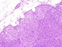
|
|
Image ID:1675 |
|
Source of Image:Ward JM |
|
Pathologist:Mikaelian I |
|
Method / Stain:H&E |
|
|
Image Caption:Solid mammary adenocarcinoma: the neoplasm is composed of solid areas of closely-packed polygonal neoplastic cells with indistinct cell borders. There are, at the periphery of the neoplasm, small clusters of neoplastic cells that have a paler cytoplasm, and a larger and more hypochromatic nucleus than neoplastic cells in the rest of the neoplasm.
|
|
|
|
Image ID:1677 |
|
Source of Image:Ward JM |
|
Pathologist:Mikaelian I |
|
Method / Stain:H&E |
|
|
Image Caption:Mammary gland: the mammary gland is replaced by multiple, nodular, unencapsulated, well-delineated, densely cellular neoplasms. These neoplasm often display central liquefactive necrosis and cavitation (*).
|
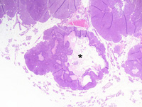
|
|
Image ID:1672 |
|
Source of Image:Ward JM |
|
Pathologist:Mikaelian I |
|
Method / Stain:H&E |
|
|
|
| MTB ID |
Tumor Name |
Organ(s) Affected |
Treatment Type |
Agents |
Strain Name |
Strain Sex |
Reproductive Status |
Tumor Frequency |
Age at Necropsy |
Description |
Reference |
| MTB:29232 |
Mammary gland adenocarcinoma - solid |
Mammary gland |
None (spontaneous) |
|
|
Female |
reproductive status not specified |
observed |
unknown |
Adenocarcinoma, solid, mammary gland |
J:94320 |
|
Image Caption:Solid mammary adenocarcinoma: a narrow rim of neoplastic cells survives at the periphery of the blood vessels located in the areas of liquefactive necrosis. The rest of the neoplasm is composed of solid areas separated by a small amount of fibrous stroma. There is prominent nuclear palissading at the periphery of neoplastic lobules.
|
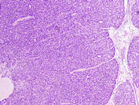
|
|
Image ID:1684 |
|
Source of Image:Ward JM |
|
Pathologist:Mikaelian I |
|
Method / Stain:H&E |
|
|
Image Caption:Solid mammary adenocarcinoma: this photomicrograph illustrates the morphology of the periphery of the neoplasm. Neoplastic cells, in this area, are large, have a moderate amount of pale acidophilic cytoplasm, and have a medium-sized hypochromatic nucleus. Mitotic activity is elevated. Neoplastic cells occasionally form inconspicuous lumens (*).
|
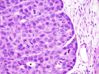
|
|
Image ID:1686 |
|
Source of Image:Ward JM |
|
Pathologist:Mikaelian I |
|
Method / Stain:H&E |
|
|
Image Caption:Solid mammary adenocarcinoma: the neoplasm is composed of two types of neoplastic cells. First, at the periphery of the neoplasm, there are small ill-defined clusters of large neoplastic cells with a pale cytoplasm and an hypochromatic nucleus. Second, accounting for most of the neoplasm, there are medium-sized polygonal cells with indistinct cell borders and a moderate amount of acidophilic cytoplasm. Anisokaryosis and anisocytosis are mild. Neoplastic cells occasionally form inconspicuous secondary lumens (arrowhead).
|
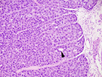
|
|
Image ID:1685 |
|
Source of Image:Ward JM |
|
Pathologist:Mikaelian I |
|
Method / Stain:H&E |
|
|
Image Caption:Solid mammary adenocarcinoma: a narrow rim of neoplastic cells survives at the periphery of the blood vessels located in the areas of liquefactive necrosis. The rest of the neoplasm is composed of solid areas separated by a small amount of fibrous stroma.
|
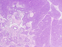
|
|
Image ID:1683 |
|
Source of Image:Ward JM |
|
Pathologist:Mikaelian I |
|
Method / Stain:H&E |
|
|
Image Caption:Mammary gland: the mammary gland is effaced by a large multilobular neoplasm with large central areas of liquefactive necrosis (*).
|
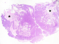
|
|
Image ID:1681 |
|
Source of Image:Ward JM |
|
Pathologist:Mikaelian I |
|
Method / Stain:H&E |
|
|
Image Caption:Mammary gland: the mammary gland is effaced by a large multilobular neoplasm with large central areas of liquefactive necrosis (*).
|
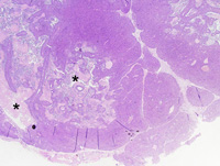
|
|
Image ID:1682 |
|
Source of Image:Ward JM |
|
Pathologist:Mikaelian I |
|
Method / Stain:H&E |
|
|
|
| MTB ID |
Tumor Name |
Organ(s) Affected |
Treatment Type |
Agents |
Strain Name |
Strain Sex |
Reproductive Status |
Tumor Frequency |
Age at Necropsy |
Description |
Reference |
| MTB:29233 |
Mammary gland adenocarcinoma - solid |
Mammary gland |
None (spontaneous) |
|
|
Female |
reproductive status not specified |
observed |
unknown |
Adenocarcinoma, solid, mammary gland |
J:94320 |
|
Image Caption:Solid mammary adenocarcinoma: only a thin rim of viable neoplastic cells persist. Other neoplastic cells have undergone liquefactive necrosis.
|
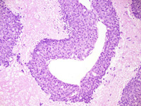
|
|
Image ID:1692 |
|
Source of Image:Ward JM |
|
Pathologist:Mikaelian I |
|
Method / Stain:H&E |
|
|
Image Caption:Mammary adenocarcinoma: the neoplasm is composed of solid areas of neoplastic cells with large coalescing areas of liquefactive necrosis. Neoplastic cells are closely-packed, polygonal, with indistinct cell borders and a moderate amount of acidophilic cytoplasm. The nucleus is central, oval, medium-sized and normochromatic. Anisokaryosis and anisocytosis are mild.
|
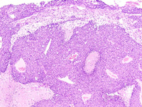
|
|
Image ID:1689 |
|
Source of Image:Ward JM |
|
Pathologist:Mikaelian I |
|
Method / Stain:H&E |
|
|
Image Caption:Mammary adenocarcinoma: the neoplasm is composed of solid areas of neoplastic cells with large coalescing areas of liquefactive necrosis. Neoplastic cells are closely-packed, polygonal, with indistinct cell borders and a moderate amount of acidophilic cytoplasm. The nucleus is central, oval, medium-sized and normochromatic. Anisokaryosis and anisocytosis are mild.
|
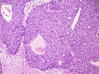
|
|
Image ID:1690 |
|
Source of Image:Ward JM |
|
Pathologist:Mikaelian I |
|
Method / Stain:H&E |
|
|
Image Caption:Mammary gland: the mammary parenchyma is entirely effaced by a densely cellular multilobular neoplasm with large central areas of liquefactive necrosis. In many places, viable neoplastic cells persists only as a narrow rim surrounding blood vessels.
|
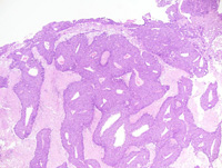
|
|
Image ID:1688 |
|
Source of Image:Ward JM |
|
Pathologist:Mikaelian I |
|
Method / Stain:H&E |
|
|
Image Caption:Solid mammary adenocarcinoma: neoplastic cells occasionally form small secondary lumens (*). Liquefactive necrosis (N) is prominent in areas located far away from blood vessels.
|
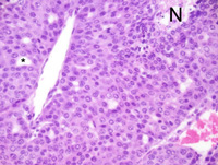
|
|
Image ID:1691 |
|
Source of Image:Ward JM |
|
Pathologist:Mikaelian I |
|
Method / Stain:H&E |
|
|
Image Caption:Mammary gland: the mammary parenchyma is entirely effaced by a densely cellular multilobular neoplasm with large central areas of liquefactive necrosis. In many places, viable neoplastic cells persists only as a narrow rim surrounding blood vessels.
|
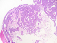
|
|
Image ID:1687 |
|
Source of Image:Ward JM |
|
Pathologist:Mikaelian I |
|
Method / Stain:H&E |
|
|
|
| MTB ID |
Tumor Name |
Organ(s) Affected |
Treatment Type |
Agents |
Strain Name |
Strain Sex |
Reproductive Status |
Tumor Frequency |
Age at Necropsy |
Description |
Reference |
| MTB:29234 |
Mammary gland adenocarcinoma - mixed |
Mammary gland |
None (spontaneous) |
|
|
Female |
reproductive status not specified |
observed |
unknown |
Adenocarcinoma, solid, comedo, with microinvasion, mammary gland |
J:94320 |
|
Image Caption:Mammary gland: the mammary gland is effaced by a large, nodular, multilobular, unencapsulated, densely cellular, neoplastic mass. This mass is composed of large coalescing lobules separated by a scant amount of loose fibrovascular stroma with large ectatic vascular spaces (*). Necrosis and cavitation are common in most neoplastic lobules. The portion of the neoplasm located at the center of the photomicrograph is composed of large polygonal cells with distinct cell borders, a large amount of acidophilic cytoplasm, and medium-size hypochromatic nuclei. The portions of the neoplasm located at the periphery of this photomicrograph are composed of crowded small cells with indistinct cell borders, a small amount of acidophilic cytoplasm, and a slightly smaller and more hypochromatic nucleus. Anisokaryosis and anisocytosis are mild. Mitoses are numerous.
|
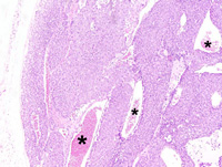
|
|
Image ID:1695 |
|
Source of Image:Ward JM |
|
Pathologist:Mikaelian I |
|
Method / Stain:H&E |
|
|
Image Caption:Mammary gland: the mammary gland is effaced by a large, nodular, multilobular, unencapsulated, densely cellular, neoplastic mass. This mass is composed of large coalescing lobules separated by a scant amount of loose fibrovascular stroma. Necrosis and cavitation are common in most neoplastic lobules.
|
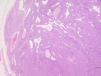
|
|
Image ID:1694 |
|
Source of Image:Ward JM |
|
Pathologist:Mikaelian I |
|
Method / Stain:H&E |
|
|
Image Caption:Mammary gland: the portion of the neoplasm located at the center of the photomicrograph is composed of large polygonal cells with distinct cell borders, a large amount of acidophilic cytoplasm, and medium-size hypochromatic nuclei. The portions of the neoplasm located at the periphery of this photomicrograph are composed of crowded small cells with indistinct cell borders, a small amount of acidophilic cytoplasm, and a slightly smaller and more hypochromatic nucleus. Anisokaryosis and anisocytosis are mild. Mitoses are numerous.
|
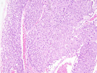
|
|
Image ID:1696 |
|
Source of Image:Ward JM |
|
Pathologist:Mikaelian I |
|
Method / Stain:H&E |
|
|
Image Caption:Solid mammary carcinoma with microinvasion: this photomicrograph illustrates an area of invasion of the stroma. Invasion occurs in the form of clusters (arrow) or individual cells (arrowhead).
|
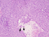
|
|
Image ID:1700 |
|
Source of Image:Ward JM |
|
Pathologist:Mikaelian I |
|
Method / Stain:H&E |
|
|
Image Caption:Solid mammary carcinoma: this photomicrograph illustrates a portion of the neoplasm where neoplastic cells are large, although small neoplastic cells (*) are also present.
|
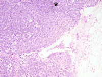
|
|
Image ID:1699 |
|
Source of Image:Ward JM |
|
Pathologist:Mikaelian I |
|
Method / Stain:H&E |
|
|
Image Caption:Solid mammary adenocarcinoma: this photomicrograph illustrates a portion of the neoplasm composed of large neoplastic cells (on the right of the photomicrograph) separated by a large vascular space from a portion of the neoplasm composed of smaller cells with less cytoplasm, and smaller and slightly more hyperchromatic nucleus. Mitoses are more numerous in the portion of the neoplasm composed of large neoplastic cells.
|
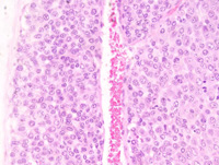
|
|
Image ID:1697 |
|
Source of Image:Ward JM |
|
Pathologist:Mikaelian I |
|
Method / Stain:H&E |
|
|
Image Caption:Mammary gland: the mammary gland is effaced by a large, nodular, multilobular, unencapsulated, densely cellular, neoplastic mass. This mass is composed of large coalescing lobules separated by a scant amount of loose fibrovascular stroma. Necrosis and cavitation are common in most neoplastic lobules.
|
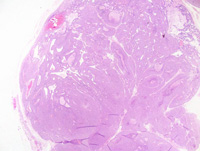
|
|
Image ID:1693 |
|
Source of Image:Ward JM |
|
Pathologist:Mikaelian I |
|
Method / Stain:H&E |
|
|
Image Caption:Solid mammary carcinoma with microinvasion: this high power photomicrograph illustrates an area of invasion of the stroma. Invasion occurs in the form of clusters or individual cells (arrowhead). Invasion in this model is not associated with epithelial to mesenchymal transition.
|
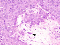
|
|
Image ID:1701 |
|
Source of Image:Ward JM |
|
Pathologist:Mikaelian I |
|
Method / Stain:H&E |
|
|
Image Caption:Solid mammary carcinoma: this photomicrograph illustrates an asymmetrical mitotic figure (arrowhead). Asymmetrical figures generally are not observed in mice transgenic for neu. However, mice transgenic for neu and with Trp53 mutations often develop tumors with prominent asymmetrical mitotic figures. A few neoplastic cells at the right of the photomicrograph contain lipid vacuoles. A few neoplastic cells at the left of the photomicrograph are undergoing single cell necrosis.
|
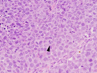
|
|
Image ID:1698 |
|
Source of Image:Ward JM |
|
Pathologist:Mikaelian I |
|
Method / Stain:H&E |
|
|
|
| MTB ID |
Tumor Name |
Organ(s) Affected |
Treatment Type |
Agents |
Strain Name |
Strain Sex |
Reproductive Status |
Tumor Frequency |
Age at Necropsy |
Description |
Reference |
| MTB:29236 |
Mammary gland adenocarcinoma |
Lung |
None (spontaneous) |
|
|
Female |
reproductive status not specified |
observed |
unknown |
Adenocarcinoma, mammary gland, metastatic to the lung |
J:94320 |
|
Image Caption:Lung: a small metastasis of a mammary tumor is present. This metastasis is predominantly composed of cell located within the lumen of a small artery (neoplastic embolus), but also of neoplastic cells that have escaped the lumen of the artery to invade the pulmonary interstitial (true metastasis).
|
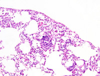
|
|
Image ID:1703 |
|
Source of Image:Ward JM |
|
Pathologist:Mikaelian I |
|
Method / Stain:H&E |
|
|
Image Caption:Lung: a small metastasis of a mammary tumor is present.
|
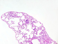
|
|
Image ID:1702 |
|
Source of Image:Ward JM |
|
Pathologist:Mikaelian I |
|
Method / Stain:H&E |
|
|
Image Caption:Lung: a small metastasis of a mammary tumor is present. This metastasis is predominantly composed of cell located within the lumen of a small artery (neoplastic embolus), but also of neoplastic cells that have escaped the lumen of the artery to invade the pulmonary interstitial (true metastasis). The endothelium of the artery (arrowhead) is still discernible. Neoplastic cells are polygonal, with ill-defined cell borders and a moderate amount of homogeneous acidophilic cytoplasm. Anisocytosis and anisokaryosis are mild. Nearby alveoli contain macrophages the cytoplasm of which is distended by acidophilic crystals (acidophilic crystals pneumonia).
|
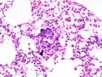
|
|
Image ID:1704 |
|
Source of Image:Ward JM |
|
Pathologist:Mikaelian I |
|
Method / Stain:H&E |
|
|
|
| MTB ID |
Tumor Name |
Organ(s) Affected |
Treatment Type |
Agents |
Strain Name |
Strain Sex |
Reproductive Status |
Tumor Frequency |
Age at Necropsy |
Description |
Reference |
| MTB:29237 |
Mammary gland adenoacanthoma |
Mammary gland |
None (spontaneous) |
|
|
Female |
reproductive status not specified |
observed |
unknown |
Adenocarcinoma, glandular, with keratinization (adenoacanthoma), mammary gland |
J:94320 |
|
Image Caption:Mammary gland: the neoplasm is composed of interconnected ducts lined by a one cell-thick epithelium (predominantly) and trabeculae supported by a small amount of fibrovascular stroma. There are multiple small areas of abrupt keratinization that result into the formation of small rafts of "ghost" epithelial cells, a feature reminicent of keratinization in pilomatricomas. A small number of neutrophils, lymphocytes and plasma cells is present in the stroma.
|
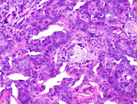
|
|
Image ID:1708 |
|
Source of Image:Ward JM |
|
Pathologist:Mikaelian I |
|
Method / Stain:H&E |
|
|
Image Caption:Mammary gland: the neoplasm is composed of interconnected ducts lined by a one cell-thick epithelium (predominantly) and trabeculae supported by a small amount of fibrovascular stroma. There are multiple small areas of keratinization that result into the formation of small "keratin pearls".
|
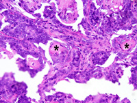
|
|
Image ID:1709 |
|
Source of Image:Ward JM |
|
Pathologist:Mikaelian I |
|
Method / Stain:H&E |
|
|
Image Caption:Mammary gland: the neoplasm is composed of interconnected ducts lined by a one cell-thick epithelium (predominantly) and trabeculae supported by a small amount of fibrovascular stroma. There are multiple small areas of keratinization characterized by the presence of keratohyalin granules (arrowhead) and that result into the formation of small "keratin pearls". Some neoplastic ducts have ruptured and cornified material has leaked into the stroma, causing a mild granulomatous reaction (arrow).
|
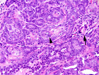
|
|
Image ID:1710 |
|
Source of Image:Ward JM |
|
Pathologist:Mikaelian I |
|
Method / Stain:H&E |
|
|
Image Caption:Mammary gland: the mammary parenchyma has been effaced by a densely cellular, nodular, unencapsulated neoplasm. This neoplasm is composed of interconnected ducts supported by a small amount of fibrovascular stroma.
|
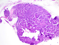
|
|
Image ID:1705 |
|
Source of Image:Ward JM |
|
Pathologist:Mikaelian I |
|
Method / Stain:H&E |
|
|
Image Caption:Mammary gland: the neoplasm is composed of interconnected ducts lined by a one cell-thick epithelium (predominantly) and trabeculae supported by a small amount of fibrovascular stroma. There are multiple small areas of abrupt keratinization that result into the formation of small rafts of "ghost" epithelial cells (arrowhead), a feature reminicent of keratinization in pilomatricomas.
|
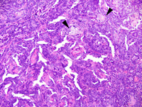
|
|
Image ID:1707 |
|
Source of Image:Ward JM |
|
Pathologist:Mikaelian I |
|
Method / Stain:H&E |
|
|
Image Caption:Mammary gland: the mammary parenchyma has been effaced by a densely cellular, nodular, unencapsulated neoplasm. This neoplasm is composed of interconnected ducts lined by a one cell-thick epithelium (predominantly) and trabeculae supported by a small amount of fibrovascular stroma.
|
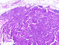
|
|
Image ID:1706 |
|
Source of Image:Ward JM |
|
Pathologist:Mikaelian I |
|
Method / Stain:H&E |
|
|
|
| MTB ID |
Tumor Name |
Organ(s) Affected |
Treatment Type |
Agents |
Strain Name |
Strain Sex |
Reproductive Status |
Tumor Frequency |
Age at Necropsy |
Description |
Reference |
| MTB:29238 |
Mammary gland adenoma |
Mammary gland |
Chemical/Drug |
pituitary isograft, 7,12-dimethylbenz[a]anthracene (DMBA) |
|
Female |
reproductive status not specified |
observed |
unknown |
Adenoma, glandular, with prominent atypia, and diffuse hyperplasia with atypia, mammary gland |
J:94320 |
|
Image Caption:Mammary adenoma: this photomicrograph of the periphery of the neoplasm illustrates the radiating glandular pattern with gradual transition into cords and trabeculae. Neoplastic cells are medium-sized, polygonal to cuboidal, with indistinct cell borders and a moderate amount of homogeneously eosinophilic cytoplasm. The nucleus is basal, round, and normochromatic. Anisocytosis and anisokaryosis overal are mild, although a few cells have a very large nucleus (arrowhead). There is prominent hyperplasia and atypia in the nearby mammary parenchyma.
|
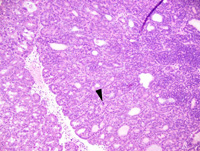
|
|
Image ID:1717 |
|
Source of Image:Ward JM |
|
Pathologist:Mikaelian I |
|
Method / Stain:H&E |
|
|
Image Caption:Mammary adenoma: this photomicrograph illustrates the glandular pattern that was present in the peripheral portions of the neoplasm. A few neoplastic cells have very large nuclei (arrowhead). Most glands are mildly ectatic and they are filed with an inspissated proteinaceous fluid.
|
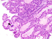
|
|
Image ID:1718 |
|
Source of Image:Ward JM |
|
Pathologist:Mikaelian I |
|
Method / Stain:H&E |
|
|
Image Caption:Mammary gland: the microadenoma is composed of closely-packed glands lined by a one cell-thick low columnar epithelium and supported by a small amount of fibrovascular stroma. Neoplastic cells are low columnar, with indistinct cell borders and a moderate amount of acidophilic cytoplasm that occasionally contains a few lipid vacuoles. The nucleus is basal, round, medium-sized and normochromatic. Anisokaryosis is moderate. Anisocytosis is mild. A few mitoses, including asymmetrical mitoses (arrowhead) are noted.
|
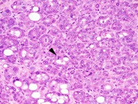
|
|
Image ID:1714 |
|
Source of Image:Ward JM |
|
Pathologist:Mikaelian I |
|
Method / Stain:H&E |
|
|
Image Caption:Mammary adenoma: this photomicrograph illustrates the trabecular pattern that was present in the central areas of the neoplasm. Neoplastic cells are crowded.
|
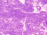
|
|
Image ID:1719 |
|
Source of Image:Ward JM |
|
Pathologist:Mikaelian I |
|
Method / Stain:H&E |
|
|
Image Caption:Mammary gland: the neoplasm is small, encapsulated, densely cellular, and expensile. It is composed of radiating glands with, at the center of the neoplasm, trabeculae and cords of neoplastic cells. The glands are lined by a one cell-thick cuboidal epithelium. There is a small amount of loose fibrovascular stroma with a mild lymphocytic and plasmacytic infiltrate that is most abundant at the center of the neoplasm.
|

|
|
Image ID:1716 |
|
Source of Image:Ward JM |
|
Pathologist:Mikaelian I |
|
Method / Stain:H&E |
|
|
Image Caption:Mammary gland: there is diffuse hyperplasia of the mammary gland with mild to moderate diffuse lactation. In addition, at the center of the tissue, there is a small, encapsulated, densely cellular, expensile neoplastic nodule (arrowheads; early stage of a glandular adenoma).
|
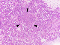
|
|
Image ID:1712 |
|
Source of Image:Ward JM |
|
Pathologist:Mikaelian I |
|
Method / Stain:H&E |
|
|
Image Caption:Mammary gland: there is diffuse hyperplasia of the mammary gland with mild to moderate diffuse lactation. In addition, at the center of the tissue, there is a small, encapsulated, densely cellular, expensile neoplastic nodule (early stage of a glandular adenoma). The nodule is composed of closely-packed glands lined by a one cell-thick low columnar epithelium and supported by a small amount of fibrovascular stroma. Neoplastic cells are low columnar, with indistinct cell borders and a moderate amount of acidophilic cytoplasm that occasionally contains a few lipid vacuoles. The nucleus is basal, round, medium-sized and normochromatic. Anisokaryosis is moderate. Anisocytosis is mild. A few mitoses are noted.
|
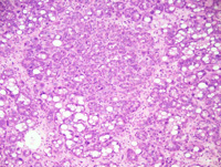
|
|
Image ID:1713 |
|
Source of Image:Ward JM |
|
Pathologist:Mikaelian I |
|
Method / Stain:H&E |
|
|
Image Caption:Mammary gland: there is diffuse hyperplasia of the mammary gland with mild to moderate diffuse lactation. In addition, at the center of the tissue, there is a small, encapsulated, densely cellular, expensile neoplastic nodule (glandular adenoma).
|
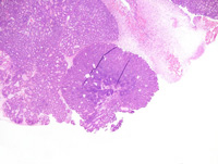
|
|
Image ID:1715 |
|
Source of Image:Ward JM |
|
Pathologist:Mikaelian I |
|
Method / Stain:H&E |
|
|
Image Caption:Mammary gland: there is diffuse hyperplasia of the mammary gland with mild to moderate diffuse lactation. In addition, at the center of the tissue, there is a small, encapsulated, densely cellular, expensile neoplastic nodule (early stage of a glandular adenoma).
|
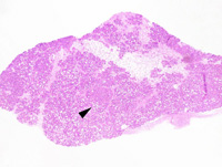
|
|
Image ID:1711 |
|
Source of Image:Ward JM |
|
Pathologist:Mikaelian I |
|
Method / Stain:H&E |
|
|
|
| MTB ID |
Tumor Name |
Organ(s) Affected |
Treatment Type |
Agents |
Strain Name |
Strain Sex |
Reproductive Status |
Tumor Frequency |
Age at Necropsy |
Description |
Reference |
| MTB:29239 |
Mammary gland hyperplasia - diffuse |
Mammary gland |
None (spontaneous) |
|
|
Female |
reproductive status not specified |
observed |
unknown |
Hyperplasia, diffuse, with squamous metaplasia, mammary gland |
J:94320 |
|
Image Caption:Diffuse mammary gland hyperplasia: there is marked diffuse hyperplasia of the mammary gland. Cells often pile-up to 2 cells-thick in a few glands and ducts. Many epithelial cells contain lipid vacuoles and they occasionally contain proteinaceous droplets. A few cells have large nuclei (arrowhead). There is mild interstitial lymphocytic and plasmacytic inflammation.
|
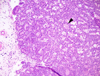
|
|
Image ID:1723 |
|
Source of Image:Ward JM |
|
Pathologist:Mikaelian I |
|
Method / Stain:H&E |
|
|
Image Caption:Mammary gland: there is severe diffuse hyperplasia of the mammary gland. All mammary acini and ducts are lined by a cuboidal epithelium with lipid and proteinaceous secretion. Most of these structures are mildly ectatic and contain an inspissated proteinaceous material. A mammary duct is ectatic, shows prominent cornification, and is surrounded by mild fibrosis and interstitial lymphocytic and plasmacytic inflammation.
|
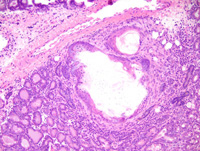
|
|
Image ID:1721 |
|
Source of Image:Ward JM |
|
Pathologist:Mikaelian I |
|
Method / Stain:H&E |
|
|
Image Caption:Diffuse mammary gland hyperplasia: there is marked diffuse hyperplasia of the mammary gland. Many epithelial cells contain lipid vacuoles and they occasionally contain proteinaceous droplets. There is moderate anisokaryosis. There is mild interstitial lymphocytic and plasmacytic inflammation.
|
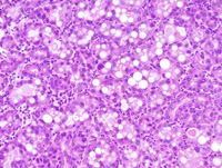
|
|
Image ID:1724 |
|
Source of Image:Ward JM |
|
Pathologist:Mikaelian I |
|
Method / Stain:H&E |
|
|
Image Caption:Mammary gland: there is severe diffuse hyperplasia of the mammary gland. All mammary acini and ducts are lined by a cuboidal epithelium with lipid and proteinaceous secretion. Most of these structures are mildly ectatic and contain an inspissated proteinaceous material. A mammary duct is ectatic, shows prominent cornification, and is surrounded by mild fibrosis and interstitial lymphocytic and plasmacytic inflammation.
|
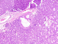
|
|
Image ID:1720 |
|
Source of Image:Ward JM |
|
Pathologist:Mikaelian I |
|
Method / Stain:H&E |
|
|
Image Caption:Ductal squamous metaplasia of the mammary gland: a mammary duct is ectatic, shows prominent cornification, and is surrounded by mild fibrosis and interstitial lymphocytic and plasmacytic inflammation.
|
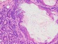
|
|
Image ID:1722 |
|
Source of Image:Ward JM |
|
Pathologist:Mikaelian I |
|
Method / Stain:H&E |
|
|
|
| MTB ID |
Tumor Name |
Organ(s) Affected |
Treatment Type |
Agents |
Strain Name |
Strain Sex |
Reproductive Status |
Tumor Frequency |
Age at Necropsy |
Description |
Reference |
| MTB:29240 |
Mammary gland adenocarcinoma |
Mammary gland |
None (spontaneous) |
|
|
Female |
reproductive status not specified |
observed |
unknown |
Adenocarcinoma, glandular, with prominent fibrosis and granulocytic inflammation, mammary gland |
J:94320 |
|
Image Caption:Mammary gland: this is a portion of a moderately to densely cellular neoplasm. This neoplasm is composed of glands and tubules lined by a one cell-thick cuboidal epithelium supported by an abundant amount of fibrovascular stroma. The stroma is infiltrated by a moderate number of granulocytes, lymphocytes and plasma cells. Neoplastic cells are cuboidal, medium-size, with indistinct cell borders and a moderate amount of strongly acidophilic cytoplasm that often contains lipid vacuoles. The nucleus is basal, oval, medium-size to large, and generally hyperchromatic. Anisokaryosis and anisocytosis are moderate. A few mitoses are detected.
|
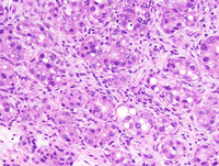
|
|
Image ID:1729 |
|
Source of Image:Ward JM |
|
Pathologist:Mikaelian I |
|
Method / Stain:H&E |
|
|
Image Caption:Mammary gland: this is a portion of a moderately to densely cellular neoplasm. This neoplasm is composed of glands and tubules supported by an abundant amount of fibrovascular stroma.
|
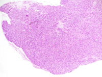
|
|
Image ID:1726 |
|
Source of Image:Ward JM |
|
Pathologist:Mikaelian I |
|
Method / Stain:H&E |
|
|
Image Caption:Glandular mammary adenocarcinoma: this photomicrograph illustrates a portion of the neoplasm where atypia is prominent and where a binucleated neoplastic cell is present.
|
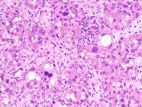
|
|
Image ID:1731 |
|
Source of Image:Ward JM |
|
Pathologist:Mikaelian I |
|
Method / Stain:H&E |
|
|
Image Caption:Mammary gland: this is a portion of a moderately to densely cellular neoplasm. This neoplasm is composed of glands and tubules lined by a one cell-thick cuboidal epithelium supported by an abundant amount of fibrovascular stroma. The stroma is infiltrated by a moderate number of granulocytes, lymphocytes and plasma cells. Neoplastic cells are cuboidal, medium-size, with indistinct cell borders and a moderate amount of strongly acidophilic cytoplasm that often contains lipid vacuoles. The nucleus is basal, oval, medium-size to large, and generally hyperchromatic. Anisokaryosis and anisocytosis are moderate.
|
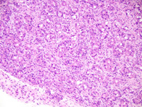
|
|
Image ID:1728 |
|
Source of Image:Ward JM |
|
Pathologist:Mikaelian I |
|
Method / Stain:H&E |
|
|
Image Caption:Mammary gland: this is a portion of a moderately to densely cellular neoplasm. This neoplasm is composed of glands and tubules lined by a one cell-thick cuboidal epithelium supported by an abundant amount of fibrovascular stroma. The stroma is infiltrated by a moderate number of granulocytes, lymphocytes and plasma cells.
|
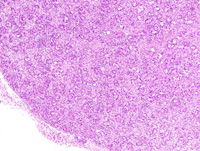
|
|
Image ID:1727 |
|
Source of Image:Ward JM |
|
Pathologist:Mikaelian I |
|
Method / Stain:H&E |
|
|
Image Caption:Glandular mammary adenocarcinoma: this photomicrograph illustrates a portion of the neoplasm where atypia is more prominent and where an asymmetrical mitosis is present.
|
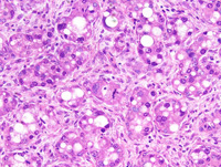
|
|
Image ID:1730 |
|
Source of Image:Ward JM |
|
Pathologist:Mikaelian I |
|
Method / Stain:H&E |
|
|
Image Caption:Mammary gland: this is a portion of a moderately to densely cellular neoplasm. This neoplasm is composed of glands and tubules supported by an abundant amount of fibrovascular stroma.
|
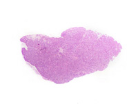
|
|
Image ID:1725 |
|
Source of Image:Ward JM |
|
Pathologist:Mikaelian I |
|
Method / Stain:H&E |
|
|
|
| MTB ID |
Tumor Name |
Organ(s) Affected |
Treatment Type |
Agents |
Strain Name |
Strain Sex |
Reproductive Status |
Tumor Frequency |
Age at Necropsy |
Description |
Reference |
| MTB:29241 |
Mammary gland adenocarcinoma - solid |
Mammary gland |
None (spontaneous) |
|
|
Female |
reproductive status not specified |
observed |
unknown |
Adenocarcinoma, solid, glandular, mammary gland |
J:94320 |
|
Image Caption:Solid mammary carcinoma: this photomicrograph illustrates a predominantly solid portion of the neoplasm. A few secondary lumens are present. Mitotic rate is elevated. Some mitoses are asymmetrical.
|
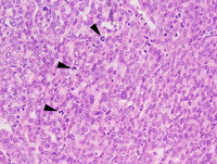
|
|
Image ID:1732 |
|
Source of Image:Ward JM |
|
Pathologist:Mikaelian I |
|
Method / Stain:H&E |
|
|
Image Caption:Mammary gland: the photomicrograph represents a portion of a densely cellular neoplasm. The neoplasm is composed of closely-packed lobules separated by a scant amount of fibrovascular stroma. Neoplastic lobules are mostly composed of solid areas with the formation of a few secondary lumens.
|
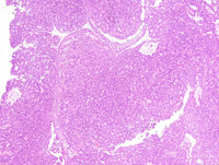
|
|
Image ID:1735 |
|
Source of Image:Ward JM |
|
Pathologist:Mikaelian I |
|
Method / Stain:H&E |
|
|
Image Caption:Mammary gland: the photomicrograph represents a portion of a densely cellular neoplasm. The neoplasm is composed of closely packed lobules separated by a scant amount of fibrovascular stroma. Neoplastic lobules are mostly composed of solid areas with the formation of a few secondary lumens. Neoplastic cells are polygonal, with indistinct cell borders and a moderate amount of homogeneous acidophilic cytoplasm. The nucleus is central, medium-size, round to oval, and normochromatic. Anisocytosis and anisokaryosis are mild. Mitoses are numerous.
|
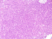
|
|
Image ID:1736 |
|
Source of Image:Ward JM |
|
Pathologist:Mikaelian I |
|
Method / Stain:H&E |
|
|
Image Caption:Mammary gland: the photomicrograph represents a portion of a densely cellular neoplasm.
|
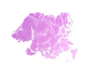
|
|
Image ID:1733 |
|
Source of Image:Ward JM |
|
Pathologist:Mikaelian I |
|
Method / Stain:H&E |
|
|
Image Caption:Mammary gland: the photomicrograph represents a portion of a densely cellular neoplasm. The neoplasm is composed of closely-packed lobules separated by a scant amount of fibrovascular stroma.
|
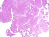
|
|
Image ID:1734 |
|
Source of Image:Ward JM |
|
Pathologist:Mikaelian I |
|
Method / Stain:H&E |
|
|
Image Caption:Solid mammary adenocarcinoma: this photomicrograph illustrates the morphology of a portion of the neoplasm with a solid phenotype. A few secondary lumens are present. Neoplastic cells are polygonal, with indistinct cell borders and a moderate amount of homogeneous acidophilic cytoplasm. The nucleus is central, medium-size, round to oval, and normochromatic. Anisocytosis and anisokaryosis are mild. Mitoses are numerous. There is no evidence of myoepithelial differentiation at the periphery of the neoplastic lobule.
|
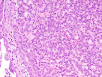
|
|
Image ID:1737 |
|
Source of Image:Ward JM |
|
Pathologist:Mikaelian I |
|
Method / Stain:H&E |
|
|
|
| MTB ID |
Tumor Name |
Organ(s) Affected |
Treatment Type |
Agents |
Strain Name |
Strain Sex |
Reproductive Status |
Tumor Frequency |
Age at Necropsy |
Description |
Reference |
| MTB:29242 |
Mammary gland adenocarcinoma - mixed |
Mammary gland |
None (spontaneous) |
|
|
Female |
reproductive status not specified |
observed |
unknown |
Adenocarcinoma, solid, comedo, mammary gland |
J:94320 |
|
Image Caption:Mammary gland: neoplastic cells are medium-sized, with indistinct cell borders and a moderate amount of amphophilic to basophilic cytoplasm. The nucleus is central, oval, medium-size, and slightly hypochromatic. Anisokaryosis and anisocytosis are mild. Mitoses are very numerous and occasionally asymmetrical. There is mild fibrosis and pleocellular inflammation at the periphery of neoplastic nests.
|
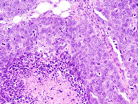
|
|
Image ID:1742 |
|
Source of Image:Ward JM |
|
Pathologist:Mikaelian I |
|
Method / Stain:H&E |
|
|
Image Caption:Mammary gland: the mammary gland is expanded by an invasive neoplasm characterized by the formation of large solid nests of neoplastic cells with various degrees of central necrosis and cavitation. Neoplastic cells are medium-sized, with indistinct cell borders and a moderate amount of amphophilic to basophilic cytoplasm. The nucleus is central, oval, medium-size, and slightly hypochromatic. Anisokaryosis and anisocytosis are mild. Mitoses are very numerous. There is mild fibrosis and pleocellular inflammation at the periphery of neoplastic nests.
|
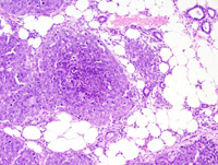
|
|
Image ID:1740 |
|
Source of Image:Ward JM |
|
Pathologist:Mikaelian I |
|
Method / Stain:H&E |
|
|
Image Caption:Mammary gland: the mammary gland is expanded by a nodular, ill-defined, invasive, unencapsulated, densely cellular neoplasm. The neoplasm is composed of very thick trabeculae, solid areas and nests that invade the pre-existing mammary stroma. Prominent areas of cavitation are present at the center of the largest trabeculae and solid areas.
|
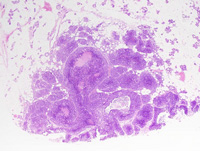
|
|
Image ID:1738 |
|
Source of Image:Ward JM |
|
Pathologist:Mikaelian I |
|
Method / Stain:H&E |
|
|
Image Caption:Mammary gland: neoplastic cells are medium-sized, with indistinct cell borders and a moderate amount of amphophilic to basophilic cytoplasm. The nucleus is central, oval, medium-size, and slightly hypochromatic. Anisokaryosis and anisocytosis are mild. Mitoses are very numerous and occasionally asymmetrical. There is mild fibrosis and pleocellular inflammation at the periphery of neoplastic nests.
|
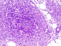
|
|
Image ID:1741 |
|
Source of Image:Ward JM |
|
Pathologist:Mikaelian I |
|
Method / Stain:H&E |
|
|
Image Caption:Mammary gland: the mammary gland is expanded by a nodular, ill-defined, invasive, unencapsulated, densely cellular neoplasm. The neoplasm is composed of very thick trabeculae, solid areas and nests that invade the pre-existing mammary stroma. Prominent areas of cavitation are present at the center of the largest trabeculae and solid areas.
|
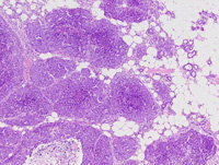
|
|
Image ID:1739 |
|
Source of Image:Ward JM |
|
Pathologist:Mikaelian I |
|
Method / Stain:H&E |
|
|
|
| MTB ID |
Tumor Name |
Organ(s) Affected |
Treatment Type |
Agents |
Strain Name |
Strain Sex |
Reproductive Status |
Tumor Frequency |
Age at Necropsy |
Description |
Reference |
| MTB:29266 |
Mammary gland adenocarcinoma - mixed |
Mammary gland |
None (spontaneous) |
|
|
Female |
reproductive status not specified |
observed |
unknown |
Adenocarcinoma, trabecular, solid, mammary gland |
J:94320 |
|
Image Caption:Mammary gland: the neoplasm is composed of closely-packed trabeculae that merge into solid areas and that are separated by a scant amount of fibrovascular stroma with a few entrapped adipocytes and a moderate lymphocytic and plasmacytic inflammation. Neoplastic cells are polygonal, with indistinct cell borders and a moderate amount of strongly amphophilic cytoplasm. The nucleus is central, oval, medium-sized, normochromatic, and with a coarsely clumped chromatin. Anisokaryosis and anisocytosis are mild. Mitotic rate is very high.
|
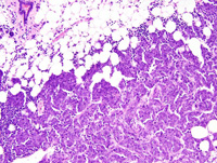
|
|
Image ID:1746 |
|
Source of Image:Ward JM |
|
Pathologist:Mikaelian I |
|
Method / Stain:H&E |
|
|
Image Caption:Mammary gland: the neoplasm is composed of closely-packed trabeculae that are 2-4 cells-thick at the periphery of the neoplasm, and up to 10 cells-thick at the center of the neoplasm. These trabeculae are separated by a scant amount of fibrovascular stroma and a moderate number of adipocytes have been entrapped in the neoplasm. A few tubules are present at the periphery of the neoplasm and are interpreted as non-neoplastic mammary tubules entrapped by the neoplasm. There is a moderate lymphocytic and plasmacytic inflammation at the periphery of the neoplasm.
|
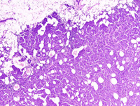
|
|
Image ID:1745 |
|
Source of Image:Ward JM |
|
Pathologist:Mikaelian I |
|
Method / Stain:H&E |
|
|
Image Caption:Mammary gland: the neoplasm is composed of closely-packed trabeculae that merge into solid areas and that are separated by a scant amount of fibrovascular stroma with a few entrapped adipocytes and a moderate lymphocytic and plasmacytic inflammation. Neoplastic cells are polygonal, with indistinct cell borders and a moderate amount of strongly amphophilic cytoplasm. The nucleus is central, oval, medium-sized, normochromatic, with a coarsely clumped chromatin and 1-3 small basophilic nucleoli. Anisokaryosis and anisocytosis are mild. Mitotic rate is very high and a few asymmetrical mitoses are identified (arrowhead).
|
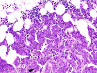
|
|
Image ID:1747 |
|
Source of Image:Ward JM |
|
Pathologist:Mikaelian I |
|
Method / Stain:H&E |
|
|
Image Caption:Solid mammary carcinoma: this photomicrograph illustrates the pattern of the neoplasm toward its' center. Neoplastic cells do not form lumens. A moderate number of adipocytes are entrapped within the neoplasm.
|
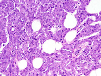
|
|
Image ID:1748 |
|
Source of Image:Ward JM |
|
Pathologist:Mikaelian I |
|
Method / Stain:H&E |
|
|
Image Caption:Mammary gland: this is a portion of a nodular, densely cellular, well-delineated, invasive neoplasm. Areas of necrosis are present at the center of the neoplasm (at the bottom of this photomicrograph).
|
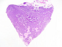
|
|
Image ID:1743 |
|
Source of Image:Ward JM |
|
Pathologist:Mikaelian I |
|
Method / Stain:H&E |
|
|
Image Caption:Mammary gland: the neoplasm is composed of closely-packed trabeculae that are 2-4 cells-thick at the periphery of the neoplasm, and up to 10 cells-thick at the center of the neoplasm. These trabeculae are separated by a scant amount of fibrovascular stroma and a moderate number of adipocytes have been entrapped in the neoplasm.
|
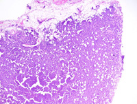
|
|
Image ID:1744 |
|
Source of Image:Ward JM |
|
Pathologist:Mikaelian I |
|
Method / Stain:H&E |
|
|
|
| MTB ID |
Tumor Name |
Organ(s) Affected |
Treatment Type |
Agents |
Strain Name |
Strain Sex |
Reproductive Status |
Tumor Frequency |
Age at Necropsy |
Description |
Reference |
| MTB:29267 |
Mammary gland adenocarcinoma |
Mammary gland |
None (spontaneous) |
|
|
Female |
reproductive status not specified |
observed |
unknown |
Adenocarcinoma, glandular, with prominent coagulation necrosis, mammary gland |
J:94320 |
|
Image Caption:Mammary gland: the mammary gland is replaced by a densely cellular neoplasm composed of closely-packed glands and branching ducts lined by a 1-2 cells-thick cuboidal epithelium and separated by a small amount of fibrovascular stroma. There are larg areas of coagulation necrosis.
|
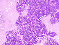
|
|
Image ID:1750 |
|
Source of Image:Ward JM |
|
Pathologist:Mikaelian I |
|
Method / Stain:H&E |
|
|
Image Caption:Mammary gland: the mammary gland is replaced by a densely cellular neoplasm composed of closely-packed glands. Approximately 60% of the neoplasm has undergone coagulation necrosis.
|
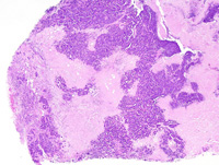
|
|
Image ID:1749 |
|
Source of Image:Ward JM |
|
Pathologist:Mikaelian I |
|
Method / Stain:H&E |
|
|
Image Caption:Mammary gland: the mammary gland is replaced by a densely cellular neoplasm composed of closely-packed glands and branching ducts lined by a 1-2 cells-thick cuboidal epithelium and separated by a small amount of fibrovascular stroma. Some ducts are moderately ectatic and filled by a strongly acidophilic amorphous material and desquamated neoplastic cells. Neoplastic cells are cuboidal, with indistinct cell borders and a moderate amount of amphophilic cytoplasm. The nucleus is central, round to oval, occasionally oriented in the long axis of the cell, medium-size, hyperchromatic, with a coarsely clumped chromatin and 1-4 small basophilic nucleoli. Anisokaryosis and anisocytosis are mild. Mitoses are numerous.
|
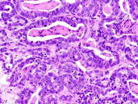
|
|
Image ID:1752 |
|
Source of Image:Ward JM |
|
Pathologist:Mikaelian I |
|
Method / Stain:H&E |
|
|
Image Caption:Mammary gland: the mammary gland is replaced by a densely cellular neoplasm composed of closely-packed glands and branching ducts lined by a 1-2 cells-thick cuboidal epithelium and separated by a small amount of fibrovascular stroma. Some ducts are moderately ectatic and filled by a strongly acidophilic amorphous material and desquamated neoplastic cells. There are larg areas of coagulation necrosis.
|
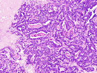
|
|
Image ID:1751 |
|
Source of Image:Ward JM |
|
Pathologist:Mikaelian I |
|
Method / Stain:H&E |
|
|
|
| MTB ID |
Tumor Name |
Organ(s) Affected |
Treatment Type |
Agents |
Strain Name |
Strain Sex |
Reproductive Status |
Tumor Frequency |
Age at Necropsy |
Description |
Reference |
| MTB:29268 |
Mammary gland adenocarcinoma - mixed |
Mammary gland |
None (spontaneous) |
|
|
Female |
reproductive status not specified |
observed |
unknown |
Adenocarcinoma, trabecular, solid, with prominent fibrosis and coagulation necrosis, mammary gland |
J:94320 |
|
Image Caption:Trabecular mammary adenocarcinoma: neoplastic cells form interconnected trabeculae that are 2-3 cells thick and that are separated by a moderate amount of fibrovascular stroma with a prominent pleocellular infiltrate. Neoplastic cells show prominent nuclear palissading at the periphery of the trabeculae. Luminal spaces are not detected. Neoplastic cells are cuboidal to low columnar, with indistinct cell borders and a small to moderate amount of amphophilic cytoplasm. The nucleus is basal, oval, medium-size, normochromatic, with a punctate chromatin and 1-2 small basophilic nucleoli. Numerous areas of necrosis are present.
|
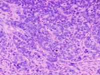
|
|
Image ID:1757 |
|
Source of Image:Ward JM |
|
Pathologist:Mikaelian I |
|
Method / Stain:H&E |
|
|
Image Caption:Trabecular mammary adenocarcinoma: neoplastic cells form interconnected trabeculae that are 2-3 cells thick and that are separated by a moderate amount of fibrovascular stroma with a prominent pleocellular infiltrate. Neoplastic cells show prominent nuclear palissading at the periphery of the trabeculae. Luminal spaces are not detected. Neoplastic cells are cuboidal to low columnar, with indistinct cell borders and a small to moderate amount of amphophilic cytoplasm. The nucleus is basal, oval and medium-size. Numerous areas of necrosis are present.
|
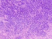
|
|
Image ID:1756 |
|
Source of Image:Ward JM |
|
Pathologist:Mikaelian I |
|
Method / Stain:H&E |
|
|
Image Caption:Trabecular mammary adenocarcinoma: neoplastic cells form interconnected trabeculae that are 2-3 cells thick and that are separated by a moderate amount of fibrovascular stroma with a prominent pleocellular infiltrate. Neoplastic cells show prominent nuclear palissading at the periphery of the trabeculae. Luminal spaces are not detected. Neoplastic cells are cuboidal to low columnar, with indistinct cell borders and a small to moderate amount of amphophilic cytoplasm. The nucleus is basal, oval, medium-size and normochromatic. There are numerous mitoses.
|
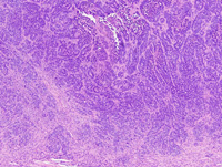
|
|
Image ID:1755 |
|
Source of Image:Ward JM |
|
Pathologist:Mikaelian I |
|
Method / Stain:H&E |
|
|
Image Caption:Mammary gland: this is a portion of a densely cellular, unencapsulated, invasive neoplasm that invades the nearby muscles. The neoplasm is composed of interconnected trabeculae that are 2-3 cells-thick and that are separated by a moderate amount of dense fibrovascular stroma.
|
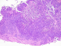
|
|
Image ID:1754 |
|
Source of Image:Ward JM |
|
Pathologist:Mikaelian I |
|
Method / Stain:H&E |
|
|
Image Caption:Mammary gland: this is a portion of a densely cellular, unencapsulated, invasive neoplasm that invades the nearby muscles (arrowhead). There is a moderate pleocellular inflammatory infiltrate throughout the neoplasm. There are large areas of coagulation necrosis (*) at the center of the neoplasm.
|
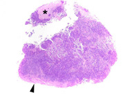
|
|
Image ID:1753 |
|
Source of Image:Ward JM |
|
Pathologist:Mikaelian I |
|
Method / Stain:H&E |
|
|
Image Caption:Trabecular mammary adenocarcinoma: this photomicrograph illustrates an area of coagulation necrosis (at the center of the photomicrograph) and nuclear palissading at the periphery of the trabeculae (in the upper left corner of the photomicrograph). Neoplastic cells occasionally form small acini. Luminal spaces are not formed.
|
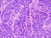
|
|
Image ID:1758 |
|
Source of Image:Ward JM |
|
Pathologist:Mikaelian I |
|
Method / Stain:H&E |
|
|
|
| MTB ID |
Tumor Name |
Organ(s) Affected |
Treatment Type |
Agents |
Strain Name |
Strain Sex |
Reproductive Status |
Tumor Frequency |
Age at Necropsy |
Description |
Reference |
| MTB:29269 |
Mammary gland adenocarcinoma - solid |
Mammary gland |
None (spontaneous) |
|
|
Female |
reproductive status not specified |
observed |
unknown |
Adenocarcinoma, solid, with epithelial to mesenchymal transition, mammary gland |
J:94320 |
|
Image Caption:Solid mammary carcinoma: neoplastic lobules are composed of closely-packed cells and often show central areas of liquefactive necrosis (comedo pattern). There is prominent nuclear palissading at the periphery of neoplastic lobules. Neoplastic cells are medium-size, polygonal, with indistinct cell borders and a small amount of acidophilic cytoplasm. The nucleus is central, oval, normochromatic, and with a coarsely clumped chromatin. Anisokaryosis and anisocytosis are mild. There are numerous mitoses.
|
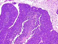
|
|
Image ID:1762 |
|
Source of Image:Ward JM |
|
Pathologist:Mikaelian I |
|
Method / Stain:H&E |
|
|
Image Caption:Solid mammary carcinoma: neoplastic lobules are composed of closely-packed cells and often show central areas of liquefactive necrosis (comedo pattern). There is prominent nuclear palissading at the periphery of neoplastic lobules. Neoplastic cells are medium-size, polygonal, with indistinct cell borders and a small amount of acidophilic cytoplasm. The nucleus is central, oval, normochromatic, and with a coarsely clumped chromatin. Anisokaryosis and anisocytosis are mild. There are numerous mitoses.
|
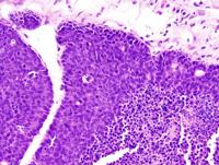
|
|
Image ID:1763 |
|
Source of Image:Ward JM |
|
Pathologist:Mikaelian I |
|
Method / Stain:H&E |
|
|
Image Caption:Epithelial to mesenchymal transition in a solid carcinoma: this high magnification picture illustrates an area of epithelial to mesenchymal transition: neoplastic cells have an epithelial phenotype in the lower and lower right portions of the photomicrograph. The have a mesenchymal phenotype in the upper left portions of the photomicrograph. Concomitant with epithelial to mesenchymal transitions, the pattern of the neoplasm changes from a solid/trabecular pattern to a streaming/whirling pattern.
|
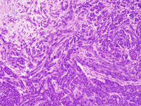
|
|
Image ID:1765 |
|
Source of Image:Ward JM |
|
Pathologist:Mikaelian I |
|
Method / Stain:H&E |
|
|
Image Caption:Epithelial to mesenchymal transition in a solid carcinoma: neoplastic cells form solid areas, are polygonal and have an epithelial phenotype in the lower right corner of the photomicrograph (E). Neoplastic cells are found as individual cells with a mesenchymal phenotypye embedded in a large amount of fibrovascular stroma in the upper left portions of the photomicrograph (M). Neoplastic cells are fusiform and form thick trabeculae in the area located between the area composed of epithelial neoplastic cells and the area composed of spindloid neoplastic cells.
|
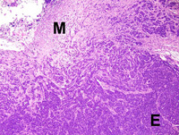
|
|
Image ID:1764 |
|
Source of Image:Ward JM |
|
Pathologist:Mikaelian I |
|
Method / Stain:H&E |
|
|
Image Caption:Mammary gland: this is a portion of a large mammary neoplasm composed of large lobules separated by thick fibrovascular septae. Numerous lobules are cavitated or have large central areas of liquefactive necrosis. An area of epithelial to mesenchymal transition (arrowhead) is present.
|
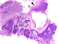
|
|
Image ID:1759 |
|
Source of Image:Ward JM |
|
Pathologist:Mikaelian I |
|
Method / Stain:H&E |
|
|
Image Caption:Mammary gland: this is a portion of a large mammary neoplasm composed of large lobules separated by thick fibrovascular septae. Numerous lobules are cavitated or have large central areas of liquefactive necrosis.
|
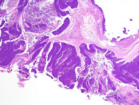
|
|
Image ID:1760 |
|
Source of Image:Ward JM |
|
Pathologist:Mikaelian I |
|
Method / Stain:H&E |
|
|
Image Caption:Solid mammary carcinoma: neoplastic lobules are composed of closely-packed cells and often show central areas of liquefactive necrosis (comedo pattern). There is prominent nuclear palissading at the periphery of neoplastic lobules.
|
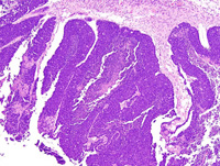
|
|
Image ID:1761 |
|
Source of Image:Ward JM |
|
Pathologist:Mikaelian I |
|
Method / Stain:H&E |
|
|
|
| MTB ID |
Tumor Name |
Organ(s) Affected |
Treatment Type |
Agents |
Strain Name |
Strain Sex |
Reproductive Status |
Tumor Frequency |
Age at Necropsy |
Description |
Reference |
| MTB:29271 |
Mammary gland adenocarcinoma - mixed |
Lung |
None (spontaneous) |
|
|
Female |
reproductive status not specified |
observed |
unknown |
Adenocarcinoma of mammary origin, glandular, solid, comedo, metastatic to the lung |
J:94320 |
|
Image Caption:Lung: two small metastases of a mammary neoplasm are present in the lung. These neoplasm are associated with mild atelectasis of the adjacent pulmonary parenchyma.
|
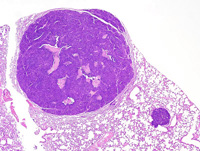
|
|
Image ID:1766 |
|
Source of Image:Ward JM |
|
Pathologist:Mikaelian I |
|
Method / Stain:H&E |
|
|
Image Caption:Lung: a pulmonary metastasis of a mammary adenocarcinoma is composed of large lobules separated by a scant amount of fibrovascular stroma. Neoplastic lobules are solid and neoplastic cells form numerous secondary lumens. Numerous large areas of liquefactive necrosis are present at the center of neoplastic lobules. There is prominent nuclear palissading at the periphery of neoplastic lobules.
|
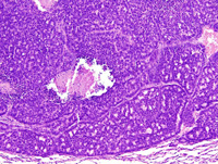
|
|
Image ID:1768 |
|
Source of Image:Ward JM |
|
Pathologist:Mikaelian I |
|
Method / Stain:H&E |
|
|
Image Caption:Lung: a pulmonary metastasis of a mammary adenocarcinoma is composed of large lobules separated by a scant amount of fibrovascular stroma. Neoplastic lobules are solid and neoplastic cells form secondary lumens (arrowheads). Numerous large areas of liquefactive necrosis are present at the center of neoplastic lobules. There is prominent nuclear palissading at the periphery of neoplastic lobules. Neoplastic cells are polygonal to columnar, with indistinct cell borders and a moderate amount of amphophilic cytoplasm that often contains a few small optically empty vacuoles (lipids?). The nucleus is central, oriented in the long axis of neoplastic cells, oval, medium-size and hyperchromatic. Anisokaryosis and anisocytosis are mild. Mitoses are numerous.
|
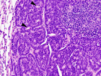
|
|
Image ID:1770 |
|
Source of Image:Ward JM |
|
Pathologist:Mikaelian I |
|
Method / Stain:H&E |
|
|
Image Caption:Lung: a pulmonary metastasis of a mammary adenocarcinoma is composed of large lobules separated by a scant amount of fibrovascular stroma. Neoplastic lobules are solid and neoplastic cells form numerous secondary lumens. Numerous large areas of liquefactive necrosis are present at the center of neoplastic lobules. There is prominent nuclear palissading at the periphery of neoplastic lobules. Neoplastic cells are polygonal to columnar, with indistinct cell borders and a moderate amount of amphophilic cytoplasm that often contains a few small optically empty vacuoles (lipids?). The nucleus is central, oriented in the long axis of neoplastic cells, oval, medium-size and hyperchromatic. Anisokaryosis and anisocytosis are mild. Mitoses are numerous.
|
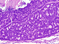
|
|
Image ID:1769 |
|
Source of Image:Ward JM |
|
Pathologist:Mikaelian I |
|
Method / Stain:H&E |
|
|
Image Caption:Lung: a pulmonary metastasis of a mammary adenocarcinoma is composed of large lobules separated by a scant amount of fibrovascular stroma. Neoplastic lobules are solid and neoplastic cells form numerous secondary lumens. Numerous large areas of liquefactive necrosis are present at the center of neoplastic lobules. There is prominent nuclear palissading at the periphery of neoplastic lobules.
|
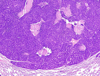
|
|
Image ID:1767 |
|
Source of Image:Ward JM |
|
Pathologist:Mikaelian I |
|
Method / Stain:H&E |
|
|
|
| MTB ID |
Tumor Name |
Organ(s) Affected |
Treatment Type |
Agents |
Strain Name |
Strain Sex |
Reproductive Status |
Tumor Frequency |
Age at Necropsy |
Description |
Reference |
| MTB:29273 |
Mammary gland adenocarcinoma - solid |
Lung |
None (spontaneous) |
|
|
Female |
reproductive status not specified |
observed |
unknown |
Mammary adenocarcinoma, solid, metastatic and embolic to the lungs |
J:94320 |
|
Image Caption:Lung: the photomicrograph represents a pulmonary metastasis of a mammary neoplasm. It is composed of small coalescing nests separated by a scant amount of fibrovascular stroma. Neoplastic cells are cuboidal, polygonal or oat-shaped, with ill-defined cell borders and a small amount of amphophilic cytoplasm. The nucleus is central, oval, normochromatic, with a finely stippled chromatin and 1-5 small basophilic nucleoli. Anisokaryosis and anisocytosis are mild. Numerous neoplastic cells undergo single cell necrosis, especially at the center of neoplastic nests.
|
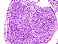
|
|
Image ID:1776 |
|
Source of Image:Ward JM |
|
Pathologist:Mikaelian I |
|
Method / Stain:H&E |
|
|
Image Caption:Lung: numerous nodular neoplasms are scattered throughout the pulmonary parenchyma. These nodules represent metastases of a mammary carcinoma. The metastases are composed of coalescing nests of various sizes. Secondary lumens are formed in some of these nests.
|
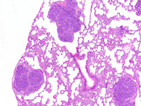
|
|
Image ID:1774 |
|
Source of Image:Ward JM |
|
Pathologist:Mikaelian I |
|
Method / Stain:H&E |
|
|
Image Caption:Lung: the photomicrograph represents a pulmonary metastasis of a mammary neoplasm. There is prominent invasion of the media of a pulmonary artery by neoplastic cells (*). Mitoses are numerous.
|
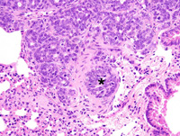
|
|
Image ID:1777 |
|
Source of Image:Ward JM |
|
Pathologist:Mikaelian I |
|
Method / Stain:H&E |
|
|
Image Caption:Lung: the photomicrograph represents a mammary neoplasm that has embolized in a pulmonary artery. The neoplasm has adhered to the wall of the blood vessel and has started to invade the intima and media of the artery. Mitoses and cells undergoing single cell necrosis are numerous. The center of the embolus has undergone liquefactive necrosis.
|
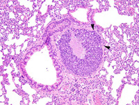
|
|
Image ID:1778 |
|
Source of Image:Ward JM |
|
Pathologist:Mikaelian I |
|
Method / Stain:H&E |
|
|
Image Caption:Lung: numerous nodular neoplasms are scattered throughout the pulmonary parenchyma. These nodules represent metastases of a mammary carcinoma.
|
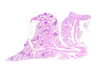
|
|
Image ID:1771 |
|
Source of Image:Ward JM |
|
Pathologist:Mikaelian I |
|
Method / Stain:H&E |
|
|
Image Caption:Lung: a pulmonary metastasis of a mammary neoplasm is present at the center of the photomicrograph. It is composed of small coalescing nests separated by a scant amount of fibrovascular stroma. Neoplastic cells are cuboidal, polygonal or oat-shaped, with ill-defined cell borders and a small amount of amphophilic cytoplasm. The nucleus is central, oval, normochromatic, with a finely stippled chromatin and 1-5 small basophilic nucleoli. Anisokaryosis and anisocytosis are mild. Mitoses are numerous.
|
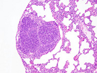
|
|
Image ID:1775 |
|
Source of Image:Ward JM |
|
Pathologist:Mikaelian I |
|
Method / Stain:H&E |
|
|
Image Caption:Lung: numerous nodular neoplasms are scattered throughout the pulmonary parenchyma. These nodules represent metastases of a mammary carcinoma.
|
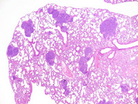
|
|
Image ID:1773 |
|
Source of Image:Ward JM |
|
Pathologist:Mikaelian I |
|
Method / Stain:H&E |
|
|
Image Caption:Lung: numerous nodular neoplasms are scattered throughout the pulmonary parenchyma. These nodules represent metastases of a mammary carcinoma.
|
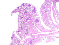
|
|
Image ID:1772 |
|
Source of Image:Ward JM |
|
Pathologist:Mikaelian I |
|
Method / Stain:H&E |
|
|
Image Caption:Lung: the photomicrograph represents a mammary neoplasm that has embolized in a pulmonary artery. The neoplasm has adhered to the wall of the blood vessel and has started to invade the intima and media of the artery. Mitoses and cells undergoing single cell necrosis are numerous. The center of the embolus has undergone liquefactive necrosis.
|
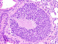
|
|
Image ID:1779 |
|
Source of Image:Ward JM |
|
Pathologist:Mikaelian I |
|
Method / Stain:H&E |
|
|
|
| MTB ID |
Tumor Name |
Organ(s) Affected |
Treatment Type |
Agents |
Strain Name |
Strain Sex |
Reproductive Status |
Tumor Frequency |
Age at Necropsy |
Description |
Reference |
| MTB:29274 |
Mammary gland carcinoma in situ |
Mammary gland |
None (spontaneous) |
|
|
Female |
reproductive status not specified |
observed |
unknown |
Carcinoma in situ and atypical lobular hyperplasia, mammary gland |
J:94320 |
|
Image Caption:Mammary gland: this photomicrograph illustrates a duct that is distended by large globules of inspissated proteinaceous material. There is mild to moderate fibrosis in the adjacent connective tissue.
|
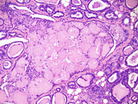
|
|
Image ID:1785 |
|
Source of Image:Ward JM |
|
Pathologist:Mikaelian I |
|
Method / Stain:H&E |
|
|
Image Caption:Mammary gland: the overall architecture of the mammary gland is maintained. Most interlobular ducts and a few intralobular ducts are moderately distended by a proteinaceous material. All intralobular ducts and acini are hypercellular and lined by a 1-3 cells-thick epithelium that occasionally forms minute papillae.
|
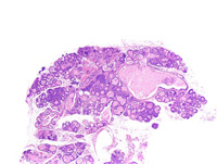
|
|
Image ID:1781 |
|
Source of Image:Ward JM |
|
Pathologist:Mikaelian I |
|
Method / Stain:H&E |
|
|
Image Caption:Mammary gland: the overall architecture of the mammary gland is maintained. Most interlobular ducts and a few intralobular ducts are moderately distended by a proteinaceous material. All intralobular ducts and acini are hypercellular and lined by a 1-3 cells-thick epithelium that occasionally forms minute papillae.
|
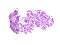
|
|
Image ID:1780 |
|
Source of Image:Ward JM |
|
Pathologist:Mikaelian I |
|
Method / Stain:H&E |
|
|
Image Caption:Mammary gland: this photomicrograph illustrates acini where epithelial cells pile-up disorderly up to 3-4 cells-thick. Other changes consist in lipid vacuoles in the cytoplasm of a few epithelial cells, desquamated epithelial cells and macrophages intermingled with a proteinaceous material in the lumen of all acini and intralobular ducts, an elevated mitotic rate, and the presence of numerous epithelial cells undergoing single cell necrosis.
|
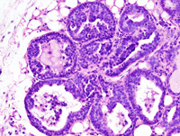
|
|
Image ID:1788 |
|
Source of Image:Ward JM |
|
Pathologist:Mikaelian I |
|
Method / Stain:H&E |
|
|
Image Caption:Mammary gland: the overall architecture of the mammary gland is maintained. Most interlobular ducts and a few intralobular ducts are moderately distended by a proteinaceous material that contains a few desquamated epithelial clels and macrophages. All intralobular ducts and acini are hypercellular and lined by a 1-3 cells-thick epithelium that occasionally forms minute papillae. There is mild to moderate interstitial fibrosis and pleocellular inflammation.
|
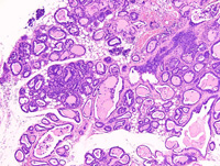
|
|
Image ID:1782 |
|
Source of Image:Ward JM |
|
Pathologist:Mikaelian I |
|
Method / Stain:H&E |
|
|
Image Caption:Mammary gland: this photomicrograph illustrates a duct that is distended by large globules of inspissated proteinaceous material. The globules of inspissated proteinaceous material are surrounded by a one cell-thick layer of basket-shaped cells. There is mild to moderate fibrosis in the adjacent connective tissue.
|
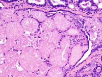
|
|
Image ID:1786 |
|
Source of Image:Ward JM |
|
Pathologist:Mikaelian I |
|
Method / Stain:H&E |
|
|
Image Caption:Mammary gland: this photomicrograph illustrates acini where epithelial cells pile-up disorderly up to 3-4 cells-thick. Other changes consist in lipid vacuoles in the cytoplasm of a few epithelial cells, desquamated epithelial cells and macrophages intermingled with a proteinaceous material in the lumen of all acini and intralobular ducts, an elevated mitotic rate, and the presence of numerous epithelial cells undergoing single cell necrosis.
|
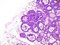
|
|
Image ID:1787 |
|
Source of Image:Ward JM |
|
Pathologist:Mikaelian I |
|
Method / Stain:H&E |
|
|
Image Caption:Mammary gland: the overall architecture of the mammary gland is maintained. Most interlobular ducts and a few intralobular ducts are moderately distended by a proteinaceous material that contains a few desquamated epithelial clels and macrophages. All intralobular ducts and acini are hypercellular and lined by a 1-3 cells-thick epithelium. In this field, epithelial cells often contain cytoplasmic lipid vacuoles.
|
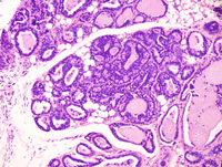
|
|
Image ID:1783 |
|
Source of Image:Ward JM |
|
Pathologist:Mikaelian I |
|
Method / Stain:H&E |
|
|
Image Caption:Mammary gland: the overall architecture of the mammary gland is maintained. Most interlobular ducts and a few intralobular ducts are moderately distended by a proteinaceous material that contains a few desquamated epithelial clels and macrophages. All intralobular ducts and acini are hypercellular and lined by a 1-3 cells-thick epithelium. In this field, epithelial cells often contain cytoplasmic lipid vacuoles. Mitoses are numerous.
|
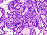
|
|
Image ID:1784 |
|
Source of Image:Ward JM |
|
Pathologist:Mikaelian I |
|
Method / Stain:H&E |
|
|
|
| MTB ID |
Tumor Name |
Organ(s) Affected |
Treatment Type |
Agents |
Strain Name |
Strain Sex |
Reproductive Status |
Tumor Frequency |
Age at Necropsy |
Description |
Reference |
| MTB:29275 |
Mammary gland hyperplasia - atypical - lobular |
Mammary gland |
None (spontaneous) |
|
|
Female |
reproductive status not specified |
observed |
unknown |
Carcinoma in situ and atypical lobular hyperplasia, mammary gland |
J:94320 |
|
Image Caption:Mammary gland: the overall architecture of the mammary gland is maintained. Most interlobular ducts and a few intralobular ducts are moderately distended by a proteinaceous material. All intralobular ducts and acini are hypercellular and lined by a 1-3 cells-thick epithelium that occasionally forms minute papillae.
|
|
|
|
Image ID:1781 |
|
Source of Image:Ward JM |
|
Pathologist:Mikaelian I |
|
Method / Stain:H&E |
|
|
Image Caption:Mammary gland: the overall architecture of the mammary gland is maintained. Most interlobular ducts and a few intralobular ducts are moderately distended by a proteinaceous material. All intralobular ducts and acini are hypercellular and lined by a 1-3 cells-thick epithelium that occasionally forms minute papillae.
|
|
|
|
Image ID:1780 |
|
Source of Image:Ward JM |
|
Pathologist:Mikaelian I |
|
Method / Stain:H&E |
|
|
Image Caption:Mammary gland: this photomicrograph illustrates acini where epithelial cells pile-up disorderly up to 3-4 cells-thick. Other changes consist in lipid vacuoles in the cytoplasm of a few epithelial cells, desquamated epithelial cells and macrophages intermingled with a proteinaceous material in the lumen of all acini and intralobular ducts, an elevated mitotic rate, and the presence of numerous epithelial cells undergoing single cell necrosis.
|
|
|
|
Image ID:1788 |
|
Source of Image:Ward JM |
|
Pathologist:Mikaelian I |
|
Method / Stain:H&E |
|
|
Image Caption:Mammary gland: the overall architecture of the mammary gland is maintained. Most interlobular ducts and a few intralobular ducts are moderately distended by a proteinaceous material that contains a few desquamated epithelial clels and macrophages. All intralobular ducts and acini are hypercellular and lined by a 1-3 cells-thick epithelium that occasionally forms minute papillae. There is mild to moderate interstitial fibrosis and pleocellular inflammation.
|
|
|
|
Image ID:1782 |
|
Source of Image:Ward JM |
|
Pathologist:Mikaelian I |
|
Method / Stain:H&E |
|
|
Image Caption:Mammary gland: this photomicrograph illustrates a duct that is distended by large globules of inspissated proteinaceous material. There is mild to moderate fibrosis in the adjacent connective tissue.
|
|
|
|
Image ID:1785 |
|
Source of Image:Ward JM |
|
Pathologist:Mikaelian I |
|
Method / Stain:H&E |
|
|
Image Caption:Mammary gland: this photomicrograph illustrates a duct that is distended by large globules of inspissated proteinaceous material. The globules of inspissated proteinaceous material are surrounded by a one cell-thick layer of basket-shaped cells. There is mild to moderate fibrosis in the adjacent connective tissue.
|
|
|
|
Image ID:1786 |
|
Source of Image:Ward JM |
|
Pathologist:Mikaelian I |
|
Method / Stain:H&E |
|
|
Image Caption:Mammary gland: this photomicrograph illustrates acini where epithelial cells pile-up disorderly up to 3-4 cells-thick. Other changes consist in lipid vacuoles in the cytoplasm of a few epithelial cells, desquamated epithelial cells and macrophages intermingled with a proteinaceous material in the lumen of all acini and intralobular ducts, an elevated mitotic rate, and the presence of numerous epithelial cells undergoing single cell necrosis.
|
|
|
|
Image ID:1787 |
|
Source of Image:Ward JM |
|
Pathologist:Mikaelian I |
|
Method / Stain:H&E |
|
|
Image Caption:Mammary gland: the overall architecture of the mammary gland is maintained. Most interlobular ducts and a few intralobular ducts are moderately distended by a proteinaceous material that contains a few desquamated epithelial clels and macrophages. All intralobular ducts and acini are hypercellular and lined by a 1-3 cells-thick epithelium. In this field, epithelial cells often contain cytoplasmic lipid vacuoles.
|
|
|
|
Image ID:1783 |
|
Source of Image:Ward JM |
|
Pathologist:Mikaelian I |
|
Method / Stain:H&E |
|
|
Image Caption:Mammary gland: the overall architecture of the mammary gland is maintained. Most interlobular ducts and a few intralobular ducts are moderately distended by a proteinaceous material that contains a few desquamated epithelial clels and macrophages. All intralobular ducts and acini are hypercellular and lined by a 1-3 cells-thick epithelium. In this field, epithelial cells often contain cytoplasmic lipid vacuoles. Mitoses are numerous.
|
|
|
|
Image ID:1784 |
|
Source of Image:Ward JM |
|
Pathologist:Mikaelian I |
|
Method / Stain:H&E |
|
|
|
| MTB ID |
Tumor Name |
Organ(s) Affected |
Treatment Type |
Agents |
Strain Name |
Strain Sex |
Reproductive Status |
Tumor Frequency |
Age at Necropsy |
Description |
Reference |
| MTB:29276 |
Mammary gland adenocarcinoma - mixed |
Mammary gland |
None (spontaneous) |
|
|
Female |
reproductive status not specified |
observed |
unknown |
Adenocarcinoma, papillary, solid, mammary gland |
J:94320 |
|
Image Caption:Mammary adenocarcinoma: this photomicrograph focuses on the solid portions of the neoplasm. The center of this area has undergone liquefactive necrosis. This area is composed of nests and solid areas. Slender fibrovascular septae (arrwoheads) subdivide the solid areas. Numerous neoplastic cells undergo single cell necrosis and mitotic rate is high.
|
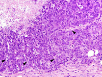
|
|
Image ID:1796 |
|
Source of Image:Ward JM |
|
Pathologist:Mikaelian I |
|
Method / Stain:H&E |
|
|
Image Caption:Mammary adenocarcinoma: this photomicrograph focuses on the papillary portions of the neoplasm. Neoplastic papillae have a slender fibrovascular core and are lined by a 2-15 cells-thick epithelium.
|
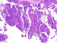
|
|
Image ID:1791 |
|
Source of Image:Ward JM |
|
Pathologist:Mikaelian I |
|
Method / Stain:H&E |
|
|
Image Caption:Mammary adenocarcinoma: this photomicrograph focuses on the solid portions of the neoplasm. The center of this area has undergone liquefactive necrosis. This area is composed of nests and solid areas. Slender fibrovascular septae subdivide the solid areas.
|
|
|
Image ID:1795 |
|
Source of Image:Ward JM |
|
Pathologist:Mikaelian I |
|
Method / Stain:H&E |
|
|
Image Caption:Mammary adenocarcinoma: this photomicrograph focuses on the papillary portions of the neoplasm. Neoplastic papillae have a slender fibrovascular core and are lined by a 2-15 cells-thick epithelium. Neoplastic cells are columnar to polygonal, with ill-defined cell borders and a small amount of amphophilic cytoplasm. The nucleus is oval to round, oriented in the long axis of the cell for columnar neoplastic cells, and slightly hyperchromatic. Numerous neoplastic cells undergo single cell necrosis and mitotic figures are numerous.
|
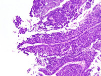
|
|
Image ID:1792 |
|
Source of Image:Ward JM |
|
Pathologist:Mikaelian I |
|
Method / Stain:H&E |
|
|
Image Caption:Mammary adenocarcinoma: this photomicrograph focuses on the solid portions of the neoplasm. The center of this solid area has undergone liquefactive necrosis.
|
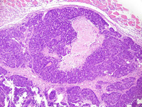
|
|
Image ID:1794 |
|
Source of Image:Ward JM |
|
Pathologist:Mikaelian I |
|
Method / Stain:H&E |
|
|
Image Caption:Mammary adenocarcinoma: this photomicrograph focuses on the papillary portions of the neoplasm. Neoplastic papillae have a slender fibrovascular core and are lined by a 2-15 cells-thick epithelium. Neoplastic cells are columnar (cells adherent to the basement membrane) to polygonal (suprabasal cells), with ill-defined cell borders and a small amount of amphophilic cytoplasm. The nucleus is oval to round, oriented in the long axis of the cell for columnar neoplastic cells, slightly hyperchromatic, and with a clumped chromatin. Numerous neoplastic cells undergo single cell necrosis and mitotic figures are numerous. Most mitoses are asymmetrical (the mitotic spindle is oriented at right angle with the basement membrane).
|
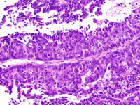
|
|
Image ID:1793 |
|
Source of Image:Ward JM |
|
Pathologist:Mikaelian I |
|
Method / Stain:H&E |
|
|
Image Caption:Mammary gland: the mammary gland is expanded by a large, nodular, well-delineated, encapsulated, densely cellular neoplasm that is comprised of papillary portions (lower portions of the neoplasm) and solid portions (upper portions of the neoplasm) with central liquefactive necrosis.
|
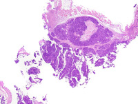
|
|
Image ID:1790 |
|
Source of Image:Ward JM |
|
Pathologist:Mikaelian I |
|
Method / Stain:H&E |
|
|
Image Caption:Mammary gland: the mammary gland is expanded by a large, nodular, well-delineated, encapsulated, densely cellular neoplasm that is comprised of papillary portions (lower portions of the neoplasm) and solid portions (upper portions of the neoplasm) with central liquefactive necrosis.
|
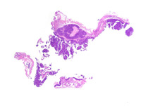
|
|
Image ID:1789 |
|
Source of Image:Ward JM |
|
Pathologist:Mikaelian I |
|
Method / Stain:H&E |
|
|
|
| MTB ID |
Tumor Name |
Organ(s) Affected |
Treatment Type |
Agents |
Strain Name |
Strain Sex |
Reproductive Status |
Tumor Frequency |
Age at Necropsy |
Description |
Reference |
| MTB:29277 |
Mammary gland carcinoma in situ - ductal |
Mammary gland |
None (spontaneous) |
|
|
Female |
reproductive status not specified |
observed |
unknown |
Ductal carcinoma in situ, mammary gland |
J:94320 |
|
Image Caption:Ductal carcinoma in situ: this photomicrograph illustrates the morphology of an interlobular duct that is distended by neoplastic cells. Neoplastic cells have not ruptured the basement membrane of the interlobular mammary duct.
|
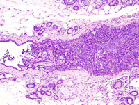
|
|
Image ID:1802 |
|
Source of Image:Ward JM |
|
Pathologist:Mikaelian I |
|
Method / Stain:H&E |
|
|
Image Caption:Mammary gland: the architecture of the mammary gland is maintained. All the acini and intralobular ducts of a mammary lobule are filled with and slightly distended by solid masses of epithelial cells. There is moderate interstitial fibrosis and pleocellular inflammation. Neoplastic cells are polygonal, with indistinct cell borders and a moderate amount of amphophilic cytoplasm. Numerous neoplastic cells undergo single cell necrosis.
|
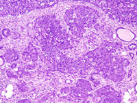
|
|
Image ID:1799 |
|
Source of Image:Ward JM |
|
Pathologist:Mikaelian I |
|
Method / Stain:H&E |
|
|
Image Caption:Mammary gland: the architecture of the mammary gland is maintained. The interlobular duct leading to a mammary lobule, and all the acini and intralobular ducts of this mammary lobule are filled with and slightly distended by solid masses of epithelial cells. There is moderate interstitial fibrosis and pleocellular inflammation. The ducts and acini of adjacent mammary lobules are moderately distended by an inspissated proteinaceous fluid, are lined by a prominent epithelium, and are surrounded by mild to moderate fibrosis.
|
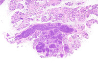
|
|
Image ID:1797 |
|
Source of Image:Ward JM |
|
Pathologist:Mikaelian I |
|
Method / Stain:H&E |
|
|
Image Caption:Mammary gland: the architecture of the mammary gland is maintained. The interlobular duct leading to a mammary lobule, and all the acini and intralobular ducts of a mammary lobule are filled with and slightly distended by solid masses of epithelial cells. There is moderate interstitial fibrosis and pleocellular inflammation. Neoplastic cells are polygonal, with indistinct cell borders and a moderate amount of amphophilic cytoplasm. Numerous neoplastic cells undergo single cell necrosis.
|
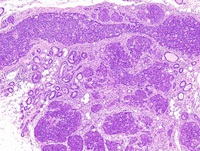
|
|
Image ID:1798 |
|
Source of Image:Ward JM |
|
Pathologist:Mikaelian I |
|
Method / Stain:H&E |
|
|
Image Caption:Ductal carcinoma in situ: this photomicrograph illustrates the morphology of an interlobular duct that is distended by neoplastic cells. Neoplastic cells have not ruptured the basement membrane of the interlobular mammary duct. Numerous neoplastic cells undergo apoptosis. A few mitoses are noted. Also, at the center of the duct, there is a small cluster of large cells (arrowheads) with better defined cell borders and a more hypochromatic than nearby cells. These features are consistent with squamous metaplasia. Myoepithelial differentiation (arrow) is present.
|
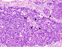
|
|
Image ID:1801 |
|
Source of Image:Ward JM |
|
Pathologist:Mikaelian I |
|
Method / Stain:H&E |
|
|
Image Caption:Ductal carcinoma in situ: there is keratinization of individual neoplastic cells (arrowheads). These cells have angular contours, a more acidophilic cytoplasm, and a larger and more hypochromatic nucleus with a larger nucleolus than nearby neoplastic cells. The other neoplastic cells are polygonal, with indistinct cell borders, a small amount of dark amphophilic cytoplasm, and a medium-size, oval, hyperchromatic nucleus with a punctate chromatin. Anisokaryosis and anisocytosis are mild. Mitosis and neoplastic cells undergoing single cell necrosis are numerous.
|
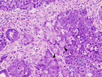
|
|
Image ID:1800 |
|
Source of Image:Ward JM |
|
Pathologist:Mikaelian I |
|
Method / Stain:H&E |
|
|
|
| MTB ID |
Tumor Name |
Organ(s) Affected |
Treatment Type |
Agents |
Strain Name |
Strain Sex |
Reproductive Status |
Tumor Frequency |
Age at Necropsy |
Description |
Reference |
| MTB:29278 |
Mammary gland hyperplasia - atypical - lobular |
Mammary gland |
None (spontaneous) |
|
|
Female |
reproductive status not specified |
observed |
unknown |
Atypical lobular hyperplasia, mammary gland |
J:94320 |
|
Image Caption:Mammary gland: the epithelium lining most acini is more prominent than normal. Most acini and ducts are mildly to moderately distended by an amorphous proteinaceous fluid. There is mild to moderate interstitial fibrosis. The mammary epithelium occasionally piles-up and shows moderate atypia. Numerous epithelial cells undergo apoptosis. A tripolar mitosis (arrowhead) is present.
|
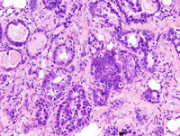
|
|
Image ID:1806 |
|
Source of Image:Ward JM |
|
Pathologist:Mikaelian I |
|
Method / Stain:H&E |
|
|
Image Caption:Mammary gland: the epithelium lining most acini and some ducts is more prominent than normal. Most acini and ducts are mildly to moderately distended by an amorphous proteinaceous fluid. There is mild to moderate interstitial fibrosis. The mammary epithelium occasionally piles-up and shows moderate atypia. Numerous epithelial cells undergo apoptosis.
|
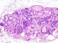
|
|
Image ID:1805 |
|
Source of Image:Ward JM |
|
Pathologist:Mikaelian I |
|
Method / Stain:H&E |
|
|
Image Caption:Mammary gland: the lobulation of the mammary gland is exaggerated. The epithelium lining most acini and some ducts is more prominent than normal. Most acini and ducts are mildly to moderately distended by an amorphous proteinaceous fluid. There is mild to moderate interstitial fibrosis.
|
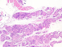
|
|
Image ID:1803 |
|
Source of Image:Ward JM |
|
Pathologist:Mikaelian I |
|
Method / Stain:H&E |
|
|
Image Caption:Mammary gland: the epithelium lining most acini and some ducts is more prominent than normal. Most acini and ducts are mildly to moderately distended by an amorphous proteinaceous fluid. There is mild to moderate interstitial fibrosis.
|
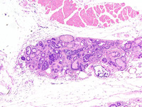
|
|
Image ID:1804 |
|
Source of Image:Ward JM |
|
Pathologist:Mikaelian I |
|
Method / Stain:H&E |
|
|
|
| MTB ID |
Tumor Name |
Organ(s) Affected |
Treatment Type |
Agents |
Strain Name |
Strain Sex |
Reproductive Status |
Tumor Frequency |
Age at Necropsy |
Description |
Reference |
| MTB:29279 |
Mammary gland hyperplasia - atypical - lobular |
Mammary gland |
None (spontaneous) |
|
|
Female |
reproductive status not specified |
observed |
unknown |
Atypical lobular hyperplasia, mammary gland |
J:94320 |
|
Image Caption:Atypical lobular hyperplasia: all alveoli are moderately ectatic, lined by a very prominent epithelium, and surrounded by mild to moderate interstitial fibrosis and pleocellular inflammation. Neoplastic cells seldom pile-up, and never pile-up more than 3 cells-thick.
|
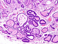
|
|
Image ID:1809 |
|
Source of Image:Ward JM |
|
Pathologist:Mikaelian I |
|
Method / Stain:H&E |
|
|
Image Caption:Mammary gland: the lobularity of the mammary gland is exagerated and there is moderate interstitial fibrosis. The epithelium lining most ducts and acini is prominent and the nucleo-cytoplasmic ration is elevated.
|
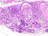
|
|
Image ID:1808 |
|
Source of Image:Ward JM |
|
Pathologist:Mikaelian I |
|
Method / Stain:H&E |
|
|
Image Caption:Mammary gland: the lobularity of the mammary gland is exagerated and there is moderate interstitial fibrosis. The epithelium lining most ducts and acini is prominent and the nucleo-cytoplasmic ration is elevated.
|
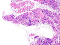
|
|
Image ID:1807 |
|
Source of Image:Ward JM |
|
Pathologist:Mikaelian I |
|
Method / Stain:H&E |
|
|
Image Caption:Atypical lobular hyperplasia: mammary alveoli are lined by a one cell-thick low columnar epithelium with a few minute places where cells pile-up up to 2 cells-thick. The cells have a medium-size, oval, and hyperchromatic nucleus. Mitotic figures and apoptotic bodies are numerous. There is mild to moderate interstitial edema, with mild interstitial fibrosis and pleocellular inflammation.
|
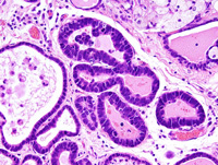
|
|
Image ID:1810 |
|
Source of Image:Ward JM |
|
Pathologist:Mikaelian I |
|
Method / Stain:H&E |
|
|
|
| MTB ID |
Tumor Name |
Organ(s) Affected |
Treatment Type |
Agents |
Strain Name |
Strain Sex |
Reproductive Status |
Tumor Frequency |
Age at Necropsy |
Description |
Reference |
| MTB:29280 |
Mammary gland carcinoma in situ - ductal |
Mammary gland |
None (spontaneous) |
|
|
Female |
reproductive status not specified |
observed |
unknown |
Ductal carcinoma in situ, mammary gland |
J:94320 |
|
Image Caption:Mammary gland: the lumen of the ducts and acini is moderately expanded by solid masses of epithelial cells. There is moderate interstitial fibrosis.
|
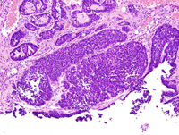
|
|
Image ID:1812 |
|
Source of Image:Ward JM |
|
Pathologist:Mikaelian I |
|
Method / Stain:H&E |
|
|
Image Caption:Mammary gland: the lumen of the ducts and acini is moderately expanded by solid masses of epithelial cells. There is moderate interstitial fibrosis. The cells are polygonal with ill-defined cell borders, a moderate amount of amphophilic cytoplasm, and a medium-size hyperchromatic nucleus. There are numerous mitotic figures.
|
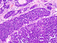
|
|
Image ID:1813 |
|
Source of Image:Ward JM |
|
Pathologist:Mikaelian I |
|
Method / Stain:H&E |
|
|
Image Caption:Mammary gland: the lumen of the ducts and acini is moderately expanded by solid masses of epithelial cells. There is moderate interstitial fibrosis.
|
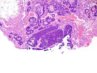
|
|
Image ID:1811 |
|
Source of Image:Ward JM |
|
Pathologist:Mikaelian I |
|
Method / Stain:H&E |
|
|
|
| MTB ID |
Tumor Name |
Organ(s) Affected |
Treatment Type |
Agents |
Strain Name |
Strain Sex |
Reproductive Status |
Tumor Frequency |
Age at Necropsy |
Description |
Reference |
| MTB:29281 |
Mammary gland adenocarcinoma - trabecular |
Mammary gland |
None (spontaneous) |
|
|
Female |
reproductive status not specified |
observed |
240 days |
Adenocarcinoma, glandular and trabecular, mammary gland |
J:94320 |
|
Image Caption:Mammary gland: the mammary gland is expanded by a nodular, well-delineated, locally invasive, and densely cellular neoplasm. This neoplasm is composed of glands and 3-6 cells-thick trabeculae supported by a moderate amount of fibrovascular stroma with a mild lymphocytic and plasmacytic inflammation. There is a moderate lymphocytic and plasmacytic inflammation at the periphery of the neoplasm.
|
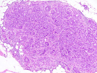
|
|
Image ID:1815 |
|
Source of Image:Ward JM |
|
Pathologist:Mikaelian I |
|
Method / Stain:H&E |
|
|
Image Caption:Mammary gland: the neoplasm is composed of glands and 3-6 cells-thick trabeculae supported by a moderate amount of fibrovascular stroma with a mild lymphocytic and plasmacytic inflammation. Neoplastic cells are polygonal to low columnar, with indistinct cell borders and a moderate amount of amphophilic cytoplasm. The nucleus is central, oval, medium-sized, hypochromatic and with 1-3 medium-size to large amphophilic nucleoli. Anisocytosis and anisokaryosis are mild to moderate. Mitoses are numerous. Numerous neoplastic cells undergo apoptosis.
|
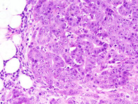
|
|
Image ID:1817 |
|
Source of Image:Ward JM |
|
Pathologist:Mikaelian I |
|
Method / Stain:H&E |
|
|
Image Caption:Mammary gland: the neoplasm is locally invasive. It is composed of glands and 3-6 cells-thick trabeculae supported by a moderate amount of fibrovascular stroma with a mild lymphocytic and plasmacytic inflammation. Neoplastic cells are polygonal to low columnar, with indistinct cell borders and a moderate amount of amphophilic cytoplasm. The nucleus is central, oval, medium-sized, hypochromatic and with 1-3 medium-size to large amphophilic nucleoli. Anisocytosis and anisokaryosis are mild to moderate. Mitoses are numerous. Numerous neoplastic cells undergo apoptosis.
|
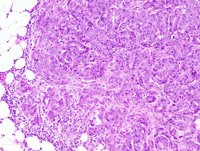
|
|
Image ID:1816 |
|
Source of Image:Ward JM |
|
Pathologist:Mikaelian I |
|
Method / Stain:H&E |
|
|
Image Caption:Mammary gland: two small neoplasms are present in the mammary gland. These neoplasms are nodular, well-delineated, locally invasive, and densely cellular.
|
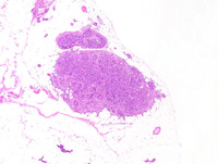
|
|
Image ID:1814 |
|
Source of Image:Ward JM |
|
Pathologist:Mikaelian I |
|
Method / Stain:H&E |
|
|
|
| MTB ID |
Tumor Name |
Organ(s) Affected |
Treatment Type |
Agents |
Strain Name |
Strain Sex |
Reproductive Status |
Tumor Frequency |
Age at Necropsy |
Description |
Reference |
| MTB:29284 |
Mammary gland adenocarcinoma - solid |
Mammary gland |
None (spontaneous) |
|
|
Female |
reproductive status not specified |
observed |
240 days |
Adenocarcinoma, solid, glandular, with epithelial to mesenchymal transition, mammary gland |
J:94320 |
|
Image Caption:Glandular mammary adenocarcinoma with epithelial to mesenchymal transition: this photomicrograph focuses on an area of epithelial to mesenchymal transition. A few glands lined by a one cell-thick cuboidal epithelium persist at the center of the area of epithelial to mesenchymal transition. Epithelial to mesenchymal transition is characterized by neoplastic cells acquiring a spindloid to epithelioid phenotype and blending into the stroma. Cells that have undergone epithelial to mesenchymal transition have distinct cell borders and a moderate to large amount of strongly acidophilic cytoplasm. The nucleus is oval, normochromatic, and with 1-2 small basophilic nucleoli. Cells with an epithelial phenotype are polygonal, with indistinct cell borders, and a small to moderate amount of amphophilic cytoplasm. The nucleus is central, round, slightly hyperchromatic and with a punctate chromatin.
|
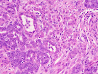
|
|
Image ID:1822 |
|
Source of Image:Ward JM |
|
Pathologist:Mikaelian I |
|
Method / Stain:H&E |
|
|
Image Caption:Glandular mammary adenocarcinoma with epithelial to mesenchymal transition: this photomicrograph focuses on an area of epithelial to mesenchymal transition. A few glands lined by a one cell-thick cuboidal epithelium persist at the center of the area of epithelial to mesenchymal transition. Epithelial to mesenchymal transition is characterized by neoplastic cells acquiring a spindloid to epithelioid phenotype and blending into the stroma.
|
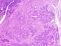
|
|
Image ID:1820 |
|
Source of Image:Ward JM |
|
Pathologist:Mikaelian I |
|
Method / Stain:H&E |
|
|
Image Caption:Glandular mammary adenocarcinoma with epithelial to mesenchymal transition: large portions of the neoplasm in this field have a glandular (epithelial) pattern. However, epithelial to mesenchymal transition (*) is prominent in the upper right corner of the photomicrograph. The glandular portions are composed of small glands embedded in a small amount of fibrovascular stroma. The spindloid portions are composed of individual neoplastic cells, small packets and small bundles embedded in moderate amount of fibrovascular stroma.
|
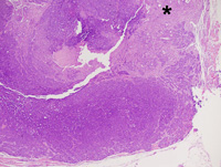
|
|
Image ID:1819 |
|
Source of Image:Ward JM |
|
Pathologist:Mikaelian I |
|
Method / Stain:H&E |
|
|
Image Caption:Glandular mammary adenocarcinoma with epithelial to mesenchymal transition: this photomicrograph represents a portion of the neoplasm that predominantly is composed of neoplastic cells with an epithelial phenotype forming glands. In addition, there is an area of epithelial to mesenchymal transition (upper left corner of the photomicrograph). Neoplastic cells with a spindloid phenotype can hardly be differentiated from stromal cells.
|
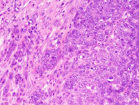
|
|
Image ID:1825 |
|
Source of Image:Ward JM |
|
Pathologist:Mikaelian I |
|
Method / Stain:H&E |
|
|
Image Caption:Mammary gland: the mammary gland is expanded by a large, nodular, unencapsulated, densely cellular neoplasm with a prominent central area of necrosis and cavitation. Most of the neoplasm has an epithelial phenotype. However, large portions of the neoplasm (?40%) show areas of epithelial to mesenchymal transition (upper right portions of the neoplasm).
|
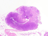
|
|
Image ID:1818 |
|
Source of Image:Ward JM |
|
Pathologist:Mikaelian I |
|
Method / Stain:H&E |
|
|
Image Caption:Glandular mammary adenocarcinoma with epithelial to mesenchymal transition: this photomicrograph represents a portion of the neoplasm that predominantly is composed of neoplastic cells with an epithelial phenotype forming glands. In addition, there is an area of epithelial to mesenchymal transition (*).
|
|
|
Image ID:1823 |
|
Source of Image:Ward JM |
|
Pathologist:Mikaelian I |
|
Method / Stain:H&E |
|
|
Image Caption:Glandular mammary adenocarcinoma with epithelial to mesenchymal transition: this photomicrograph focuses on an area of epithelial to mesenchymal transition. A few glands lined by a one cell-thick cuboidal epithelium persist at the center of the area of epithelial to mesenchymal transition. Epithelial to mesenchymal transition is characterized by neoplastic cells acquiring a spindloid to epithelioid phenotype and blending into the stroma.
|
|
|
Image ID:1821 |
|
Source of Image:Ward JM |
|
Pathologist:Mikaelian I |
|
Method / Stain:H&E |
|
|
Image Caption:Glandular mammary adenocarcinoma with epithelial to mesenchymal transition: this photomicrograph represents a portion of the neoplasm that predominantly is composed of neoplastic cells with an epithelial phenotype forming glands. In addition, there is an area of epithelial to mesenchymal transition (*).
|
|
|
Image ID:1824 |
|
Source of Image:Ward JM |
|
Pathologist:Mikaelian I |
|
Method / Stain:H&E |
|
|
|
| MTB ID |
Tumor Name |
Organ(s) Affected |
Treatment Type |
Agents |
Strain Name |
Strain Sex |
Reproductive Status |
Tumor Frequency |
Age at Necropsy |
Description |
Reference |
| MTB:29288 |
Mammary gland adenocarcinoma - papillary |
Mammary gland |
None (spontaneous) |
|
|
Female |
reproductive status not specified |
observed |
unknown |
Adenocarcinoma, glandular and papillary, with prominent coagulation necrosis, mammary gland |
J:94320 |
|
Image Caption:Glandular mammary adenocarcinoma: the neoplasm is composed of closely-packed papillae and glands supported by a scant amount of fibrovascular stroma. There are large coalescing areas of coagulation necrosis at the center of the neoplasm. Neoplastic cells form glands lined by a one cell-thick low columnar to cuboidal epithelium supported by a scant amount of fibrovascular stroma. Many neoplastic glands are dilated by desquamated and degenerated neoplastic cells. Neoplastic often pile-up disorderly up to 5 cells-thick. Neoplastic cells are cuboidal to columnar with indistinct cell borders and a moderate amount of strongly amphophilic cytoplasm. The nucleus is central, oval, and slightly hyperchromatic.
|
|
|
Image ID:1830 |
|
Source of Image:Ward JM |
|
Pathologist:Mikaelian I |
|
Method / Stain:H&E |
|
|
Image Caption:Glandular mammary adenocarcinoma: the neoplasm is composed of closely-packed papillae and glands supported by a scant amount of fibrovascular stroma. There are large coalescing areas of coagulation necrosis at the center of the neoplasm. Neoplastic cells form glands lined by a one cell-thick low columnar to cuboidal epithelium supported by a scant amount of fibrovascular stroma. Many neoplastic glands are dilated by desquamated and degenerated neoplastic cells.
|
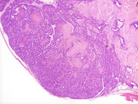
|
|
Image ID:1828 |
|
Source of Image:Ward JM |
|
Pathologist:Mikaelian I |
|
Method / Stain:H&E |
|
|
Image Caption:Glandular mammary adenocarcinoma: the neoplasm is composed of closely-packed papillae and glands supported by a scant amount of fibrovascular stroma. There are large coalescing areas of coagulation necrosis at the center of the neoplasm. Neoplastic cells form glands lined by a one cell-thick low columnar to cuboidal epithelium supported by a scant amount of fibrovascular stroma. Many neoplastic glands are dilated by desquamated and degenerated neoplastic cells. Neoplastic often pile-up disorderly up to 5 cells-thick. Neoplastic cells are cuboidal to columnar with indistinct cell borders and a moderate amount of strongly amphophilic cytoplasm. The nucleus is central, oval, and slightly hyperchromatic. Numerous neoplastic cells undergo single cell necrosis.
|
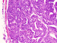
|
|
Image ID:1831 |
|
Source of Image:Ward JM |
|
Pathologist:Mikaelian I |
|
Method / Stain:H&E |
|
|
Image Caption:Mammary gland: the mammary gland is expanded by a very large, unencapsulated, expensile, multilobular neoplastic mass. Most of the neoplasm has undergone coagulation necrosis and only the peripheral portions survive, as well as a few neoplastic glands at the vicinity of the blood vessels towards the center of the neoplasm.
|
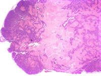
|
|
Image ID:1826 |
|
Source of Image:Ward JM |
|
Pathologist:Mikaelian I |
|
Method / Stain:H&E |
|
|
Image Caption:Glandular mammary adenocarcinoma: the neoplasm is composed of closely-packed glands and papillae supported by a scant amount of fibrovascular stroma. There are large coalescing areas of coagulation necrosis at the center of the neoplasm.
|
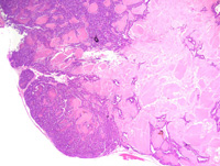
|
|
Image ID:1827 |
|
Source of Image:Ward JM |
|
Pathologist:Mikaelian I |
|
Method / Stain:H&E |
|
|
Image Caption:Glandular mammary adenocarcinoma: the neoplasm is composed of closely-packed papillae and glands supported by a scant amount of fibrovascular stroma. There are large coalescing areas of coagulation necrosis at the center of the neoplasm. Neoplastic cells form glands lined by a one cell-thick low columnar to cuboidal epithelium supported by a scant amount of fibrovascular stroma. Many neoplastic glands are dilated by desquamated and degenerated neoplastic cells. Neoplastic often pile-up disorderly up to 5 cells-thick. Neoplastic cells are cuboidal to columnar with indistinct cell borders and a moderate amount of strongly amphophilic cytoplasm. The nucleus is central, oval, and slightly hyperchromatic.
|
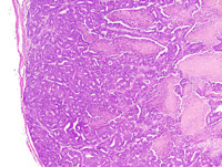
|
|
Image ID:1829 |
|
Source of Image:Ward JM |
|
Pathologist:Mikaelian I |
|
Method / Stain:H&E |
|
|
|
| MTB ID |
Tumor Name |
Organ(s) Affected |
Treatment Type |
Agents |
Strain Name |
Strain Sex |
Reproductive Status |
Tumor Frequency |
Age at Necropsy |
Description |
Reference |
| MTB:29290 |
Mammary gland adenocarcinoma - mixed |
Mammary gland |
None (spontaneous) |
|
|
Female |
reproductive status not specified |
observed |
unknown |
Adenocarcinoma, solid, comedo, mammary gland |
J:94320 |
|
Image Caption:Mammary gland: the mammary gland is effaced by a nodular, partly encapsulated, locally invasive, densely cellular neoplasm. This neoplasm is composed of solid areas with central cavitation (comedo pattern). Areas suggestive of the early stages of epithelial to mesenchymal transition are present at the periphery of the neoplasm (arrowhead).
|
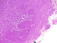
|
|
Image ID:1834 |
|
Source of Image:Ward JM |
|
Pathologist:Mikaelian I |
|
Method / Stain:H&E |
|
|
Image Caption:Central portions of the neoplasm (solid with early stage of pseudoglandular pattern): the neoplasm is composed of very thick coalescing trabeculae separated by a small amount of loose fibrovascular stroma. Numerous neoplastic cells undergo single cell necrosis which results into the formation of small punched-out areas in the neoplastic trabeculae, a pattern known as "pseudoglandular pattern". Mitotic rate is high.
|
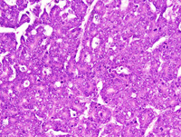
|
|
Image ID:1838 |
|
Source of Image:Ward JM |
|
Pathologist:Mikaelian I |
|
Method / Stain:H&E |
|
|
Image Caption:Peripheral portions of the neoplasm (trabecular area): neoplastic cells form trabeculae, cords and occasionally glands with an inconspicuous lumen (*) separated by a small to moderate amount of fibrovascular stroma. Neoplastic cells are polygonal, with indistinct cell borders and a moderate amount of acidophilic cytoplasm. The nucleus is central, round to oval, slightly hypochromatic, with a stippled chromatin and 1-3 large nuclei. Anisokaryosis is moderate. Anisocytosis is mild. Numerous neoplastic cells undergo single cell necrosis. A few mitoses are present.
|
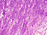
|
|
Image ID:1837 |
|
Source of Image:Ward JM |
|
Pathologist:Mikaelian I |
|
Method / Stain:H&E |
|
|
Image Caption:Mammary gland: the neoplasm is composed of solid areas with central cavitation (comedo pattern, left portions of the photomicrograph) and solid areas (center of the photomicrograph). Neoplastic cells form trabeculae and become spindloid towards the periphery of the neoplasm. However, they do not form bundles and fascicles. Apoptosis is prominent in the solid portions of the neoplasm.
|
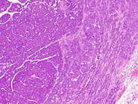
|
|
Image ID:1835 |
|
Source of Image:Ward JM |
|
Pathologist:Mikaelian I |
|
Method / Stain:H&E |
|
|
Image Caption:Central portions of the neoplasm (solid and comedo patterns): the neoplasm is composed of very thick coalescing trabeculae and nests separated by a small amount of loose fibrovascular stroma. Numerous neoplastic cells undergo single cell necrosis which results into the cavitation of the nests and trabeculae, a pattern known as the comedo pattern.
|
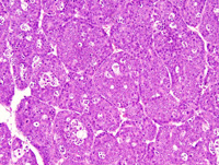
|
|
Image ID:1839 |
|
Source of Image:Ward JM |
|
Pathologist:Mikaelian I |
|
Method / Stain:H&E |
|
|
Image Caption:Peripheral portions of the neoplasm (solid/trabecular area): the neoplasm is composed of solid areas (left portions of the photomicrograph). Neoplastic cells form trabeculae and become spindloid towards the periphery of the neoplasm. However, they do not form bundles and fascicles. Apoptosis is prominent in the solid portions of the neoplasm.
|
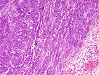
|
|
Image ID:1836 |
|
Source of Image:Ward JM |
|
Pathologist:Mikaelian I |
|
Method / Stain:H&E |
|
|
Image Caption:Mammary gland: the mammary gland is effaced by a nodular, partly encapsulated, locally invasive, densely cellular neoplasm. This neoplasm is composed of solid areas with central cavitation (comedo pattern). Areas suggestive of the early stages of epithelial to mesenchymal transition are present at the periphery of the neoplasm (arrowhead).
|
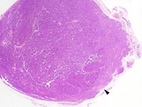
|
|
Image ID:1833 |
|
Source of Image:Ward JM |
|
Pathologist:Mikaelian I |
|
Method / Stain:H&E |
|
|
Image Caption:Mammary gland: the mammary gland is effaced by a nodular, partly encapsulated, locally invasive, densely cellular neoplasm. This neoplasm is composed of solid areas with central cavitation (comedo pattern; towards the center). Areas suggestive of the early stages of epithelial to mesenchymal transition are present at the periphery of the neoplasm (arrowhead).
|
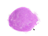
|
|
Image ID:1832 |
|
Source of Image:Ward JM |
|
Pathologist:Mikaelian I |
|
Method / Stain:H&E |
|
|
|
| MTB ID |
Tumor Name |
Organ(s) Affected |
Treatment Type |
Agents |
Strain Name |
Strain Sex |
Reproductive Status |
Tumor Frequency |
Age at Necropsy |
Description |
Reference |
| MTB:29295 |
Mammary gland adenocarcinoma - solid |
Mammary gland |
None (spontaneous) |
|
|
Female |
reproductive status not specified |
observed |
unknown |
Adenocarcinoma, solid, mammary gland |
J:94320 |
|
Image Caption:Solid mammary carcinoma: the neoplasm is composed of large solid areas supported by a scant amount of fibrovascular stroma. Numerous neoplastic cells undergo apoptosis. Neoplastic cells are polygonal, with indistinct cell borders and a moderate amount of strongly amphophilic cytoplasm. The nucleus is central, oval, medium-size, slightly hypochromatic, with a clumped chromatin and 1-3 large acidophilic nucleoli Anisokaryosis and anisocytosis are moderate. Mitoses are numerous. Many neoplastic cells undergo apoptosis.
|
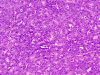
|
|
Image ID:1847 |
|
Source of Image:Ward JM |
|
Pathologist:Mikaelian I |
|
Method / Stain:H&E |
|
|
Image Caption:Solid mammary carcinoma: the neoplasm is composed of large solid areas supported by a scant amount of fibrovascular stroma. Numerous neoplastic cells undergo apoptosis. Neoplastic cells are polygonal, with indistinct cell borders and a moderate amount of strongly amphophilic cytoplasm. The nucleus is central, oval, medium-size and slightly hypochromatic. Mitoses are numerous. Many neoplastic cells undergo apoptosis.
|
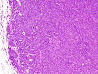
|
|
Image ID:1844 |
|
Source of Image:Ward JM |
|
Pathologist:Mikaelian I |
|
Method / Stain:H&E |
|
|
Image Caption:Mammary gland: the mammary gland is expanded by a large, multilobular, well-delineated, densely cellular neoplasm. Numerous small areas of necrosis and cavitation are scattered throughout the neoplasm. The neoplasm is mostly composed of solid areas supported by a small amount of fibrovascular stroma. However, the presence of numerous areas of cavitation confer a pseudoglandular pattern to the neoplasm.
|
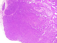
|
|
Image ID:1842 |
|
Source of Image:Ward JM |
|
Pathologist:Mikaelian I |
|
Method / Stain:H&E |
|
|
Image Caption:Mammary gland: the mammary gland is expanded by a large, multilobular, well-delineated, densely cellular neoplasm. Numerous small areas of necrosis and cavitation are scattered throughout the neoplasm.
|
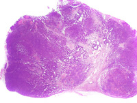
|
|
Image ID:1840 |
|
Source of Image:Ward JM |
|
Pathologist:Mikaelian I |
|
Method / Stain:H&E |
|
|
Image Caption:Solid mammary carcinoma: the neoplasm is composed of large solid areas supported by a scant amount of fibrovascular stroma. Numerous neoplastic cells undergo apoptosis.
|
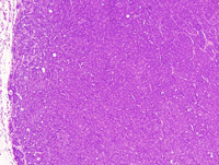
|
|
Image ID:1843 |
|
Source of Image:Ward JM |
|
Pathologist:Mikaelian I |
|
Method / Stain:H&E |
|
|
Image Caption:Solid mammary carcinoma: the neoplasm is composed of large solid areas supported by a scant amount of fibrovascular stroma. Numerous neoplastic cells undergo apoptosis. Neoplastic cells are polygonal, with indistinct cell borders and a moderate amount of strongly amphophilic cytoplasm. The nucleus is central, oval, medium-size, slightly hypochromatic, with a clumped chromatin and 1-3 large acidophilic nucleoli Anisokaryosis and anisocytosis are moderate. Mitoses are numerous. Many neoplastic cells undergo apoptosis.
|
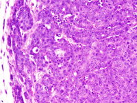
|
|
Image ID:1845 |
|
Source of Image:Ward JM |
|
Pathologist:Mikaelian I |
|
Method / Stain:H&E |
|
|
Image Caption:Mammary gland: the mammary gland is expanded by a large, multilobular, well-delineated, densely cellular neoplasm. Numerous small areas of necrosis and cavitation are scattered throughout the neoplasm.
|
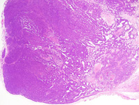
|
|
Image ID:1841 |
|
Source of Image:Ward JM |
|
Pathologist:Mikaelian I |
|
Method / Stain:H&E |
|
|
Image Caption:Solid mammary carcinoma: the neoplasm is composed of large solid areas supported by a scant amount of fibrovascular stroma. Numerous neoplastic cells undergo apoptosis. Neoplastic cells are polygonal, with indistinct cell borders and a moderate amount of strongly amphophilic cytoplasm. The nucleus is central, oval, medium-size, slightly hypochromatic, with a clumped chromatin and 1-3 large acidophilic nucleoli Anisokaryosis and anisocytosis are moderate. Mitoses are numerous. Many neoplastic cells undergo apoptosis.
|
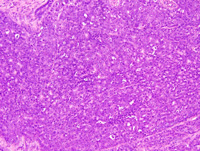
|
|
Image ID:1846 |
|
Source of Image:Ward JM |
|
Pathologist:Mikaelian I |
|
Method / Stain:H&E |
|
|
|
| MTB ID |
Tumor Name |
Organ(s) Affected |
Treatment Type |
Agents |
Strain Name |
Strain Sex |
Reproductive Status |
Tumor Frequency |
Age at Necropsy |
Description |
Reference |
| MTB:29296 |
Mammary gland adenocarcinoma - mixed |
Mammary gland |
None (spontaneous) |
|
|
Female |
reproductive status not specified |
observed |
unknown |
Adenocarcinoma, glandular, acinar, cystic, solid with secondary lumens, mammary gland |
J:94320 |
|
Image Caption:Mammary gland: this photomicrograph illustrates a cystic neoplastic lobule lined by a 5-10 cells thick epithelium that forms a few small secondary lumens. Neoplastic cells are cuboidal to polygonal, with indistinct cell borders and a small amount of strongly amphophilic cytoplasm. The nucleus is central, oval, normochromatic, with a clumped chromatin and occasionally a small basophilic nucleolus. Anisokaryosis and anisocytosis are mild. Mitoses are numerous. A skeletal muscle fiber (M) is entrapped in the neoplasm.
|
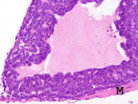
|
|
Image ID:1853 |
|
Source of Image:Ward JM |
|
Pathologist:Mikaelian I |
|
Method / Stain:H&E |
|
|
Image Caption:Mammary adenocarcinoma: this photomicrograph represents a portion of the neoplasm with an acinar pattern. There is mild multifocal lactational activity.
|
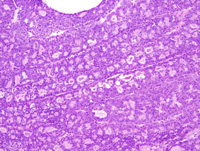
|
|
Image ID:1855 |
|
Source of Image:Ward JM |
|
Pathologist:Mikaelian I |
|
Method / Stain:H&E |
|
|
Image Caption:Mammary adenocarcinoma: this photomicrograph illustrates a portion of the neoplasm that is cystic and presents a few papillary projections (upper right corner). The other portions of the neoplasm is composed of closely-packed glands.
|
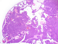
|
|
Image ID:1849 |
|
Source of Image:Ward JM |
|
Pathologist:Mikaelian I |
|
Method / Stain:H&E |
|
|
Image Caption:Mammary gland: the architecture of the mammary gland is effaced by a multilobular, densely cellular, polycystic neoplasm.
|
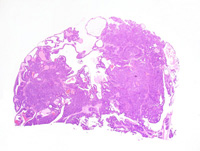
|
|
Image ID:1848 |
|
Source of Image:Ward JM |
|
Pathologist:Mikaelian I |
|
Method / Stain:H&E |
|
|
Image Caption:Mammary gland: this photomicrograph illustrates a cystic neoplastic lobule lined by a 5-10 cells thick epithelium that forms a few small secondary lumens. Neoplastic cells are cuboidal to polygonal, with indistinct cell borders and a small amount of strongly amphophilic cytoplasm. The nucleus is central, oval, and normochromatic. Mitoses are numerous.
|
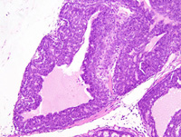
|
|
Image ID:1852 |
|
Source of Image:Ward JM |
|
Pathologist:Mikaelian I |
|
Method / Stain:H&E |
|
|
Image Caption:Mammary adenocarcinoma: this photomicrograph represents a portion of the neoplasm with an acinar pattern. There is mild multifocal lactational activity.
|
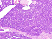
|
|
Image ID:1854 |
|
Source of Image:Ward JM |
|
Pathologist:Mikaelian I |
|
Method / Stain:H&E |
|
|
Image Caption:Mammary gland: at its' periphery, the neoplasm formed hollow frond-like structures that contained a proteinaceous fluid with a few degenerated neoplastic cells. In these areas, the basal layers of the neoplastic epithelium clearly showed nuclear palissading.
|
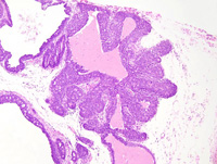
|
|
Image ID:1857 |
|
Source of Image:Ward JM |
|
Pathologist:Mikaelian I |
|
Method / Stain:H&E |
|
|
Image Caption:Mammary gland: the architecture of the mammary gland is effaced by a multilobular, densely cellular, polycystic neoplasm. The neoplasm is composed of coalescing lobules separated by thick fibrovascular septae. Neoplastic lobules are composed of closely-packed glands. Some of the glands are cystic and they are filled by a moderate amount of proteinaceous fluid.
|
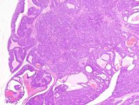
|
|
Image ID:1850 |
|
Source of Image:Ward JM |
|
Pathologist:Mikaelian I |
|
Method / Stain:H&E |
|
|
Image Caption:Mammary gland: the neoplasm is composed of coalescing lobules separated by thick fibrovascular septae. The center of the lobules is generally cystic and occasionally contains tissues that have undergone coagulation necrosis and hemorrhage (*). Neoplastic lobules are composed of closely-packed glands generally lined by a one cell-thick cuboidal epithelium. Neoplastic cell occasionally pile-up disorderly (up to 4-5 cells-thick) but retain the ability to form secondary lumens.
|
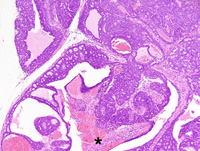
|
|
Image ID:1851 |
|
Source of Image:Ward JM |
|
Pathologist:Mikaelian I |
|
Method / Stain:H&E |
|
|
Image Caption:Mammary gland: at its' periphery, the neoplasm formed hollow frond-like structures that contained a proteinaceous fluid with a few degenerated neoplastic cells. In these areas, the basal layers of the neoplastic epithelium clearly showed nuclear palissading. This type of nuclear palissading is reminiscent of myoepithelial differentiation that is observed in type P tumors. However, unlike type P tumors, this neoplasm does not display ductal differentiation nor areas of squamous differentiation.
|
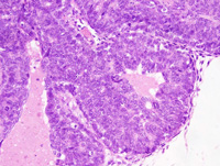
|
|
Image ID:1859 |
|
Source of Image:Ward JM |
|
Pathologist:Mikaelian I |
|
Method / Stain:H&E |
|
|
Image Caption:Mammary adenocarcinoma: this photomicrograph represents a portion of the neoplasm with an acinar pattern. There is mild multifocal lactational activity that is characterized by the presence of a small amount of proteinaceous fluid in the lumen of neoplastic acini.
|
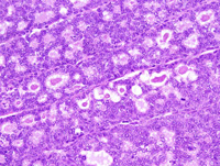
|
|
Image ID:1856 |
|
Source of Image:Ward JM |
|
Pathologist:Mikaelian I |
|
Method / Stain:H&E |
|
|
Image Caption:Mammary gland: at its' periphery, the neoplasm formed hollow frond-like structures that contained a proteinaceous fluid with a few degenerated neoplastic cells. In these areas, the basal layers of the neoplastic epithelium clearly showed nuclear palissading. This type of nuclear palissading is reminiscent of myoepithelial differentiation that is observed in type P tumors. However, unlike type P tumors, this neoplasm does not display ductal differentiation nor areas of squamous differentiation.
|
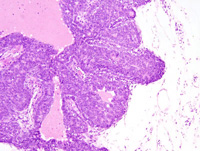
|
|
Image ID:1858 |
|
Source of Image:Ward JM |
|
Pathologist:Mikaelian I |
|
Method / Stain:H&E |
|
|
|
| MTB ID |
Tumor Name |
Organ(s) Affected |
Treatment Type |
Agents |
Strain Name |
Strain Sex |
Reproductive Status |
Tumor Frequency |
Age at Necropsy |
Description |
Reference |
| MTB:29297 |
Mammary gland carcinoma - adenosquamous |
Mammary gland |
None (spontaneous) |
|
|
Female |
reproductive status not specified |
observed |
unknown |
Carcinoma, adenosquamous, and squamous metaplasia, mammary gland |
J:94320 |
|
Image Caption:Mammary gland: hyperplasia, squamous metaplasia with the formation or large rafts of "ghost" epithelial cells, interstitial fibrosis, and interstitial lymphocytic and plasmacytic inflammation are prominent in this mammary lobule.
|
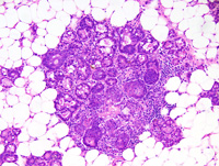
|
|
Image ID:1864 |
|
Source of Image:Ward JM |
|
Pathologist:Mikaelian I |
|
Method / Stain:H&E |
|
|
Image Caption:Adenosquamous carcinoma: this photomicrograph illustrates the granulomatous, lymphoplasmacytic and fibrosing inflammatory reaction associated with rupture of neoplastic glands and leakage of cornified debris in the panniculus. Multinucleated giant cells (arrowhead) of histiocytic origin surround some rafts of "ghost" epithelial cells.
|
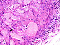
|
|
Image ID:1873 |
|
Source of Image:Ward JM |
|
Pathologist:Mikaelian I |
|
Method / Stain:H&E |
|
|
Image Caption:Adenosquamous carcinoma, glandular portions: this photomicrograph illustrates the glandular portions of an adenosquamous carcinoma. Epithelial cells contain prominent lipid vacuoles.
|
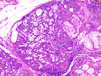
|
|
Image ID:1871 |
|
Source of Image:Ward JM |
|
Pathologist:Mikaelian I |
|
Method / Stain:H&E |
|
|
Image Caption:Adenosquamous carcinoma, glandular portions: this photomicrograph illustrates the glandular portions of an adenosquamous carcinoma. Epithelial cells contain prominent lipid vacuoles. Keratinization results into the formation of keratin pearls (arrowhead).
|
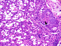
|
|
Image ID:1872 |
|
Source of Image:Ward JM |
|
Pathologist:Mikaelian I |
|
Method / Stain:H&E |
|
|
Image Caption:Mammary gland: multiple small (<1 mm in diameter) nodules are scattered throughout the mammary gland. The largest lobules are composed of glands packed with cornified debris and lined by a squamous epithelium.
|
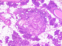
|
|
Image ID:1861 |
|
Source of Image:Ward JM |
|
Pathologist:Mikaelian I |
|
Method / Stain:H&E |
|
|
Image Caption:Mammary gland: this photomicrograph represents an area of squamous metaplasia with dysplasia (upper right portions of the photomicrograph) and an area of lobular hyperplasia of the mammary gland (at the center and in the lower right corner of the photomicrograph).
|
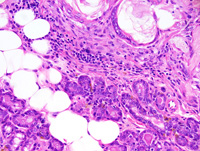
|
|
Image ID:1863 |
|
Source of Image:Ward JM |
|
Pathologist:Mikaelian I |
|
Method / Stain:H&E |
|
|
Image Caption:Adenosquamous carcinoma: the neoplasm is connected to a large interlobular duct (*). The neoplasm is multilobular, well-delineated, locally invasive, moderately cellular and contains large rafts of "ghost" epithelial cells (E) that are surrounded by a prominent granulomatous and sclerosing inflammation. Other portions of the neoplasm are composed of closely-packed glands (G).
|
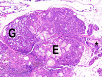
|
|
Image ID:1867 |
|
Source of Image:Ward JM |
|
Pathologist:Mikaelian I |
|
Method / Stain:H&E |
|
|
Image Caption:Mammary gland: most of the mammary gland shows moderate lobular hyperplasia which is characterized by an increase in the number of mammary acini which are mildly ectatic (lower left corner of the photomicrograph). In addition, multifocally, entire lobules show squamous metaplasia of their epithelium (upper right corner of the photomicrograph). The acini in these areas are filled with large rafts of "ghost" epithelial cells and they often rupture, resulting in the formation of a foreign-body type granulomatous reaction. There is also moderate interstitial fibrosis and lymphoplasmacytic inflammation surrounding the mammary lobules that have undergone squamous metaplasia.
|
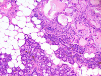
|
|
Image ID:1862 |
|
Source of Image:Ward JM |
|
Pathologist:Mikaelian I |
|
Method / Stain:H&E |
|
|
Image Caption:Mammary gland: hyperplasia, squamous metaplasia with the formation or large rafts of "ghost" epithelial cells, interstitial fibrosis, and interstitial lymphocytic and plasmacytic inflammation are prominent in this mammary lobule. In addition, a few neutrophils have transmigrated through the metaplastic mammary epithelium and are intermingled with the cornified debris that often expand the lumen of mammary acini.
|
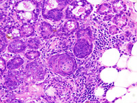
|
|
Image ID:1865 |
|
Source of Image:Ward JM |
|
Pathologist:Mikaelian I |
|
Method / Stain:H&E |
|
|
Image Caption:Mammary gland: multiple small (<1 mm in diameter) nodules are scattered throughout the mammary gland. All mammary lobules are prominent, indicating moderate diffuse mammary lobular hyperplasia.
|
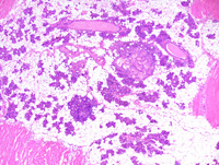
|
|
Image ID:1860 |
|
Source of Image:Ward JM |
|
Pathologist:Mikaelian I |
|
Method / Stain:H&E |
|
|
Image Caption:Adenosquamous carcinoma: this photomicrograph illustrates the area of transition between the neoplasm and the interlobular duct (*).
|
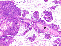
|
|
Image ID:1868 |
|
Source of Image:Ward JM |
|
Pathologist:Mikaelian I |
|
Method / Stain:H&E |
|
|
Image Caption:Mammary gland: this photomicrograph represents an adenosquamous carcinoma of the mammary gland. The neoplasm is connected to a large interlobular duct (*). The neoplasm is multilobular, well-delineated, locally invasive, moderately cellular and contains large rafts of "ghost" epithelial cells that are surrounded by a prominent granulomatous and sclerosing inflammation.
|
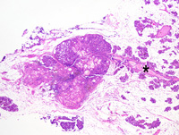
|
|
Image ID:1866 |
|
Source of Image:Ward JM |
|
Pathologist:Mikaelian I |
|
Method / Stain:H&E |
|
|
Image Caption:Adenosquamous carcinoma: this photomicrograph illustrates the area of transition between the neoplasm and the interlobular duct (*). There is prominent squamous metaplasia in the interlobular duct. Cornification is abrupt and results into the formation of large rafts of "ghost" epithelial cells.
|
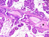
|
|
Image ID:1869 |
|
Source of Image:Ward JM |
|
Pathologist:Mikaelian I |
|
Method / Stain:H&E |
|
|
Image Caption:Interlobular duct at the vicinity of an adenosquamous carcinoma: there is prominent squamous metaplasia in the interlobular duct. Cornification is abrupt and results into the formation of large rafts of "ghost" epithelial cells.
|
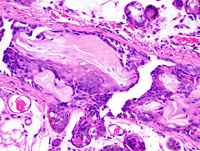
|
|
Image ID:1870 |
|
Source of Image:Ward JM |
|
Pathologist:Mikaelian I |
|
Method / Stain:H&E |
|
|
|
| MTB ID |
Tumor Name |
Organ(s) Affected |
Treatment Type |
Agents |
Strain Name |
Strain Sex |
Reproductive Status |
Tumor Frequency |
Age at Necropsy |
Description |
Reference |
| MTB:29298 |
Mammary gland metaplasia - squamous |
Mammary gland |
None (spontaneous) |
|
|
Female |
reproductive status not specified |
observed |
unknown |
Carcinoma, adenosquamous, and squamous metaplasia, mammary gland |
J:94320 |
|
Image Caption:Mammary gland: this photomicrograph represents an area of squamous metaplasia with dysplasia (upper right portions of the photomicrograph) and an area of lobular hyperplasia of the mammary gland (at the center and in the lower right corner of the photomicrograph).
|
|
|
|
Image ID:1863 |
|
Source of Image:Ward JM |
|
Pathologist:Mikaelian I |
|
Method / Stain:H&E |
|
|
Image Caption:Mammary gland: hyperplasia, squamous metaplasia with the formation or large rafts of "ghost" epithelial cells, interstitial fibrosis, and interstitial lymphocytic and plasmacytic inflammation are prominent in this mammary lobule.
|
|
|
|
Image ID:1864 |
|
Source of Image:Ward JM |
|
Pathologist:Mikaelian I |
|
Method / Stain:H&E |
|
|
Image Caption:Mammary gland: multiple small (<1 mm in diameter) nodules are scattered throughout the mammary gland. All mammary lobules are prominent, indicating moderate diffuse mammary lobular hyperplasia.
|
|
|
|
Image ID:1860 |
|
Source of Image:Ward JM |
|
Pathologist:Mikaelian I |
|
Method / Stain:H&E |
|
|
Image Caption:Adenosquamous carcinoma: this photomicrograph illustrates the area of transition between the neoplasm and the interlobular duct (*).
|
|
|
|
Image ID:1868 |
|
Source of Image:Ward JM |
|
Pathologist:Mikaelian I |
|
Method / Stain:H&E |
|
|
Image Caption:Mammary gland: this photomicrograph represents an adenosquamous carcinoma of the mammary gland. The neoplasm is connected to a large interlobular duct (*). The neoplasm is multilobular, well-delineated, locally invasive, moderately cellular and contains large rafts of "ghost" epithelial cells that are surrounded by a prominent granulomatous and sclerosing inflammation.
|
|
|
|
Image ID:1866 |
|
Source of Image:Ward JM |
|
Pathologist:Mikaelian I |
|
Method / Stain:H&E |
|
|
Image Caption:Adenosquamous carcinoma: this photomicrograph illustrates the area of transition between the neoplasm and the interlobular duct (*). There is prominent squamous metaplasia in the interlobular duct. Cornification is abrupt and results into the formation of large rafts of "ghost" epithelial cells.
|
|
|
|
Image ID:1869 |
|
Source of Image:Ward JM |
|
Pathologist:Mikaelian I |
|
Method / Stain:H&E |
|
|
Image Caption:Adenosquamous carcinoma, glandular portions: this photomicrograph illustrates the glandular portions of an adenosquamous carcinoma. Epithelial cells contain prominent lipid vacuoles. Keratinization results into the formation of keratin pearls (arrowhead).
|
|
|
|
Image ID:1872 |
|
Source of Image:Ward JM |
|
Pathologist:Mikaelian I |
|
Method / Stain:H&E |
|
|
Image Caption:Adenosquamous carcinoma: this photomicrograph illustrates the granulomatous, lymphoplasmacytic and fibrosing inflammatory reaction associated with rupture of neoplastic glands and leakage of cornified debris in the panniculus. Multinucleated giant cells (arrowhead) of histiocytic origin surround some rafts of "ghost" epithelial cells.
|
|
|
|
Image ID:1873 |
|
Source of Image:Ward JM |
|
Pathologist:Mikaelian I |
|
Method / Stain:H&E |
|
|
Image Caption:Adenosquamous carcinoma, glandular portions: this photomicrograph illustrates the glandular portions of an adenosquamous carcinoma. Epithelial cells contain prominent lipid vacuoles.
|
|
|
|
Image ID:1871 |
|
Source of Image:Ward JM |
|
Pathologist:Mikaelian I |
|
Method / Stain:H&E |
|
|
Image Caption:Mammary gland: most of the mammary gland shows moderate lobular hyperplasia which is characterized by an increase in the number of mammary acini which are mildly ectatic (lower left corner of the photomicrograph). In addition, multifocally, entire lobules show squamous metaplasia of their epithelium (upper right corner of the photomicrograph). The acini in these areas are filled with large rafts of "ghost" epithelial cells and they often rupture, resulting in the formation of a foreign-body type granulomatous reaction. There is also moderate interstitial fibrosis and lymphoplasmacytic inflammation surrounding the mammary lobules that have undergone squamous metaplasia.
|
|
|
|
Image ID:1862 |
|
Source of Image:Ward JM |
|
Pathologist:Mikaelian I |
|
Method / Stain:H&E |
|
|
Image Caption:Interlobular duct at the vicinity of an adenosquamous carcinoma: there is prominent squamous metaplasia in the interlobular duct. Cornification is abrupt and results into the formation of large rafts of "ghost" epithelial cells.
|
|
|
|
Image ID:1870 |
|
Source of Image:Ward JM |
|
Pathologist:Mikaelian I |
|
Method / Stain:H&E |
|
|
Image Caption:Mammary gland: hyperplasia, squamous metaplasia with the formation or large rafts of "ghost" epithelial cells, interstitial fibrosis, and interstitial lymphocytic and plasmacytic inflammation are prominent in this mammary lobule. In addition, a few neutrophils have transmigrated through the metaplastic mammary epithelium and are intermingled with the cornified debris that often expand the lumen of mammary acini.
|
|
|
|
Image ID:1865 |
|
Source of Image:Ward JM |
|
Pathologist:Mikaelian I |
|
Method / Stain:H&E |
|
|
Image Caption:Adenosquamous carcinoma: the neoplasm is connected to a large interlobular duct (*). The neoplasm is multilobular, well-delineated, locally invasive, moderately cellular and contains large rafts of "ghost" epithelial cells (E) that are surrounded by a prominent granulomatous and sclerosing inflammation. Other portions of the neoplasm are composed of closely-packed glands (G).
|
|
|
|
Image ID:1867 |
|
Source of Image:Ward JM |
|
Pathologist:Mikaelian I |
|
Method / Stain:H&E |
|
|
Image Caption:Mammary gland: multiple small (<1 mm in diameter) nodules are scattered throughout the mammary gland. The largest lobules are composed of glands packed with cornified debris and lined by a squamous epithelium.
|
|
|
|
Image ID:1861 |
|
Source of Image:Ward JM |
|
Pathologist:Mikaelian I |
|
Method / Stain:H&E |
|
|
|
| MTB ID |
Tumor Name |
Organ(s) Affected |
Treatment Type |
Agents |
Strain Name |
Strain Sex |
Reproductive Status |
Tumor Frequency |
Age at Necropsy |
Description |
Reference |
| MTB:29299 |
Mammary gland hyperplasia - lobular |
Mammary gland |
None (spontaneous) |
|
|
Female |
reproductive status not specified |
observed |
unknown |
Lobular hyperplasia and pigmentation, mammary gland |
J:94320 |
|
Image Caption:Mammary gland: the lobular architecture of the mammary gland is prominent.
|
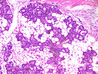
|
|
Image ID:1874 |
|
Source of Image:Ward JM |
|
Pathologist:Mikaelian I |
|
Method / Stain:H&E |
|
|
Image Caption:Mammary gland: the lobular architecture of the mammary gland is prominent. Large amounts of a golden-yellow and coarsely granular intracytoplasmic pigment are present in cells located within the epithelium or in the lumen of the mammary ducts and acini. This pigment most likely is located in macrophages rather than in epithelial cells.
|
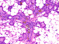
|
|
Image ID:1875 |
|
Source of Image:Ward JM |
|
Pathologist:Mikaelian I |
|
Method / Stain:H&E |
|
|
|
| MTB ID |
Tumor Name |
Organ(s) Affected |
Treatment Type |
Agents |
Strain Name |
Strain Sex |
Reproductive Status |
Tumor Frequency |
Age at Necropsy |
Description |
Reference |
| MTB:29305 |
Mammary gland carcinoma - spindle cell |
Mammary gland |
None (spontaneous) |
|
|
Female |
reproductive status not specified |
observed |
unknown |
Carcinoma, spindle cell, with epithelial areas, mammary gland |
J:94320 |
|
Image Caption:Spindle cell carcinoma: this photomicrograph illustrates an epithelial area. As exemplified on this photomicrograph, cornification may be observed in neoplasms undergoing epithelial to mesenchymal transition. Cornification is characterized by neoplastic cells acquiring a more abundant and more acidophilic cytoplasm as well as more angular contours. Their nucleus is larger, more hypochromatic, and with a larger nucleolus than other neoplastic cells.
|
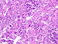
|
|
Image ID:1894 |
|
Source of Image:Ward JM |
|
Pathologist:Mikaelian I |
|
Method / Stain:H&E |
|
|
Image Caption:Spindle cell carcinoma: this photomicrograph illustrates the presence of a few cells with an epithelial phenotype entrapped in a area that is otherwise exclusively composed of cells with a mesenchymal phenotype.
|
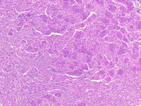
|
|
Image ID:1886 |
|
Source of Image:Ward JM |
|
Pathologist:Mikaelian I |
|
Method / Stain:H&E |
|
|
Image Caption:Spindle cell carcinoma: this photomicrograph illustrates an epithelial area. As exemplified on this photomicrograph, cornification may be observed in neoplasms undergoing epithelial to mesenchymal transition. Cornification is characterized by neoplastic cells acquiring a more abundant and more acidophilic cytoplasm as well as more angular contours. Their nucleus is larger, more hypochromatic, and with a larger nucleolus than other neoplastic cells.
|
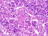
|
|
Image ID:1893 |
|
Source of Image:Ward JM |
|
Pathologist:Mikaelian I |
|
Method / Stain:H&E |
|
|
Image Caption:Spindle cell carcinoma: this photomicrograph illustrates the presence of a few cells with an epithelial phenotype entrapped in a area that is otherwise exclusively composed of cells with a mesenchymal phenotype. There is gradual transition between the cells with the epithelial phenotype and the cells with the mesenchymal phenotype.
|
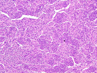
|
|
Image ID:1887 |
|
Source of Image:Ward JM |
|
Pathologist:Mikaelian I |
|
Method / Stain:H&E |
|
|
Image Caption:Spindle cell carcinoma: this photomicrograph illustrates an epithelial area. As exemplified on this photomicrograph, cornification may be observed in neoplasms undergoing epithelial to mesenchymal transition. Cornification is characterized by neoplastic cells acquiring a more abundant and more acidophilic cytoplasm as well as more angular contours. Their nucleus is larger, more hypochromatic, and with a larger nucleolus than other neoplastic cells.
|
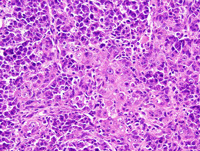
|
|
Image ID:1891 |
|
Source of Image:Ward JM |
|
Pathologist:Mikaelian I |
|
Method / Stain:H&E |
|
|
Image Caption:Spindle cell carcinoma: this photomicrograph illustrates the presence of a few cells with an epithelial phenotype entrapped in a area that is otherwise exclusively composed of cells with a mesenchymal phenotype. There is gradual transition between the cells with the epithelial phenotype and the cells with the mesenchymal phenotype.
|
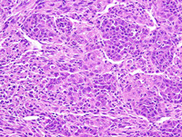
|
|
Image ID:1888 |
|
Source of Image:Ward JM |
|
Pathologist:Mikaelian I |
|
Method / Stain:H&E |
|
|
Image Caption:Spindle cell carcinoma: in addition to skeletal muscle fibers, a few adipocytes have been entrapped in the neoplasm. The adipocytes appear as optically-empty round vacuoles.
|
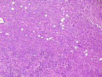
|
|
Image ID:1889 |
|
Source of Image:Ward JM |
|
Pathologist:Mikaelian I |
|
Method / Stain:H&E |
|
|
Image Caption:Spindle cell carcinoma: this photomicrograph illustrates an epithelial area. As exemplified on this photomicrograph, cornification may be observed in neoplasms undergoing epithelial to mesenchymal transition. Cornification is characterized by neoplastic cells acquiring a more abundant and more acidophilic cytoplasm as well as more angular contours. Their nucleus is larger, more hypochromatic, and with a larger nucleolus than other neoplastic cells.
|
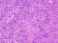
|
|
Image ID:1890 |
|
Source of Image:Ward JM |
|
Pathologist:Mikaelian I |
|
Method / Stain:H&E |
|
|
Image Caption:Spindle cell carcinoma: this high magnification photomicrograph illustrates a portion of the neoplasm where neoplastic cells have an epithelial phenotype and form trabeculae and gradually transform into cells with a mesenchymal phenotype forming fascicles. Dilated sinusoidal vessels are present in this portion of the neoplasm.
|
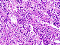
|
|
Image ID:1892 |
|
Source of Image:Ward JM |
|
Pathologist:Mikaelian I |
|
Method / Stain:H&E |
|
|
Image Caption:Spindle cell carcinoma: this photomicrograph illustrates the presence of a few cells with an epithelial phenotype entrapped in a area that is otherwise exclusively composed of cells with a mesenchymal phenotype.
|
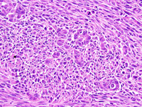
|
|
Image ID:1885 |
|
Source of Image:Ward JM |
|
Pathologist:Mikaelian I |
|
Method / Stain:H&E |
|
|
Image Caption:Spindle cell carcinoma: this photomicrograph illustrates the presence of a few cells with an epithelial phenotype entrapped in a area that is otherwise exclusively composed of cells with a mesenchymal phenotype.
|
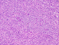
|
|
Image ID:1884 |
|
Source of Image:Ward JM |
|
Pathologist:Mikaelian I |
|
Method / Stain:H&E |
|
|
Image Caption:Mammary gland: the mammary gland is entirely effaced and greatly expanded by a nodular, well-delineated, densely cellular neoplasm. This neoplasm is predominantly composed of bundles and fascicles separated by a scant amount of fibrovascular stroma. A few epithelial areas (*) characterized by the formation of small nests and trabeculae persist at the periphery of the neoplasm.
|
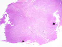
|
|
Image ID:1876 |
|
Source of Image:Ward JM |
|
Pathologist:Mikaelian I |
|
Method / Stain:H&E |
|
|
Image Caption:Mammary gland: the neoplasm is predominantly composed of bundles and fascicles separated by a scant amount of fibrovascular stroma. A few epithelial areas (*) characterized by the formation of small nests and trabeculae persist at the periphery of the neoplasm. A few fascicles of skeletal muscle fibers and individual skeletal muscle fibers (arrowheads) are entrapped in the neoplasm.
|
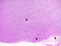
|
|
Image ID:1878 |
|
Source of Image:Ward JM |
|
Pathologist:Mikaelian I |
|
Method / Stain:H&E |
|
|
Image Caption:Mammary gland: the neoplasm is predominantly composed of bundles and fascicles separated by a scant amount of fibrovascular stroma. A few epithelial areas characterized by the formation of small nests and trabeculae persist at the periphery of the neoplasm and there is gradual transition between the epithelial and the mesenchymal phenotype. Cells with an epithelial phenotype are polygonal with distinct cell borders, a moderate amount of strongly amphophilic cytoplasm, and a round to oval medium-size nucleus. Cells with a mesenchymal phenotype are spindloid, with distinct cell borders, a small to moderate amount of acidophilic cytoplasm, and an elongated hyperchromatic nucleus.
|
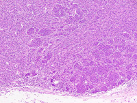
|
|
Image ID:1879 |
|
Source of Image:Ward JM |
|
Pathologist:Mikaelian I |
|
Method / Stain:H&E |
|
|
Image Caption:Mammary gland: this high magnification microphotograph focuses on the cells with an mesenchymal phenotype. These cells are organized in fascicles oriented at right angle one with another, are spindloid to fusifrom, with distinct cell borders, a small to moderate amount of acidophilic cytoplasm, and an elongated hyperchromatic nucleus. A moderate number of skeletal muscle fibers is entrapped in the neoplasm.
|
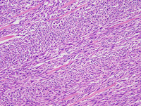
|
|
Image ID:1882 |
|
Source of Image:Ward JM |
|
Pathologist:Mikaelian I |
|
Method / Stain:H&E |
|
|
Image Caption:Mammary gland: the neoplasm is predominantly composed of bundles and fascicles separated by a scant amount of fibrovascular stroma. A few epithelial areas characterized by the formation of small nests and trabeculae persist at the periphery of the neoplasm and there is gradual transition between the epithelial and the mesenchymal phenotype. Cells with an epithelial phenotype are polygonal with distinct cell borders, a moderate amount of strongly amphophilic cytoplasm, and a round to oval medium-size nucleus. Cells with a mesenchymal phenotype are spindloid, with distinct cell borders, a small to moderate amount of acidophilic cytoplasm, and an elongated hyperchromatic nucleus. Numerous neoplastic cells undergo apoptosis.
|
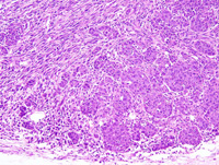
|
|
Image ID:1880 |
|
Source of Image:Ward JM |
|
Pathologist:Mikaelian I |
|
Method / Stain:H&E |
|
|
Image Caption:Mammary gland: this high magnification microphotograph focuses on the cells with an mesenchymal phenotype. These cells are organized in fascicles oriented at right angle one with another, are spindloid to fusifrom, with distinct cell borders, a small to moderate amount of acidophilic cytoplasm, and an elongated hyperchromatic nucleus. A moderate number of skeletal muscle fibers is entrapped in the neoplasm (arrowheads), and some are degenerated.
|
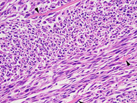
|
|
Image ID:1883 |
|
Source of Image:Ward JM |
|
Pathologist:Mikaelian I |
|
Method / Stain:H&E |
|
|
Image Caption:Mammary gland: the mammary gland is entirely effaced and greatly expanded by a nodular, well-delineated, densely cellular neoplasm. This neoplasm is predominantly composed of bundles and fascicles separated by a scant amount of fibrovascular stroma. A few epithelial areas characterized by the formation of small nests and trabeculae persist at the periphery of the neoplasm.
|
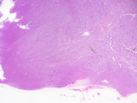
|
|
Image ID:1877 |
|
Source of Image:Ward JM |
|
Pathologist:Mikaelian I |
|
Method / Stain:H&E |
|
|
Image Caption:Mammary gland: this high magnification microphotograph focuses on the cells with an epithelial phenotype. These cells are organized in nests, are polygonal with distinct cell borders, a moderate amount of strongly amphophilic cytoplasm, and a round to oval medium-size nucleus. Numerous neoplastic cells undergo apoptosis.
|
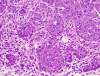
|
|
Image ID:1881 |
|
Source of Image:Ward JM |
|
Pathologist:Mikaelian I |
|
Method / Stain:H&E |
|
|
|
| MTB ID |
Tumor Name |
Organ(s) Affected |
Treatment Type |
Agents |
Strain Name |
Strain Sex |
Reproductive Status |
Tumor Frequency |
Age at Necropsy |
Description |
Reference |
| MTB:29306 |
Mammary gland adenoma - papillary |
Mammary gland |
None (spontaneous) |
|
|
Female |
reproductive status not specified |
observed |
unknown |
Papillary adenoma, intraductal, mammary gland |
J:94320 |
|
Image Caption:Mammary gland: a mammary duct is markedly ectatic. It contains a large amount of strongly acidophilic proteinaceous fluid and a few slender papillary projections lined by a two cells-thick epithelium where neoplastic cells occasionally pile-up. The occurence of two layers of cells is consistent with myoepithelial and luminal differentiation of neoplastic cells (complex papillary adenoma). Similar smaller lesions are present in the nearby mammary parenchyma.
|
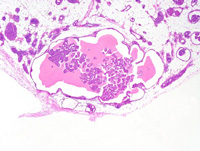
|
|
Image ID:1895 |
|
Source of Image:Ward JM |
|
Pathologist:Mikaelian I |
|
Method / Stain:H&E |
|
|
Image Caption:Mammary gland: a mammary duct is markedly ectatic. It contains a large amount of strongly acidophilic proteinaceous fluid and a few slender papillary projections lined by a two cells-thick epithelium where neoplastic cells occasionally pile-up. The occurence of two layers of cells is consistent with myoepithelial and luminal differentiation of neoplastic cells (complex papillary adenoma).
|
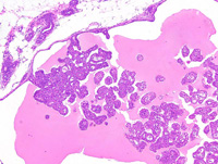
|
|
Image ID:1898 |
|
Source of Image:Ward JM |
|
Pathologist:Mikaelian I |
|
Method / Stain:H&E |
|
|
Image Caption:Mammary gland: a mammary duct is markedly ectatic. It contains a large amount of strongly acidophilic proteinaceous fluid and a few slender papillary projections lined by a two cells-thick epithelium where neoplastic cells occasionally pile-up. The occurence of two layers of cells is consistent with myoepithelial and luminal differentiation of neoplastic cells (complex papillary adenoma). Luminal neoplastic cells are cuboidal, small, with indistinct cell borders and a moderate amount of granular acidophilic cytoplasm. The nucleus is central, oval, medium-size and normochromatic. Basal neoplastic cells (arrowheads) are cuboidal, small, with indistinct cell borders, a scant amount of acidophilic cytoplasm and a small hyperchromatic nucleus. There is no evidence of atypia and mitotic rate is low.
|
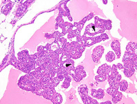
|
|
Image ID:1896 |
|
Source of Image:Ward JM |
|
Pathologist:Mikaelian I |
|
Method / Stain:H&E |
|
|
Image Caption:Mammary gland: a mammary duct is markedly ectatic. It contains a large amount of strongly acidophilic proteinaceous fluid and a few slender papillary projections lined by a two cells-thick epithelium where neoplastic cells occasionally pile-up. The occurence of two layers of cells is consistent with myoepithelial and luminal differentiation of neoplastic cells (complex papillary adenoma). Luminal neoplastic cells are cuboidal, small, with indistinct cell borders and a moderate amount of granular acidophilic cytoplasm. The nucleus is central, oval, medium-size and normochromatic. Basal neoplastic cells (arrowheads) form discontinuous rows. They are cuboidal, small, with indistinct cell borders, a scant amount of acidophilic cytoplasm and a small hyperchromatic nucleus. There is no evidence of atypia and mitotic rate is low.
|
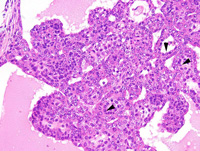
|
|
Image ID:1897 |
|
Source of Image:Ward JM |
|
Pathologist:Mikaelian I |
|
Method / Stain:H&E |
|
|
|
| MTB ID |
Tumor Name |
Organ(s) Affected |
Treatment Type |
Agents |
Strain Name |
Strain Sex |
Reproductive Status |
Tumor Frequency |
Age at Necropsy |
Description |
Reference |
| MTB:29307 |
Mammary gland hyperplasia |
Mammary gland |
None (spontaneous) |
|
|
Female |
reproductive status not specified |
observed |
unknown |
Intraductal hyperplasia, multicentric, marked, mammary gland |
J:94320 |
|
Image Caption:Mammary gland: almost all interlobular and intralobular mammary ducts contain multiple and often coalescing small cauliflower-like projections. There is no evidence of fibrosis or inflammation around these ducts. These projections are composed of a slender papillary core covered by several layers of small spindloid cells with indistinct cell borders, a scant amount of acidophilic cytoplasm, and a small oval hyperchromatic nucleus. Covering these spindloid cells is a one cell-thick layer of cuboidal to low columnar luminal cells with ill-defined cell borders, a moderate amount of acidophilic cytoplasm, and a medium-size central hypochromatic nucleus. Anisokaryosis and anisocytosis are mild. Mitoses are not detected. A few luminal cells contain lipid vacuoles.
The presence of two layers of cells indicates that the cell that gave rise to these lesions is not committed to the luminal or to the basal lineage, and hence these lesions may be categorized as "complex" lesions.
|
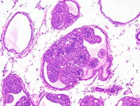
|
|
Image ID:1901 |
|
Source of Image:Ward JM |
|
Pathologist:Mikaelian I |
|
Method / Stain:H&E |
|
|
Image Caption:Mammary gland: an interlobular mammary duct is moderately expanded by a cauliflower-like proliferative process characterized by the formation of slender fibrovascular cores that support two populations of cells. First, there is a 1-5 cells-thick layer of oat-shaped cells with indistinct cell borders, a small amount of pale acidophilic cytoplasm and a small hyperchromatic oval nucleus. Second, there is a one cell-thick layer of cuboidal to low columnar cells that form conspicuous lumens filled with an amorphous proteinaceous material. These cells have indistinct cell borders, a moderate amount of acidophilic cytoplasm and a medium-size, central, normochromatic nucleus with a clumped chromatin. A few mast cells are present in the periductal connective tissue.
The presence of two layers of cells indicates that the cell that gave rise to these lesions is not committed to the luminal or to the basal lineage, and hence these lesions may be categorized as "complex" lesions.
|
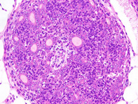
|
|
Image ID:1904 |
|
Source of Image:Ward JM |
|
Pathologist:Mikaelian I |
|
Method / Stain:H&E |
|
|
Image Caption:Mammary gland: almost all interlobular and intralobular mammary ducts contain multiple and often coalescing small cauliflower-like projections. There is no evidence of fibrosis or inflammation around these ducts.
|
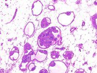
|
|
Image ID:1900 |
|
Source of Image:Ward JM |
|
Pathologist:Mikaelian I |
|
Method / Stain:H&E |
|
|
Image Caption:Mammary gland: almost all interlobular and intralobular mammary ducts contain multiple and often coalescing small cauliflower-like projections. There is no evidence of fibrosis or inflammation around these ducts.
|
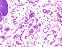
|
|
Image ID:1899 |
|
Source of Image:Ward JM |
|
Pathologist:Mikaelian I |
|
Method / Stain:H&E |
|
|
Image Caption:Mammary gland: an interlobular mammary duct is moderately expanded by a cauliflower-like proliferative process characterized by the formation of slender fibrovascular cores that support two populations of cells. First, there is a 1-5 cells-thick layer of oat-shaped cells with indistinct cell borders, a small amount of pale acidophilic cytoplasm and a small hyperchromatic oval nucleus. Second, there is a one cell-thick layer of cuboidal to low columnar cells that form conspicuous lumens filled with an amorphous proteinaceous material. These cells have indistinct cell borders, a moderate amount of acidophilic cytoplasm and a medium-size, central, normochromatic nucleus with a clumped chromatin. A few mast cells are present in the periductal connective tissue.
The presence of two layers of cells indicates that the cell that gave rise to these lesions is not committed to the luminal or to the basal lineage, and hence these lesions may be categorized as "complex" lesions.
|
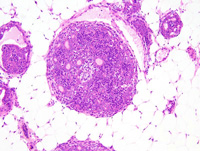
|
|
Image ID:1903 |
|
Source of Image:Ward JM |
|
Pathologist:Mikaelian I |
|
Method / Stain:H&E |
|
|
Image Caption:Mammary gland: an interlobular mammary duct contains multiple coalescing small cauliflower-like projections. These projections are composed of a slender papillary core covered by several layers of small spindloid cells with indistinct cell borders, a scant amount of acidophilic cytoplasm, and a small oval hyperchromatic nucleus. Covering these spindloid cells is a one cell-thick layer of cuboidal to low columnar luminal cells with ill-defined cell borders, a moderate amount of acidophilic cytoplasm, and a medium-size central hypochromatic nucleus. Anisokaryosis and anisocytosis are mild. Mitoses are not detected. A few cells with a luminal phenotype contain lipid vacuoles (arrowheads).
The presence of two layers of cells indicates that the cell that gave rise to these lesions is not committed to the luminal or to the basal lineage, and hence these lesions may be categorized as "complex" lesions.
|
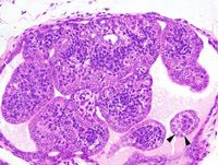
|
|
Image ID:1902 |
|
Source of Image:Ward JM |
|
Pathologist:Mikaelian I |
|
Method / Stain:H&E |
|
|
|
| MTB ID |
Tumor Name |
Organ(s) Affected |
Treatment Type |
Agents |
Strain Name |
Strain Sex |
Reproductive Status |
Tumor Frequency |
Age at Necropsy |
Description |
Reference |
| MTB:29308 |
Mammary gland carcinoma in situ - ductal |
Mammary gland |
None (spontaneous) |
|
|
Female |
reproductive status not specified |
observed |
unknown |
Intraductal hyperplasia, multicentric, marked, mammary gland |
J:94320 |
|
Image Caption:Mammary gland: almost all interlobular and intralobular mammary ducts contain multiple and often coalescing small cauliflower-like projections. There is no evidence of fibrosis or inflammation around these ducts.
|
|
|
|
Image ID:1899 |
|
Source of Image:Ward JM |
|
Pathologist:Mikaelian I |
|
Method / Stain:H&E |
|
|
Image Caption:Mammary gland: an interlobular mammary duct is moderately expanded by a cauliflower-like proliferative process characterized by the formation of slender fibrovascular cores that support two populations of cells. First, there is a 1-5 cells-thick layer of oat-shaped cells with indistinct cell borders, a small amount of pale acidophilic cytoplasm and a small hyperchromatic oval nucleus. Second, there is a one cell-thick layer of cuboidal to low columnar cells that form conspicuous lumens filled with an amorphous proteinaceous material. These cells have indistinct cell borders, a moderate amount of acidophilic cytoplasm and a medium-size, central, normochromatic nucleus with a clumped chromatin. A few mast cells are present in the periductal connective tissue.
The presence of two layers of cells indicates that the cell that gave rise to these lesions is not committed to the luminal or to the basal lineage, and hence these lesions may be categorized as "complex" lesions.
|
|
|
|
Image ID:1903 |
|
Source of Image:Ward JM |
|
Pathologist:Mikaelian I |
|
Method / Stain:H&E |
|
|
Image Caption:Mammary gland: a mammary lobule is greatly expanded. The ductal portions of the lobule are prominent and surrounded by prominent sclerosis. Cells in the ducts pile-up disorderly up to 3 cells-thick. Cells are cuboidal to polygonal, with indistinct cell borders and a moderate amount of granular and strongly acidophilic cytoplasm. The nucleus is central, oval, medium-size, normochromatic, with a punctate chromatin and one small basophilic nucleolus. Anisokaryosis and anisocytosis are mild. Mitoses are numerous.
|
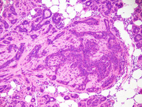
|
|
Image ID:1907 |
|
Source of Image:Ward JM |
|
Pathologist:Mikaelian I |
|
Method / Stain:H&E |
|
|
Image Caption:Mammary gland: almost all interlobular and intralobular mammary ducts contain multiple and often coalescing small cauliflower-like projections. There is no evidence of fibrosis or inflammation around these ducts. These projections are composed of a slender papillary core covered by several layers of small spindloid cells with indistinct cell borders, a scant amount of acidophilic cytoplasm, and a small oval hyperchromatic nucleus. Covering these spindloid cells is a one cell-thick layer of cuboidal to low columnar luminal cells with ill-defined cell borders, a moderate amount of acidophilic cytoplasm, and a medium-size central hypochromatic nucleus. Anisokaryosis and anisocytosis are mild. Mitoses are not detected. A few luminal cells contain lipid vacuoles.
The presence of two layers of cells indicates that the cell that gave rise to these lesions is not committed to the luminal or to the basal lineage, and hence these lesions may be categorized as "complex" lesions.
|
|
|
|
Image ID:1901 |
|
Source of Image:Ward JM |
|
Pathologist:Mikaelian I |
|
Method / Stain:H&E |
|
|
Image Caption:Mammary gland: a mammary lobule is greatly expanded. The ductal portions of the lobule are prominent and surrounded by prominent sclerosis. The acini are hyperplastic and are surrounded by lesser sclerosis.
|
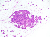
|
|
Image ID:1905 |
|
Source of Image:Ward JM |
|
Pathologist:Mikaelian I |
|
Method / Stain:H&E |
|
|
Image Caption:Mammary gland: an interlobular mammary duct contains multiple coalescing small cauliflower-like projections. These projections are composed of a slender papillary core covered by several layers of small spindloid cells with indistinct cell borders, a scant amount of acidophilic cytoplasm, and a small oval hyperchromatic nucleus. Covering these spindloid cells is a one cell-thick layer of cuboidal to low columnar luminal cells with ill-defined cell borders, a moderate amount of acidophilic cytoplasm, and a medium-size central hypochromatic nucleus. Anisokaryosis and anisocytosis are mild. Mitoses are not detected. A few cells with a luminal phenotype contain lipid vacuoles (arrowheads).
The presence of two layers of cells indicates that the cell that gave rise to these lesions is not committed to the luminal or to the basal lineage, and hence these lesions may be categorized as "complex" lesions.
|
|
|
|
Image ID:1902 |
|
Source of Image:Ward JM |
|
Pathologist:Mikaelian I |
|
Method / Stain:H&E |
|
|
Image Caption:Mammary gland: this photomicrograph focuses on the alveoli of a mammary lobule affected by ductal carcinoma in situ. There is moderate anisokaryosis. In contrast to the ducts where mitotic figures were prominent, mitoses are not found in the alveoli.
|
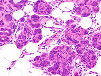
|
|
Image ID:1909 |
|
Source of Image:Ward JM |
|
Pathologist:Mikaelian I |
|
Method / Stain:H&E |
|
|
Image Caption:Mammary gland: a mammary lobule is greatly expanded. The ductal portions of the lobule are prominent and surrounded by prominent sclerosis. Cells in the ducts pile-up disorderly up to 3 cells-thick. Cells are cuboidal to polygonal, with indistinct cell borders and a moderate amount of granular and strongly acidophilic cytoplasm. The nucleus is central, oval, medium-size, normochromatic, with a punctate chromatin and one small basophilic nucleolus. Anisokaryosis and anisocytosis are mild. Mitoses are numerous. Myoepithelial differentiation is prominent (arrows). There is a focal area of squamous differentiation (arrowhead).
|
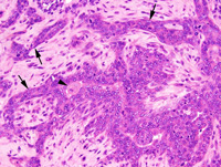
|
|
Image ID:1908 |
|
Source of Image:Ward JM |
|
Pathologist:Mikaelian I |
|
Method / Stain:H&E |
|
|
Image Caption:Mammary gland: an interlobular mammary duct is moderately expanded by a cauliflower-like proliferative process characterized by the formation of slender fibrovascular cores that support two populations of cells. First, there is a 1-5 cells-thick layer of oat-shaped cells with indistinct cell borders, a small amount of pale acidophilic cytoplasm and a small hyperchromatic oval nucleus. Second, there is a one cell-thick layer of cuboidal to low columnar cells that form conspicuous lumens filled with an amorphous proteinaceous material. These cells have indistinct cell borders, a moderate amount of acidophilic cytoplasm and a medium-size, central, normochromatic nucleus with a clumped chromatin. A few mast cells are present in the periductal connective tissue.
The presence of two layers of cells indicates that the cell that gave rise to these lesions is not committed to the luminal or to the basal lineage, and hence these lesions may be categorized as "complex" lesions.
|
|
|
|
Image ID:1904 |
|
Source of Image:Ward JM |
|
Pathologist:Mikaelian I |
|
Method / Stain:H&E |
|
|
Image Caption:Mammary gland: almost all interlobular and intralobular mammary ducts contain multiple and often coalescing small cauliflower-like projections. There is no evidence of fibrosis or inflammation around these ducts.
|
|
|
|
Image ID:1900 |
|
Source of Image:Ward JM |
|
Pathologist:Mikaelian I |
|
Method / Stain:H&E |
|
|
Image Caption:Mammary gland: a mammary lobule is greatly expanded. The ductal portions of the lobule are prominent and surrounded by prominent sclerosis. The acini are hyperplastic and are surrounded by lesser sclerosis. Cells in the ducts pile-up disorderly up to 3 cells-thick.
|
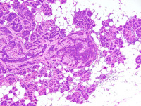
|
|
Image ID:1906 |
|
Source of Image:Ward JM |
|
Pathologist:Mikaelian I |
|
Method / Stain:H&E |
|
|
|
| MTB ID |
Tumor Name |
Organ(s) Affected |
Treatment Type |
Agents |
Strain Name |
Strain Sex |
Reproductive Status |
Tumor Frequency |
Age at Necropsy |
Description |
Reference |
| MTB:29309 |
Mammary gland adenocarcinoma - complex |
Mammary gland |
None (spontaneous) |
|
|
Female |
reproductive status not specified |
observed |
unknown |
Adenocarcinoma, complex, mammary gland |
J:94320 |
|
Image Caption:Mammary adenocarcinoma: this photomicrograph illustrates the morphology of the neoplasm towards its' center. Large blood-filled vascular spaces are present. Neoplastic cells form nests and trabeculae that often undergo cystic degeneration and are supported by a moderate amount of loose fibrovascular stroma. There are two types of neoplastic cells: (1) there are large polygonal cells with a moderate amount of pale acidophilic cytoplasm and a large central hypochromatic round to oval nucleus; (2) there are small polygonal cells with a small amount of amphophilic cytoplasm and a small hyperchromatic oval nucleus. Cells intermediate between these two phenotypes are also present. Anisokaryosis and anisocytosis are mild. Mitoses are rare.
|
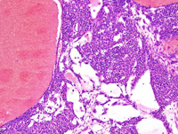
|
|
Image ID:1917 |
|
Source of Image:Ward JM |
|
Pathologist:Mikaelian I |
|
Method / Stain:H&E |
|
|
Image Caption:Mammary adenocarcinoma: this photomicrograph illustrates an area of ductal differentiation. Neoplastic cells form a two cell-thick epithelium. Luminal cells are cuboidal. Basal cells are either attenuated with a morphology reminiscent of myoepithelial cells, or they are cuboidal. Anisokaryosis and anisocytosis are mild. Mitoses are rare.
|
|
|
Image ID:1919 |
|
Source of Image:Ward JM |
|
Pathologist:Mikaelian I |
|
Method / Stain:H&E |
|
|
Image Caption:Mammary gland: the mammary gland is expanded by a large, densely cellular neoplasm. This neoplasm is composed of solid trabeculae and nests (predominantly at the periphery of the neoplasm) and cystic ducts (predominantly at the center of the neoplasm) supported by a small amount of loose fibrovascular stroma with numerous blood-filled dilated vascular spaces.
|
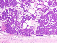
|
|
Image ID:1913 |
|
Source of Image:Ward JM |
|
Pathologist:Mikaelian I |
|
Method / Stain:H&E |
|
|
Image Caption:Mammary adenocarcinoma: this photomicrograph illustrates the morphology of the neoplasm towards its' center. Large blood-filled vascular spaces are present. Neoplastic cells form nests and trabeculae that often undergo cystic degeneration and are supported by a moderate amount of loose fibrovascular stroma. There are two types of neoplastic cells: (1) there are large polygonal cells with a moderate amount of pale acidophilic cytoplasm and a large central hypochromatic round to oval nucleus; (2) there are small polygonal cells with a small amount of amphophilic cytoplasm and a small hyperchromatic oval nucleus. Cells intermediate between these two phenotypes are also present.
|
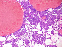
|
|
Image ID:1916 |
|
Source of Image:Ward JM |
|
Pathologist:Mikaelian I |
|
Method / Stain:H&E |
|
|
Image Caption:Mammary gland: the mammary gland is expanded by a large, densely cellular neoplasm. This neoplasm is composed of solid trabeculae and nests (predominantly at the periphery of the neoplasm) and cystic ducts (predominantly at the center of the neoplasm) supported by a small amount of loose fibrovascular stroma with numerous blood-filled dilated vascular spaces.
|
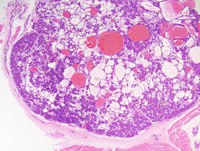
|
|
Image ID:1911 |
|
Source of Image:Ward JM |
|
Pathologist:Mikaelian I |
|
Method / Stain:H&E |
|
|
Image Caption:Mammary adenocarcinoma: this photomicrograph illustrates the morphology of the periphery of the neoplasm. The neoplasm in this area is composed of small nests, interconnected trabeculae, and areas of cystic degeneration separated by a moderate amount of loose fibrovascular stroma. There are two types of neoplastic cells: (1) there are large polygonal cells with a moderate amount of pale acidophilic cytoplasm and a large central hypochromatic round nucleus; (2) there are small polygonal cells with a small amount of amphophilic cytoplasm and a small hyperchromatic oval nucleus. Cells intermediate between these two phenotypes are also present.
|
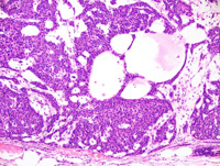
|
|
Image ID:1914 |
|
Source of Image:Ward JM |
|
Pathologist:Mikaelian I |
|
Method / Stain:H&E |
|
|
Image Caption:Mammary gland: the mammary gland is expanded by a large, densely cellular neoplasm. This neoplasm is composed of solid trabeculae and nests (predominantly at the periphery of the neoplasm) and cystic ducts (predominantly at the center of the neoplasm) supported by a small amount of fibrovascular stroma with numerous blood-filled dilated vascular spaces.
|
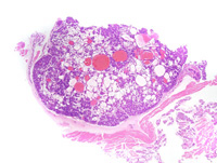
|
|
Image ID:1910 |
|
Source of Image:Ward JM |
|
Pathologist:Mikaelian I |
|
Method / Stain:H&E |
|
|
Image Caption:Mammary adenocarcinoma: this photomicrograph illustrates the morphology of the periphery of the neoplasm. The neoplasm in this area is composed of small nests, interconnected trabeculae, and areas of cystic degeneration separated by a moderate amount of loose fibrovascular stroma. There are two types of neoplastic cells: (1) there are large polygonal cells with a moderate amount of pale acidophilic cytoplasm and a large central hypochromatic round nucleus; (2) there are small polygonal cells with a small amount of amphophilic cytoplasm and a small hyperchromatic oval to coma-shaped nucleus. Cells intermediate between these two phenotypes are also present. The large cells resemble the large light cells of the developing mammary gland.
|
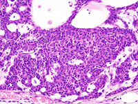
|
|
Image ID:1915 |
|
Source of Image:Ward JM |
|
Pathologist:Mikaelian I |
|
Method / Stain:H&E |
|
|
Image Caption:Mammary gland: the mammary gland is expanded by a large, densely cellular neoplasm. This neoplasm is composed of solid trabeculae and nests (predominantly at the periphery of the neoplasm) and cystic ducts (predominantly at the center of the neoplasm) supported by a small amount of loose fibrovascular stroma with numerous blood-filled dilated vascular spaces.
|
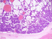
|
|
Image ID:1912 |
|
Source of Image:Ward JM |
|
Pathologist:Mikaelian I |
|
Method / Stain:H&E |
|
|
Image Caption:Mammary adenocarcinoma: this photomicrograph illustrates the morphology of the neoplasm towards its' center. Large blood-filled vascular spaces are present. Neoplastic cells form nests and trabeculae that often undergo cystic degeneration and are supported by a moderate amount of loose fibrovascular stroma. There are two types of neoplastic cells: (1) there are large polygonal cells with a moderate amount of pale acidophilic cytoplasm and a large central hypochromatic round to oval nucleus; (2) there are small polygonal cells with a small amount of amphophilic cytoplasm and a small hyperchromatic oval nucleus. Cells intermediate between these two phenotypes are also present. Anisokaryosis and anisocytosis are mild. Mitoses are rare.
|
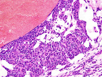
|
|
Image ID:1918 |
|
Source of Image:Ward JM |
|
Pathologist:Mikaelian I |
|
Method / Stain:H&E |
|
|
|
| MTB ID |
Tumor Name |
Organ(s) Affected |
Treatment Type |
Agents |
Strain Name |
Strain Sex |
Reproductive Status |
Tumor Frequency |
Age at Necropsy |
Description |
Reference |
| MTB:29310 |
Mammary gland hyperplasia - atypical - lobular |
Mammary gland |
None (spontaneous) |
|
|
Female |
reproductive status not specified |
observed |
unknown |
Lobular hyperplasia, atypical, mammary gland |
J:94320 |
|
Image Caption:Atypical lobular hyperplasia: this high power photomicrograph illustrates an area of ductal differentiation (arrowheads) and an area of acinar differentiation (*). The myoepithelial layer is more prominent in the area of ductal differentiation than in the area of acinar differentiation. A few mitoses are noted.
|
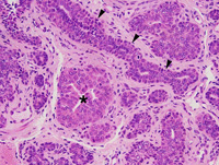
|
|
Image ID:1924 |
|
Source of Image:Ward JM |
|
Pathologist:Mikaelian I |
|
Method / Stain:H&E |
|
|
Image Caption:Mammary gland: there is prominent increased lobulation of the mammary gland. There is marked ectasia of the interlobular ducts that are filled with a proteinaceous fluid. Acini and intralobular ducts connecting to the cystic interlobular ducts are hyperplastic and there is moderate interstitial fibrosis. The mammary gland recapitulates the morphology of the pubertal mammary gland, with the formation of fronds that rapidly differentiate into ducts.
|
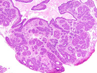
|
|
Image ID:1922 |
|
Source of Image:Ward JM |
|
Pathologist:Mikaelian I |
|
Method / Stain:H&E |
|
|
Image Caption:Mammary gland: there is prominent increased lobulation of the mammary gland. There is marked ectasia of the ducts of a mammary lobule. Acini in this lobule are hyperplastic and there is moderate interstitial fibrosis.
|
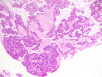
|
|
Image ID:1921 |
|
Source of Image:Ward JM |
|
Pathologist:Mikaelian I |
|
Method / Stain:H&E |
|
|
Image Caption:Atypical lobular hyperplasia: the interlobular and intralobular ducts are lined by two types of cells: (1) a one cell-thick layer of attenuated to low cuboidal cells that are reminiscent of myoepithelial cells; (2) a 2-4 cells-thick layer of cuboidal to polygonal cells. The acini form nests that are composed of cells that are larger, with a more acidophilic cytoplasm, and a larger and more hypochromatic nucleus than cells that line the ducts. Central cavitation and the formation of small slit-like lumens are common in the acinar areas. There is moderate interstitial fibrosis with mild pleocellular inflammation.
|
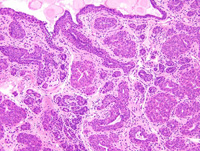
|
|
Image ID:1923 |
|
Source of Image:Ward JM |
|
Pathologist:Mikaelian I |
|
Method / Stain:H&E |
|
|
Image Caption:Mammary gland: there is prominent increased lobulation of the mammary gland. There is marked ectasia of the ducts of a mammary lobule. Acini in this lobule are hyperplastic and there is moderate interstitial fibrosis.
|
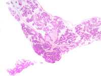
|
|
Image ID:1920 |
|
Source of Image:Ward JM |
|
Pathologist:Mikaelian I |
|
Method / Stain:H&E |
|
|
Image Caption:Atypical lobular hyperplasia: this high power photomicrograph illustrates the formation of fronds at the end of the area of acinar differentiation. These areas resemble the terminal end bud of the pubertal mammary gland. A few mitoses are noted.
|
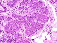
|
|
Image ID:1925 |
|
Source of Image:Ward JM |
|
Pathologist:Mikaelian I |
|
Method / Stain:H&E |
|
|
|
| MTB ID |
Tumor Name |
Organ(s) Affected |
Treatment Type |
Agents |
Strain Name |
Strain Sex |
Reproductive Status |
Tumor Frequency |
Age at Necropsy |
Description |
Reference |
| MTB:29311 |
Mammary gland adenocarcinoma |
Mammary gland |
None (spontaneous) |
|
|
Female |
reproductive status not specified |
observed |
unknown |
Adenocarcinoma, glandular, secretory, mammary gland |
J:94320 |
|
Image Caption:Mammary gland: the mammary gland is effaced by a nodular, unencapsulated, well-delineated, multilobular, densely cellular neoplasm. The glandular pattern is obvious in the peripheral portions of the neoplasm, even at this low magnification. Numerous areas of liquefactive necrosis (*) are present at the center of the neoplasm.
|
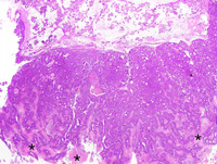
|
|
Image ID:1927 |
|
Source of Image:Ward JM |
|
Pathologist:Mikaelian I |
|
Method / Stain:H&E |
|
|
Image Caption:Mammary gland: the mammary gland is effaced by a nodular, unencapsulated, well-delineated, multilobular, densely cellular neoplasm.
|
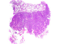
|
|
Image ID:1926 |
|
Source of Image:Ward JM |
|
Pathologist:Mikaelian I |
|
Method / Stain:H&E |
|
|
Image Caption:Mammary gland: neoplastic lobules are composed of closely-packed glands lined by a one cell-thick cuboidal epithelium. There are also a few solid areas with secondary lumens. Glands and secondary lumens show various degrees of ectasia, and some contain a small to moderate of amorphous acidophilic fluid and occasionally a few desquamated neoplastic cells. Neoplastic cells are cuboidal to polygonal, with distinct cell borders and a moderate amount of strongly amphophilic cytoplasm that occasionally contains small lipid vacuoles and hyaline droplets. The nucleus is central, medium-size to large, round to oval, slightly hypochromatic, with a finely stippled chromatin and 1-3 large basophilic nucleoli. Anisokaryosis and anisocytosis are moderate. A few mitoses are detected. Neoplastic cells occasionally pile-up disorderly.
|
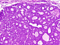
|
|
Image ID:1930 |
|
Source of Image:Ward JM |
|
Pathologist:Mikaelian I |
|
Method / Stain:H&E |
|
|
Image Caption:Mammary gland: neoplastic lobules are composed of closely-packed glands lined by a one cell-thick cuboidal epithelium. There are also a few solid areas with secondary lumens. Glands and secondary lumens show various degrees of ectasia, and some contain a small to moderate of amorphous acidophilic fluid and occasionally a few desquamated neoplastic cells. Neoplastic cells are cuboidal to polygonal, with distinct cell borders and a moderate amount of strongly amphophilic cytoplasm that occasionally contains small lipid vacuoles and hyaline droplets. The nucleus is central, medium-size to large, round to oval, slightly hypochromatic, with a finely stippled chromatin and 1-3 large basophilic nucleoli. Anisokaryosis and anisocytosis are moderate. A few mitoses are detected. Neoplastic cells occasionally pile-up disorderly.
|
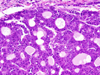
|
|
Image ID:1931 |
|
Source of Image:Ward JM |
|
Pathologist:Mikaelian I |
|
Method / Stain:H&E |
|
|
Image Caption:Mammary gland: neoplastic lobules are composed of closely-packed glands lined by a one cell-thick cuboidal epithelium. There are also a few solid areas with secondary lumens. Glands and secondary lumens show various degrees of ectasia, and some contain a small to moderate of amorphous acidophilic fluid and occasionally a few desquamated neoplastic cells. Neoplastic cells are cuboidal to polygonal, with distinct cell borders and a moderate amount of strongly amphophilic cytoplasm that occasionally contains small lipid vacuoles and hyaline droplets. The nucleus is central, medium-size to large, round to oval, slightly hypochromatic, with a finely stippled chromatin and 1-3 large basophilic nucleoli. Anisokaryosis and anisocytosis are moderate. A few mitoses are detected. Neoplastic cells occasionally pile-up disorderly.
|
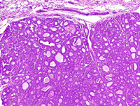
|
|
Image ID:1929 |
|
Source of Image:Ward JM |
|
Pathologist:Mikaelian I |
|
Method / Stain:H&E |
|
|
Image Caption:Mammary gland: the mammary gland is effaced by a nodular, unencapsulated, well-delineated, multilobular, densely cellular neoplasm. The glandular pattern is obvious in the peripheral portions of the neoplasm, even at this low magnification. Neoplastic lobules are composed of closely packed glands lined by a one cell-thick cuboidal epithelium. There are also a few solid areas with the formation of secondary lumens (cribriform pattern). These is a scant amount of fibrovascular stroma.
|
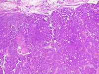
|
|
Image ID:1928 |
|
Source of Image:Ward JM |
|
Pathologist:Mikaelian I |
|
Method / Stain:H&E |
|
|
|
| MTB ID |
Tumor Name |
Organ(s) Affected |
Treatment Type |
Agents |
Strain Name |
Strain Sex |
Reproductive Status |
Tumor Frequency |
Age at Necropsy |
Description |
Reference |
| MTB:29312 |
Mammary gland adenocarcinoma - mixed |
Mammary gland |
None (spontaneous) |
|
|
Female |
reproductive status not specified |
observed |
unknown |
Adenocarcinoma, glandular, solid, cribriform, mammary gland |
J:94320 |
|
Image Caption:Mammary adenocarcinoma: this photomicrograph represents a portion of the inner portions of the neoplasm. A narrow rim of neoplastic cells survive around dilated blood vessels (V). The rest of the tissue has undergone necrosis.
|
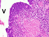
|
|
Image ID:1938 |
|
Source of Image:Ward JM |
|
Pathologist:Mikaelian I |
|
Method / Stain:H&E |
|
|
Image Caption:Mammary adenocarcinoma: in the peripheral portions of the neoplasm, neoplastic cells form small glands supported by a scant amount of fibrovascular stroma. Neoplastic cells are cuboidal to low columnar, occasionally polygonal, with distinct cell borders and a moderate amount of strongly amphophilic cytoplasm that occasionally contains a few small lipid vacuoles. The nucleus is central, oval, medium-sized and generally hyperchromatic. Anisokaryosis and anisocytosis are mild.
|
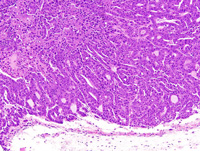
|
|
Image ID:1935 |
|
Source of Image:Ward JM |
|
Pathologist:Mikaelian I |
|
Method / Stain:H&E |
|
|
Image Caption:Mammary adenocarcinoma: in the peripheral portions of the neoplasm, neoplastic cells form small glands supported by a scant amount of fibrovascular stroma.
|
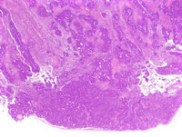
|
|
Image ID:1933 |
|
Source of Image:Ward JM |
|
Pathologist:Mikaelian I |
|
Method / Stain:H&E |
|
|
Image Caption:Mammary adenocarcinoma: in the peripheral portions of the neoplasm, neoplastic cells form small glands supported by a scant amount of fibrovascular stroma. Neoplastic cells are cuboidal to low columnar, occasionally polygonal, with distinct cell borders and a moderate amount of strongly amphophilic cytoplasm that occasionally contains a few small lipid vacuoles. The nucleus is central, oval, medium-sized and generally hyperchromatic. Anisokaryosis and anisocytosis are mild. A few mitoses (arrowheads) are detected.
|
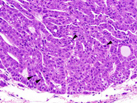
|
|
Image ID:1936 |
|
Source of Image:Ward JM |
|
Pathologist:Mikaelian I |
|
Method / Stain:H&E |
|
|
Image Caption:Mammary adenocarcinoma: in the peripheral portions of the neoplasm, neoplastic cells form small glands supported by a scant amount of fibrovascular stroma. Neoplastic cells are cuboidal to low columnar, occasionally polygonal, with distinct cell borders and a moderate amount of strongly amphophilic cytoplasm that occasionally contains a few small lipid vacuoles. The nucleus is central, oval, medium-sized and generally hyperchromatic. Anisokaryosis and anisocytosis are mild.
|
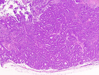
|
|
Image ID:1934 |
|
Source of Image:Ward JM |
|
Pathologist:Mikaelian I |
|
Method / Stain:H&E |
|
|
Image Caption:Mammary gland neoplasm (portion of): the mammary gland is expanded by a nodular, well-delineated, multilobular, densely cellular neoplasm. Linear areas of liquefactive necrosis (*) are present at the center of the neoplasm and form a reticular pattern.
|
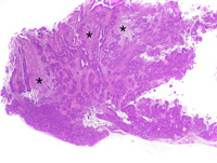
|
|
Image ID:1932 |
|
Source of Image:Ward JM |
|
Pathologist:Mikaelian I |
|
Method / Stain:H&E |
|
|
Image Caption:Mammary adenocarcinoma: this photomicrograph represents a portion of the inner portions of the neoplasm. A narrow rim of neoplastic cells survive around dilated blood vessels (V). The rest of the tissue has undergone necrosis.
|
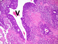
|
|
Image ID:1937 |
|
Source of Image:Ward JM |
|
Pathologist:Mikaelian I |
|
Method / Stain:H&E |
|
|
|
| MTB ID |
Tumor Name |
Organ(s) Affected |
Treatment Type |
Agents |
Strain Name |
Strain Sex |
Reproductive Status |
Tumor Frequency |
Age at Necropsy |
Description |
Reference |
| MTB:29313 |
Mammary gland adenocarcinoma - solid |
Mammary gland |
None (spontaneous) |
|
|
Female |
reproductive status not specified |
observed |
unknown |
Adenocarcinoma, solid, mammary gland |
J:94320 |
|
Image Caption:Solid mammary adenocarcinoma: the neoplasm is composed of closely-packed small polygonal cells that form solid areas supported by a scant amount of loose fibrovascular stroma. Many blood vessels within the stroma are moderately ectatic.
|
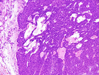
|
|
Image ID:1941 |
|
Source of Image:Ward JM |
|
Pathologist:Mikaelian I |
|
Method / Stain:H&E |
|
|
Image Caption:Mammary gland: the mammary gland is entirely effaced by a densely cellular, expensile, unencapsulated, neoplasm. This neoplasm is composed of solid areas with multiple small foci of liquefactive necrosis.
|
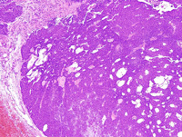
|
|
Image ID:1940 |
|
Source of Image:Ward JM |
|
Pathologist:Mikaelian I |
|
Method / Stain:H&E |
|
|
Image Caption:Mammary gland: the mammary gland is entirely effaced by a densely cellular, expensile, unencapsulated, neoplasm. This neoplasm is composed of solid areas with multiple small foci of liquefactive necrosis.
|
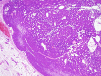
|
|
Image ID:1939 |
|
Source of Image:Ward JM |
|
Pathologist:Mikaelian I |
|
Method / Stain:H&E |
|
|
Image Caption:Solid mammary adenocarcinoma: the neoplasm is composed of closely-packed small polygonal cells that form solid areas supported by a scant amount of loose fibrovascular stroma. Many blood vessels within the stroma are moderately ectatic. Neoplastic cells are medium-sized to small, with indistinct cell borders and a small to moderate amount of strongly acidophilic cytoplasm. The nucleus is central, oval, medium-sized and hyperchromatic anisocytosis and anisokaryosis are mild.
|
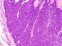
|
|
Image ID:1942 |
|
Source of Image:Ward JM |
|
Pathologist:Mikaelian I |
|
Method / Stain:H&E |
|
|
|
| MTB ID |
Tumor Name |
Organ(s) Affected |
Treatment Type |
Agents |
Strain Name |
Strain Sex |
Reproductive Status |
Tumor Frequency |
Age at Necropsy |
Description |
Reference |
| MTB:29314 |
Mammary gland adenocarcinoma - mixed |
Mammary gland |
None (spontaneous) |
|
|
Female |
reproductive status not specified |
observed |
unknown |
Adenocarcinoma, solid, trabecular, glandular, mammary gland |
J:94320 |
|
Image Caption:Mammary gland: the neoplasm is composed of nests, trabeculae, and glands separated by moderately thick fibrous septae. This photomicrograph focuses on an area where glands predominate. Neoplastic cells are cuboidal to polygonal, with ill-defined cell borders and a moderate amount of amphophilic cytoplasm. The nucleus is basal, medium-size, oval, and normochromatic. Anisokaryosis and anisocytosis are mild. Mitoses are numerous.
|
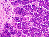
|
|
Image ID:1948 |
|
Source of Image:Ward JM |
|
Pathologist:Mikaelian I |
|
Method / Stain:H&E |
|
|
Image Caption:Mammary gland: the neoplasm is composed of nests, trabeculae, and glands separated by moderately thick fibrous septae. This photomicrograph focuses on an area where nests predominate. Necrosis is present at the center of the largest nest. A moderate number of secondary lumens is present in the solid areas.
|
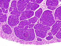
|
|
Image ID:1950 |
|
Source of Image:Ward JM |
|
Pathologist:Mikaelian I |
|
Method / Stain:H&E |
|
|
Image Caption:Mammary gland: the mammary gland is expanded by a nodular, expensile, densely cellular neoplasm. The neoplasm is composed of nests, trabeculae, and glands separated by moderately thick fibrous septae.
|
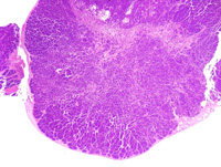
|
|
Image ID:1944 |
|
Source of Image:Ward JM |
|
Pathologist:Mikaelian I |
|
Method / Stain:H&E |
|
|
Image Caption:Mammary gland: the neoplasm is composed of nests, trabeculae, and glands separated by moderately thick fibrous septae. This photomicrograph focuses on an area where glands predominate. Neoplastic cells are cuboidal to polygonal, with ill-defined cell borders and a moderate amount of amphophilic cytoplasm. The nucleus is basal, medium-size, oval, and normochromatic. Anisokaryosis and anisocytosis are mild.
|
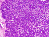
|
|
Image ID:1946 |
|
Source of Image:Ward JM |
|
Pathologist:Mikaelian I |
|
Method / Stain:H&E |
|
|
Image Caption:Mammary gland: the neoplasm is composed of nests, trabeculae, and glands separated by moderately thick fibrous septae. This photomicrograph focuses on an area where glands predominate. Neoplastic cells are cuboidal to polygonal, with ill-defined cell borders and a moderate amount of amphophilic cytoplasm. The nucleus is basal, medium-size, oval, and normochromatic. Anisokaryosis and anisocytosis are mild.
|
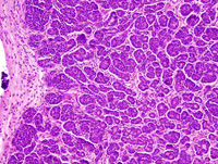
|
|
Image ID:1947 |
|
Source of Image:Ward JM |
|
Pathologist:Mikaelian I |
|
Method / Stain:H&E |
|
|
Image Caption:Mammary gland: the neoplasm is composed of nests, trabeculae, and glands separated by moderately thick fibrous septae. This photomicrograph focuses on an area where nests predominate. Necrosis is present at the center of the largest nest. A moderate number of secondary lumens is present in the solid areas (arrowheads).
|

|
|
Image ID:1951 |
|
Source of Image:Ward JM |
|
Pathologist:Mikaelian I |
|
Method / Stain:H&E |
|
|
Image Caption:Mammary gland: the mammary gland is expanded by a nodular, expensile, densely cellular neoplasm. The neoplasm is composed of nests, trabeculae, and glands separated by moderately thick fibrous septae.
|
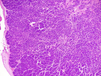
|
|
Image ID:1945 |
|
Source of Image:Ward JM |
|
Pathologist:Mikaelian I |
|
Method / Stain:H&E |
|
|
Image Caption:Mammary gland: the mammary gland is expanded by a nodular, expensile, densely cellular neoplasm.
|
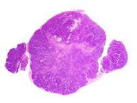
|
|
Image ID:1943 |
|
Source of Image:Ward JM |
|
Pathologist:Mikaelian I |
|
Method / Stain:H&E |
|
|
Image Caption:Mammary gland: the neoplasm is composed of nests, trabeculae, and glands separated by moderately thick fibrous septae. This photomicrograph focuses on an area where nests predominate. Necrosis is present at the center of the largest nest. A moderate number of secondary lumens is present in the solid areas.
|

|
|
Image ID:1949 |
|
Source of Image:Ward JM |
|
Pathologist:Mikaelian I |
|
Method / Stain:H&E |
|
|
|
| MTB ID |
Tumor Name |
Organ(s) Affected |
Treatment Type |
Agents |
Strain Name |
Strain Sex |
Reproductive Status |
Tumor Frequency |
Age at Necropsy |
Description |
Reference |
| MTB:29315 |
Mammary gland hyperplasia - atypical |
Mammary gland |
None (spontaneous) |
|
|
Female |
reproductive status not specified |
observed |
unknown |
Hyperplasia, atypical, mammary gland |
J:94320 |
|
Image Caption:Mammary gland: the lumen of mammary ducts (predominantly) and acini is moderately expanded by a strongly acidophilic proteinaceous material that also contains a few clusters of desquamated cells. The epithelium lining these ducts and acini is hypercellular and columnar and it forms a few minute papillary projections. Cells lining these ducts have a larger nucleus than what is expected for the normal mammary gland.
|
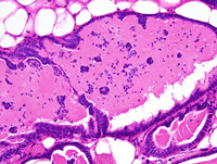
|
|
Image ID:1954 |
|
Source of Image:Ward JM |
|
Pathologist:Mikaelian I |
|
Method / Stain:H&E |
|
|
Image Caption:Mammary gland: the lumen of mammary ducts (predominantly) and acini is moderately expanded by a strongly acidophilic proteinaceous material that also contains a few clusters of desquamated cells. The epithelium lining these ducts and acini is hypercellular and columnar and it forms a few minute papillary projections.
|
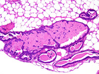
|
|
Image ID:1953 |
|
Source of Image:Ward JM |
|
Pathologist:Mikaelian I |
|
Method / Stain:H&E |
|
|
Image Caption:Mammary gland: the lumen of mammary ducts (predominantly) and acini is moderately expanded by a strongly acidophilic proteinaceous material that also contains a few clusters of desquamated cells. The epithelium lining these ducts and acini is hypercellular and columnar.
|
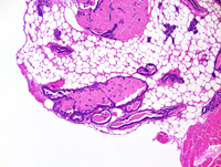
|
|
Image ID:1952 |
|
Source of Image:Ward JM |
|
Pathologist:Mikaelian I |
|
Method / Stain:H&E |
|
|
|
| MTB ID |
Tumor Name |
Organ(s) Affected |
Treatment Type |
Agents |
Strain Name |
Strain Sex |
Reproductive Status |
Tumor Frequency |
Age at Necropsy |
Description |
Reference |
| MTB:29316 |
Mammary gland carcinoma - mixed |
Mammary gland |
None (spontaneous) |
|
|
Female |
reproductive status not specified |
observed |
unknown |
Spindle cell carcinoma arising in a glandular carcinoma, mammary gland |
J:94320 |
|
Image Caption:Mammary gland: the neoplasm is composed of a few trabeculae and tubules interspersed among ill-defined bundles of neoplastic spindloid cells. There is gradual transition between some of the trabeculae and the spindloid cells.
|
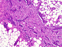
|
|
Image ID:1956 |
|
Source of Image:Ward JM |
|
Pathologist:Mikaelian I |
|
Method / Stain:H&E |
|
|
Image Caption:Mammary gland: the neoplasm is composed of a few trabeculae and tubules interspersed among ill-defined bundles of neoplastic spindloid cells. There is gradual transition between some of the trabeculae and the spindloid cells. Neoplastic cells with an epithelial phenotype are polygonal, with distinct cell borders and a moderate amount of strongly amphophilic cytoplasm. Neoplastic cells with a mesenchymal phenotype are spindloid to oat-shaped, with distinct cell borders and a moderate amount of delicately fibrillar acidophilic cytoplasm and a slightly elongated hyperchromatic nucleus. The stroma is loose and edematous in the right portion of the photomicrograph, and dense and amorphous in the left portions of the photomicrograph.
|
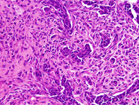
|
|
Image ID:1958 |
|
Source of Image:Ward JM |
|
Pathologist:Mikaelian I |
|
Method / Stain:H&E |
|
|
Image Caption:Mammary gland: the neoplasm is composed of a few trabeculae and tubules interspersed among ill-defined bundles of neoplastic spindloid cells. There is gradual transition between some of the trabeculae and the spindloid cells. Neoplastic cells with an epithelial phenotype are polygonal, with distinct cell borders and a moderate amount of strongly amphophilic cytoplasm. Neoplastic cells with a mesenchymal phenotype are spindloid to oat-shaped, with distinct cell borders and a moderate amount of delicately fibrillar acidophilic cytoplasm and a slightly elongated hyperchromatic nucleus.
|
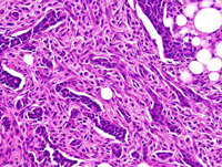
|
|
Image ID:1957 |
|
Source of Image:Ward JM |
|
Pathologist:Mikaelian I |
|
Method / Stain:H&E |
|
|
Image Caption:Mammary gland: the mammary gland is focally expanded by a stellate, unencapsulated, ill-defined, invasive neoplasm. This neoplasm is composed of a few trabeculae and tubules interspersed among ill-defined bundles of neoplastic spindloid cells.
|
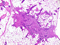
|
|
Image ID:1955 |
|
Source of Image:Ward JM |
|
Pathologist:Mikaelian I |
|
Method / Stain:H&E |
|
|
|
| MTB ID |
Tumor Name |
Organ(s) Affected |
Treatment Type |
Agents |
Strain Name |
Strain Sex |
Reproductive Status |
Tumor Frequency |
Age at Necropsy |
Description |
Reference |
| MTB:29317 |
Mammary gland adenoma |
Mammary gland |
None (spontaneous) |
|
|
Female |
reproductive status not specified |
observed |
unknown |
Adenoma, glandular, mammary gland |
J:94320 |
|
Image Caption:Mammary gland: the mammary gland is expanded by a nodular, unencapsulated, densely cellular neoplastic mass that has entrapped a moderate number of adipocytes. This mass is composed of glands supported by a small amount of fibrovascular stroma (some of which is pre-existing mammary connective tissue).
|
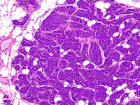
|
|
Image ID:1959 |
|
Source of Image:Ward JM |
|
Pathologist:Mikaelian I |
|
Method / Stain:H&E |
|
|
Image Caption:Mammary gland: the mammary gland is expanded by a nodular, unencapsulated, densely cellular neoplastic mass that has entrapped a moderate number of adipocytes. This mass is composed of glands supported by a small amount of fibrovascular stroma (some of which is pre-existing mammary connective tissue).
|
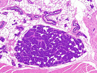
|
|
Image ID:1962 |
|
Source of Image:Ward JM |
|
Pathologist:Mikaelian I |
|
Method / Stain:H&E |
|
|
Image Caption:Mammary gland: the mammary gland is expanded by a small, nodular, unencapsulated, densely cellular neoplastic mass that has entrapped a moderate number of adipocytes. This mass is composed of glands supported by a small amount of fibrovascular stroma (some of which is pre-existing mammary connective tissue).
|
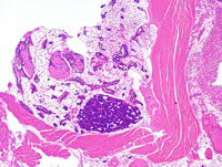
|
|
Image ID:1961 |
|
Source of Image:Ward JM |
|
Pathologist:Mikaelian I |
|
Method / Stain:H&E |
|
|
Image Caption:Mammary gland: the mammary gland is expanded by a nodular, unencapsulated, densely cellular neoplastic mass that has entrapped a moderate number of adipocytes. This mass is composed of glands supported by a small amount of fibrovascular stroma (some of which is pre-existing mammary connective tissue). The glands are contoured, anastomosed, with an inconspicuous lumen, and they are lined by a one cell-thick cuboidal to low columnar epithelium. Neoplastic cells have ill-defined cell borders, a moderate amount of strongly amphophilic cytoplasm, and a medium-size, oval, hyperchromatic nucleus oriented in the long axis of the neoplastic cell. Anisokaryosis and anisocytosis are minimal. A few mitoses are present.
|
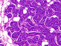
|
|
Image ID:1960 |
|
Source of Image:Ward JM |
|
Pathologist:Mikaelian I |
|
Method / Stain:H&E |
|
|
|
| MTB ID |
Tumor Name |
Organ(s) Affected |
Treatment Type |
Agents |
Strain Name |
Strain Sex |
Reproductive Status |
Tumor Frequency |
Age at Necropsy |
Description |
Reference |
| MTB:29318 |
Mammary gland adenocarcinoma - mixed |
Mammary gland |
None (spontaneous) |
|
|
Female |
reproductive status not specified |
observed |
unknown |
Adenocarcinoma, papillary, glandular, complex, mammary gland |
J:94320 |
|
Image Caption:Mammary gland: neoplastic lobules are composed of closely-packed glands and acini supported by a small to moderate amount of loose fibrovascular stroma. The papillary pattern can be nicely appreciated at this magnification. Neoplastic papillae are massive and are lined by a two cells-thick cuboidal epithelium.
|
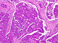
|
|
Image ID:1967 |
|
Source of Image:Ward JM |
|
Pathologist:Mikaelian I |
|
Method / Stain:H&E |
|
|
Image Caption:Mammary gland: the mammary gland is effaced by a nodular, unenencapsulated, multilobular, densely cellular neoplasm. Neoplastic lobules are composed of closely-packed glands and acini supported by a small to moderate amount of loose fibrovascular stroma.
|
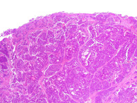
|
|
Image ID:1964 |
|
Source of Image:Ward JM |
|
Pathologist:Mikaelian I |
|
Method / Stain:H&E |
|
|
Image Caption:Mammary gland: neoplastic lobules are composed of closely-packed glands and acini supported by a small to moderate amount of loose fibrovascular stroma. The papillary pattern can be nicely appreciated at this magnification. Many of the neoplastic glands are ectatic and contain a moderate amount of amorphous acidophilic fluid.
|
|
|
Image ID:1965 |
|
Source of Image:Ward JM |
|
Pathologist:Mikaelian I |
|
Method / Stain:H&E |
|
|
Image Caption:Mammary gland: neoplastic lobules are composed of closely-packed glands and acini supported by a small to moderate amount of loose fibrovascular stroma. The papillary pattern can be nicely appreciated at this magnification. Neoplastic papillae are massive and are lined by a two cells-thick cuboidal epithelium.
|
|
|
Image ID:1966 |
|
Source of Image:Ward JM |
|
Pathologist:Mikaelian I |
|
Method / Stain:H&E |
|
|
Image Caption:Mammary gland: the mammary gland is effaced by a nodular, unenencapsulated, multilobular, densely cellular neoplasm. Neoplastic lobules are composed of closely-packed glands and acini supported by a small to moderate amount of loose fibrovascular stroma.
|
|
|
Image ID:1963 |
|
Source of Image:Ward JM |
|
Pathologist:Mikaelian I |
|
Method / Stain:H&E |
|
|
Image Caption:Mammary gland: neoplastic lobules are composed of closely-packed glands and acini supported by a small to moderate amount of loose fibrovascular stroma. The papillary pattern can be nicely appreciated at this magnification. Neoplastic papillae are massive and are lined by a two cells-thick cuboidal epithelium. The basal layer of cell is indicative of myoepithelial differentiation and is most easily identified in the papillary areas of the neoplasm (half lower right part of the photomicrographs), although it is also present in the glandular portions of the neoplasm (half upper left part of the photomicrograph). Anisokaryosis and anisocytosis are mild.
|
|
|
Image ID:1968 |
|
Source of Image:Ward JM |
|
Pathologist:Mikaelian I |
|
Method / Stain:H&E |
|
|
|
| MTB ID |
Tumor Name |
Organ(s) Affected |
Treatment Type |
Agents |
Strain Name |
Strain Sex |
Reproductive Status |
Tumor Frequency |
Age at Necropsy |
Description |
Reference |
| MTB:29319 |
Mammary gland adenocarcinoma - papillary |
Mammary gland |
None (spontaneous) |
|
|
Female |
reproductive status not specified |
observed |
unknown |
Adenocarcinoma, papillary, glandular, with squamous metaplasia and secretory activity, mammary gland |
J:94320 |
|
Image Caption:Papillary and glandular adenocarcinoma: the neoplasm is composed of glands in this area. Neoplastic cells ar ecuboidal to polygonal, with indistinct cell borders and a small to moderate amount of strongly acidophilic cytoplasm. The nucleus is central, medium-sized, oval and hyperchromatic. Anisokaryosis and anisocytosis are mild. There are numerous mitotic figures.
|
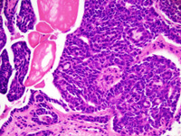
|
|
Image ID:1972 |
|
Source of Image:Ward JM |
|
Pathologist:Mikaelian I |
|
Method / Stain:H&E |
|
|
Image Caption:Papillary and glandular adenocarcinoma: the papillae and the glands are lined by a one cell-thick cuboidal to low columnar epithelium supported by a small amount of fibrovascular stroma. Neoplastic lobules are separated by thick fibrovascular septae.
|
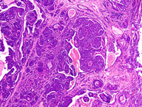
|
|
Image ID:1970 |
|
Source of Image:Ward JM |
|
Pathologist:Mikaelian I |
|
Method / Stain:H&E |
|
|
Image Caption:Mammary gland: this photomicrograph shows an area of transition between a papillary portion of the neoplasm (on the left) and of prominent squamous metaplasia (on the right). Squamous metaplasia results into the formation of large rafts of "ghost" cells.
|
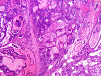
|
|
Image ID:1977 |
|
Source of Image:Ward JM |
|
Pathologist:Mikaelian I |
|
Method / Stain:H&E |
|
|
Image Caption:Papillary and glandular adenocarcinoma: the papillae and the glands are lined by a one cell-thick cuboidal to low columnar epithelium supported by a small amount of fibrovascular stroma. Neoplastic lobules are separated by thick fibrovascular septae.
|
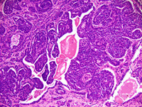
|
|
Image ID:1971 |
|
Source of Image:Ward JM |
|
Pathologist:Mikaelian I |
|
Method / Stain:H&E |
|
|
Image Caption:Papillary and glandular adenocarcinoma: this photomicrograph illustrates a papillary portion of the neoplasm. The papillae are lined by a one cell-thick cuboidal epithelium. Large ectatic blood vessels are present in the stroma.
|
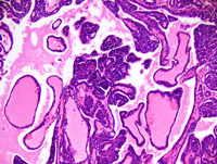
|
|
Image ID:1974 |
|
Source of Image:Ward JM |
|
Pathologist:Mikaelian I |
|
Method / Stain:H&E |
|
|
Image Caption:Mammary gland: the mammary gland is effaced by a nodular, multilobular, unencapsulated, invasive, and densely cellular neoplasm. Neoplastic lobules are separated by thick fibrovascular septae and are composed of papillae (predominantly) and glands supported by a small amount of fibrovascular stroma.
|
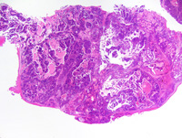
|
|
Image ID:1969 |
|
Source of Image:Ward JM |
|
Pathologist:Mikaelian I |
|
Method / Stain:H&E |
|
|
Image Caption:Papillary and glandular adenocarcinoma: this photomicrograph illustrates a papillary portion of the neoplasm. The papillae are lined by a one cell-thick cuboidal epithelium. Large ectatic blood vessels are present in the stroma.
|
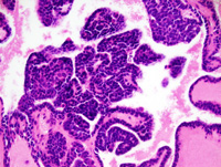
|
|
Image ID:1976 |
|
Source of Image:Ward JM |
|
Pathologist:Mikaelian I |
|
Method / Stain:H&E |
|
|
Image Caption:Papillary and glandular adenocarcinoma: this photomicrograph illustrates an area of squamous metaplasia. Squamous metaplasia is abrupt and resembles keratinization in the hair follicle.
|
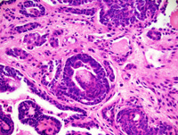
|
|
Image ID:1975 |
|
Source of Image:Ward JM |
|
Pathologist:Mikaelian I |
|
Method / Stain:H&E |
|
|
Image Caption:Mammary gland: the mammary gland is effaced by a nodular, multilobular, unencapsulated, invasive, and densely cellular neoplasm. Neoplastic lobules are separated by thick fibrovascular septae and are composed of papillae (predominantly) and glands supported by a small amount of fibrovascular stroma. Prominent lipid secretion is present in some portions of the neoplasm (*).
|
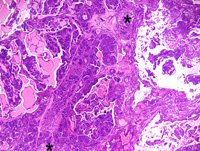
|
|
Image ID:1973 |
|
Source of Image:Ward JM |
|
Pathologist:Mikaelian I |
|
Method / Stain:H&E |
|
|
|
| MTB ID |
Tumor Name |
Organ(s) Affected |
Treatment Type |
Agents |
Strain Name |
Strain Sex |
Reproductive Status |
Tumor Frequency |
Age at Necropsy |
Description |
Reference |
| MTB:29320 |
Mammary gland hyperplasia - alveolar/ductal |
Mammary gland |
None (spontaneous) |
|
|
Female |
reproductive status not specified |
observed |
unknown |
Hyperplasia, ductal and alveolar, with squamous metaplasia, mammary gland |
J:94320 |
|
Image Caption:Papillary adenocarcinoma in an area of squamous metaplasia: squamous metaplasia results into the formation of large rafts of "ghost" cells.
|
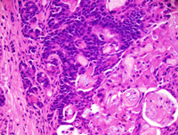
|
|
Image ID:1984 |
|
Source of Image:Ward JM |
|
Pathologist:Mikaelian I |
|
Method / Stain:H&E |
|
|
Image Caption:Mammary gland: the mammary gland is markedly diffusely enlarged by hyperplastic acini and ducts. Cells in these areas do not pile-up and do not show atypia. In addition, there is prominent abrupt cornification (C) in the lumen of a moderate number of ducts and acini, resulting in the formation of large rafts of "ghost" cells.
|
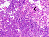
|
|
Image ID:1980 |
|
Source of Image:Ward JM |
|
Pathologist:Mikaelian I |
|
Method / Stain:H&E |
|
|
Image Caption:Papillary adenocarcinoma in an area of squamous metaplasia: squamous metaplasia results into the formation of large rafts of "ghost" cells.
|
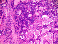
|
|
Image ID:1983 |
|
Source of Image:Ward JM |
|
Pathologist:Mikaelian I |
|
Method / Stain:H&E |
|
|
Image Caption:Mammary gland: there is a diffuse expansion of the mammary gland by closely-packed glands and ducts the lumen of which often contains a small amount of proteinaceous fluid. A moderate number of epithelial cells contains lipid droplets. There is no evidence of atypia and cells do not pile-up disorderly.
|
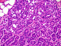
|
|
Image ID:1979 |
|
Source of Image:Ward JM |
|
Pathologist:Mikaelian I |
|
Method / Stain:H&E |
|
|
Image Caption:Hyperplasia and squamous metaplasia, mammary gland: this photomicrograph illustrates an area of transition between an area of hyperplasia of the mammary gland (on the left) and an area of squamous metaplasia (on the right). A raft of ghost epithelial cells (*) and multinucleated epithelial cells (arrowhead) are present. A few nuclei have an apical location instead of a basal location, a feature consistent with dysplasia.
|
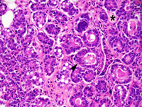
|
|
Image ID:1982 |
|
Source of Image:Ward JM |
|
Pathologist:Mikaelian I |
|
Method / Stain:H&E |
|
|
Image Caption:Mammary gland: there is a diffuse expansion of the mammary gland by closely-packed glands and ducts. A moderate number of epithelial cells contains lipid droplets. There is no evidence of atypia and cells do not pile-up disorderly.
|
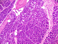
|
|
Image ID:1978 |
|
Source of Image:Ward JM |
|
Pathologist:Mikaelian I |
|
Method / Stain:H&E |
|
|
Image Caption:Mammary gland: the mammary gland is markedly diffusely enlarged by hyperplastic acini and ducts. In addition, there is prominent abrupt cornification (C) in the lumen of a moderate number of ducts and acini, resulting in the formation of large rafts of "ghost" cells.
|
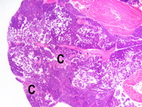
|
|
Image ID:1985 |
|
Source of Image:Ward JM |
|
Pathologist:Mikaelian I |
|
Method / Stain:H&E |
|
|
Image Caption:Mammary gland: the mammary gland is markedly diffusely enlarged by hyperplastic acini and ducts. Cells in these areas do not pile-up and do not show atypia. In addition, there is prominent abrupt cornification (C) in the lumen of a moderate number of ducts and acini, resulting in the formation of large rafts of "ghost" cells. There is a moderate interstitial lymphocytic and plasmacytic infiltration in the connective tissue surrounding the glands showing cornification.
|
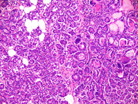
|
|
Image ID:1981 |
|
Source of Image:Ward JM |
|
Pathologist:Mikaelian I |
|
Method / Stain:H&E |
|
|
|
| MTB ID |
Tumor Name |
Organ(s) Affected |
Treatment Type |
Agents |
Strain Name |
Strain Sex |
Reproductive Status |
Tumor Frequency |
Age at Necropsy |
Description |
Reference |
| MTB:29321 |
Mammary gland carcinoma in situ |
Mammary gland |
None (spontaneous) |
|
|
Female |
reproductive status not specified |
observed |
unknown |
Carcinoma in situ and hyperplasia, mammary gland |
J:94320 |
|
Image Caption:Mammary gland: the mammary gland is markedly diffusely enlarged by markedly hyperplastic acini and ducts. In adddition, there are multifocal areas of carcinoma in situ (*). These areas are characterized by epithelial cells piling-up in the ducts and acini, dilation of the lumen of the ducts and acini, and mild to moderate interstitial fibrosis. In addition, there is prominent abrupt cornification (C) in the lumen of a moderate number of ducts and acini, resulting in the formation of large rafts of "ghost" cells.
|
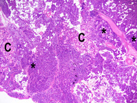
|
|
Image ID:1986 |
|
Source of Image:Ward JM |
|
Pathologist:Mikaelian I |
|
Method / Stain:H&E |
|
|
Image Caption:Mammary gland: most of the photomicrograph repesents an area of severe ductal and acinar hyperplasia. However, at the center of the photomicrograph, cells of mammary ducts pile-up, have larger nuclei than nearby cells, and there is moderate interstitial fibrosis.
|
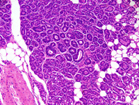
|
|
Image ID:1987 |
|
Source of Image:Ward JM |
|
Pathologist:Mikaelian I |
|
Method / Stain:H&E |
|
|
Image Caption:Mammary gland: most of the photomicrograph repesents an area of severe ductal and acinar hyperplasia. However, at the center of the photomicrograph, cells of mammary ducts pile-up, have larger nuclei than nearby cells, and there is moderate interstitial fibrosis. The nucleus of a moderate number of cells has a luminal location and there is a moderate number of mitoses.
|
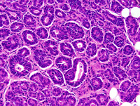
|
|
Image ID:1988 |
|
Source of Image:Ward JM |
|
Pathologist:Mikaelian I |
|
Method / Stain:H&E |
|
|
|
| MTB ID |
Tumor Name |
Organ(s) Affected |
Treatment Type |
Agents |
Strain Name |
Strain Sex |
Reproductive Status |
Tumor Frequency |
Age at Necropsy |
Description |
Reference |
| MTB:29322 |
Mammary gland hyperplasia |
Mammary gland |
None (spontaneous) |
|
|
Female |
reproductive status not specified |
observed |
unknown |
Carcinoma in situ and hyperplasia, mammary gland |
J:94320 |
|
Image Caption:Mammary gland: most of the photomicrograph repesents an area of severe ductal and acinar hyperplasia. However, at the center of the photomicrograph, cells of mammary ducts pile-up, have larger nuclei than nearby cells, and there is moderate interstitial fibrosis.
|
|
|
|
Image ID:1987 |
|
Source of Image:Ward JM |
|
Pathologist:Mikaelian I |
|
Method / Stain:H&E |
|
|
Image Caption:Mammary gland: most of the photomicrograph repesents an area of severe ductal and acinar hyperplasia. However, at the center of the photomicrograph, cells of mammary ducts pile-up, have larger nuclei than nearby cells, and there is moderate interstitial fibrosis. The nucleus of a moderate number of cells has a luminal location and there is a moderate number of mitoses.
|
|
|
|
Image ID:1988 |
|
Source of Image:Ward JM |
|
Pathologist:Mikaelian I |
|
Method / Stain:H&E |
|
|
Image Caption:Mammary gland: the mammary gland is markedly diffusely enlarged by markedly hyperplastic acini and ducts. In adddition, there are multifocal areas of carcinoma in situ (*). These areas are characterized by epithelial cells piling-up in the ducts and acini, dilation of the lumen of the ducts and acini, and mild to moderate interstitial fibrosis. In addition, there is prominent abrupt cornification (C) in the lumen of a moderate number of ducts and acini, resulting in the formation of large rafts of "ghost" cells.
|
|
|
|
Image ID:1986 |
|
Source of Image:Ward JM |
|
Pathologist:Mikaelian I |
|
Method / Stain:H&E |
|
|
|
| MTB ID |
Tumor Name |
Organ(s) Affected |
Treatment Type |
Agents |
Strain Name |
Strain Sex |
Reproductive Status |
Tumor Frequency |
Age at Necropsy |
Description |
Reference |
| MTB:29356 |
Mammary gland tumor - type P |
Mammary gland |
None (spontaneous) |
|
|
Female |
reproductive status not specified |
observed |
unknown |
Type P tumor, mammary gland |
J:94320 |
|
Image Caption:Mammary gland: the mammary gland is replaced by a densely cellular neoplasm composed of cellular areas separated by thick strands of dense fibrovascular stroma. Epithelial areas are composed of microacini and papillary projections that have developed within the lumen of structures with a ductal morphology.
|
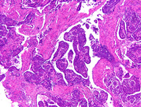
|
|
Image ID:1990 |
|
Source of Image:Ward JM |
|
Pathologist:Mikaelian I |
|
Method / Stain:H&E |
|
|
Image Caption:Type P tumor: in some portions of the neoplasm, neoplastic cells formed microacini filed with a a coagulated proteinaceous material. Neoplastic cells are small, polygonal, with indistinct cell borders and a moderate amount of strongly amphophilic cytoplasm. The nucleus is central, round to oval, medium-sized, hyperchromatic and with a coarsely clumped chromatin. Anisocytosis and anisokaryosis are mild.
|
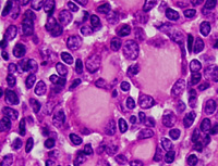
|
|
Image ID:1993 |
|
Source of Image:Ward JM |
|
Pathologist:Mikaelian I |
|
Method / Stain:H&E |
|
|
Image Caption:Mammary gland: the mammary gland is replaced by a densely cellular neoplasm composed of cellular areas separated by thick strands of dense fibrovascular stroma. Epithelial areas are composed of microacini and papillary projections that have developed within the lumen of structures with a ductal morphology.
|
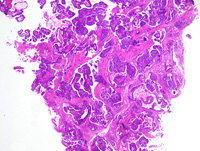
|
|
Image ID:1989 |
|
Source of Image:Ward JM |
|
Pathologist:Mikaelian I |
|
Method / Stain:H&E |
|
|
Image Caption:Mammary gland: the mammary gland is replaced by a densely cellular neoplasm composed of cellular areas separated by thick strands of dense fibrovascular stroma. Epithelial areas are composed of microacini and papillary projections that have developed within the lumen of structures with a ductal morphology.
|
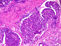
|
|
Image ID:1991 |
|
Source of Image:Ward JM |
|
Pathologist:Mikaelian I |
|
Method / Stain:H&E |
|
|
Image Caption:Mammary gland: the mammary gland is replaced by a densely cellular neoplasm composed of cellular areas separated by thick strands of dense fibrovascular stroma. Epithelial areas are composed of microacini and papillary projections that have developed within the lumen of structures with a ductal morphology.
|
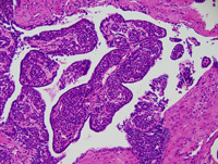
|
|
Image ID:1994 |
|
Source of Image:Ward JM |
|
Pathologist:Mikaelian I |
|
Method / Stain:H&E |
|
|
Image Caption:Type P tumor: a few large ducts lined by a pseudostratified epithelium are scattered throughout the neoplasm. Cells lining these ducts have a large amount of strongly acidophilic cytoplasm. Similar structures are also present in type P tumors which develop in mice transgenic for Wnt1.
|
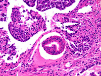
|
|
Image ID:1996 |
|
Source of Image:Ward JM |
|
Pathologist:Mikaelian I |
|
Method / Stain:H&E |
|
|
Image Caption:Mammary gland, type P tumor: in this portion of the neoplasm, neoplastic cells have formed large papillae. These papillae are lined, on their surface, by a two cell-thick epithelium resembling the mammary ductal epithelium which invaginates (arrowhead) to form terminal end bud-like structures (*).
|
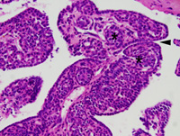
|
|
Image ID:1995 |
|
Source of Image:Ward JM |
|
Pathologist:Mikaelian I |
|
Method / Stain:H&E |
|
|
Image Caption:Type P tumor: in some portions of the neoplasm, neoplastic cells formed microacini filed with a a coagulated proteinaceous material. Neoplastic cells are small, polygonal, with indistinct cell borders and a moderate amount of strongly amphophilic cytoplasm. Anisocytosis and anisokaryosis are mild.
|
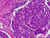
|
|
Image ID:1992 |
|
Source of Image:Ward JM |
|
Pathologist:Mikaelian I |
|
Method / Stain:H&E |
|
|
|
| MTB ID |
Tumor Name |
Organ(s) Affected |
Treatment Type |
Agents |
Strain Name |
Strain Sex |
Reproductive Status |
Tumor Frequency |
Age at Necropsy |
Description |
Reference |
| MTB:29357 |
Mammary gland tumor - type P |
Lung |
None (spontaneous) |
|
|
Female |
reproductive status not specified |
observed |
unknown |
Type P tumor, metastatic to the lung |
J:94320 |
|
Image Caption:Lung: there are multiple metastases of the type P mammary tumor. These metastases share many features with the primary tumor, including the formation of microacini and of large papillary structures in ducts resembling the mammary ducts. Minute areas of cornification (arrowhead) are present in the areas of ductal differentiation.
|
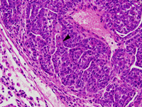
|
|
Image ID:2000 |
|
Source of Image:Ward JM |
|
Pathologist:Mikaelian I |
|
Method / Stain:H&E |
|
|
Image Caption:Lung: there are multiple metastases of the type P mammary tumor. These metastases share many features with the primary tumor, including the formation of microacini and of large papillary structures in ducts resembling the mammary ducts.
|
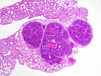
|
|
Image ID:1997 |
|
Source of Image:Ward JM |
|
Pathologist:Mikaelian I |
|
Method / Stain:H&E |
|
|
Image Caption:Lung: there are multiple metastases of the type P mammary tumor. These metastases share many features with the primary tumor, including the formation of microacini and of large papillary structures in ducts resembling the mammary ducts.
|
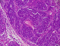
|
|
Image ID:1999 |
|
Source of Image:Ward JM |
|
Pathologist:Mikaelian I |
|
Method / Stain:H&E |
|
|
Image Caption:Lung: there are multiple metastases of the type P mammary tumor. These metastases share many features with the primary tumor, including the formation of microacini and of large papillary structures in ducts resembling the mammary ducts.
|
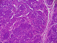
|
|
Image ID:2001 |
|
Source of Image:Ward JM |
|
Pathologist:Mikaelian I |
|
Method / Stain:H&E |
|
|
Image Caption:Lung: there are multiple metastases of the type P mammary tumor. These metastases share many features with the primary tumor, including the formation of microacini and of large papillary structures in ducts resembling the mammary ducts.
|
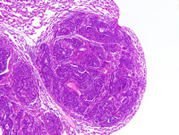
|
|
Image ID:1998 |
|
Source of Image:Ward JM |
|
Pathologist:Mikaelian I |
|
Method / Stain:H&E |
|
|
Image Caption:Lung: minute areas of cornification are present in the areas of ductal differentiation. Cells undergoing cornification often contain keratohyalin granules (basophilic granules) in their cytoplasm.
|
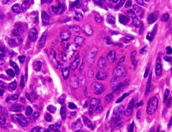
|
|
Image ID:2003 |
|
Source of Image:Ward JM |
|
Pathologist:Mikaelian I |
|
Method / Stain:H&E |
|
|
Image Caption:Lung: the metastases share many features with the primary tumor, including the formation of microacini and of large papillary structures in ducts resembling the mammary ducts. Minute areas of cornification (arrowhead) are present in the areas of ductal differentiation. Cells undergoing cornification often contain keratohyalin granules (basophilic granules) in their cytoplasm.
|
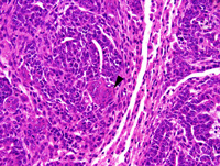
|
|
Image ID:2002 |
|
Source of Image:Ward JM |
|
Pathologist:Mikaelian I |
|
Method / Stain:H&E |
|
|
|
| MTB ID |
Tumor Name |
Organ(s) Affected |
Treatment Type |
Agents |
Strain Name |
Strain Sex |
Reproductive Status |
Tumor Frequency |
Age at Necropsy |
Description |
Reference |
| MTB:29361 |
Mammary gland adenocarcinoma - papillary |
Mammary gland |
None (spontaneous) |
|
|
Female |
reproductive status not specified |
observed |
unknown |
Adenocarcinoma, papillary, mammary gland |
J:94320 |
|
Image Caption:Mammary gland: the mammary gland is expanded by a nodular, partly encapsulated, locally invasive, densely cellular neoplasm with a prominent papillary pattern. The neoplasm is composed of closely-packed papillae lined by a one cell-thick cuboidal epithelium supported by a moderate amount of dense fibrovascular stroma. Neoplastic cells are cuboidal to polygonal, with ill-defined cell borders and a moderate amount of acidophilic cytoplasm that often contains lipid vacuoles. The nucleus is central, round, and medium-size.
|
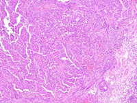
|
|
Image ID:2006 |
|
Source of Image:Ward JM |
|
Pathologist:Mikaelian I |
|
Method / Stain:H&E |
|
|
Image Caption:Papillary mammary adenocarcinoma: this high power photomicrograph illustrates a portion at the periphery of the neoplasm where most of the neoplasm has a papillary pattern, although small clusters of cells invade the periphery of the neoplasm (arrowheads). Neoplastic cells become spindloid and form cords at the center of the photomicrograph. Numerous neoplastic cells undergo apoptosis.
|
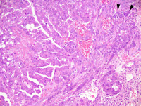
|
|
Image ID:2007 |
|
Source of Image:Ward JM |
|
Pathologist:Mikaelian I |
|
Method / Stain:H&E |
|
|
Image Caption:Mammary gland: the mammary gland is expanded by a nodular, partly encapsulated, locally invasive, densely cellular neoplasm with a prominent papillary pattern. The neoplasm is composed of closely-packed papillae lined by a one cell-thick cuboidal epithelium supported by a moderate amount of dense fibrovascular stroma.
|
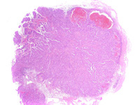
|
|
Image ID:2004 |
|
Source of Image:Ward JM |
|
Pathologist:Mikaelian I |
|
Method / Stain:H&E |
|
|
Image Caption:Papillary mammary adenocarcinoma: the neoplasm is composed of closely-packed papillae lined by a one cell-thick cuboidal epithelium supported by a moderate amount of dense fibrovascular stroma. Neoplastic cells often pile-up disorderly. Neoplastic cells are cuboidal to polygonal, with ill-defined cell borders and a moderate amount of acidophilic cytoplasm that often contains lipid vacuoles. The nucleus is central, round, medium-size, normochromatic, with a clumped chromatin and 1-2 small (generally) to medium size bacophilic nucleoli. Anisokaryosis is moderate. Anisocytosis is mild. Numerous neoplastic cells undergo apoptosis. The stroma is fibrous.
|
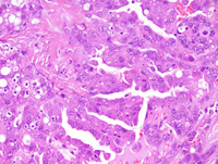
|
|
Image ID:2008 |
|
Source of Image:Ward JM |
|
Pathologist:Mikaelian I |
|
Method / Stain:H&E |
|
|
Image Caption:Mammary gland: the mammary gland is expanded by a nodular, partly encapsulated, locally invasive, densely cellular neoplasm with a prominent papillary pattern. The neoplasm is composed of closely-packed papillae lined by a one cell-thick cuboidal epithelium supported by a moderate amount of dense fibrovascular stroma.
|
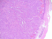
|
|
Image ID:2005 |
|
Source of Image:Ward JM |
|
Pathologist:Mikaelian I |
|
Method / Stain:H&E |
|
|
Image Caption:Papillary mammary adenocarcinoma: this high power photomicrograph of the periphery of the neoplasm illustrates an area where neoplastic cells form small trabeculae and cords and become spindloid. The stroma is infiltrated by a moderate number of lymphocytes, plasma cells, and also mast cells (arrowheads).
|
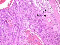
|
|
Image ID:2009 |
|
Source of Image:Ward JM |
|
Pathologist:Mikaelian I |
|
Method / Stain:H&E |
|
|
|
| MTB ID |
Tumor Name |
Organ(s) Affected |
Treatment Type |
Agents |
Strain Name |
Strain Sex |
Reproductive Status |
Tumor Frequency |
Age at Necropsy |
Description |
Reference |
| MTB:29362 |
Mammary gland adenocarcinoma - solid |
Mammary gland |
None (spontaneous) |
|
|
Female |
reproductive status not specified |
observed |
unknown |
Adenocarcinoma, solid, glandular, mammary gland |
J:94320 |
|
Image Caption:Mammary gland: this photomicrograph focuses on the center of the neoplasm where neoplastic cells form thick interconnected trabeculae that often show central liquefactive necrosis (comedo pattern). Although the pattern of the neoplasm is solid, there are numerous slit-like secondary lumens. There is a small to moderate amount of hyalinized fibrovascular stroma.
|
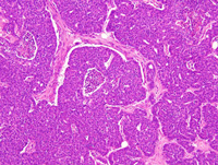
|
|
Image ID:2016 |
|
Source of Image:Ward JM |
|
Pathologist:Mikaelian I |
|
Method / Stain:H&E |
|
|
Image Caption:Mammary gland: the mammary gland is effaced by a nodular, unencapsulated, densely cellular, well-delineated neoplasm. The neoplasm is composed of solid areas with central necrosis (comedo pattern) at the center of the neoplasm, with the gradual transition to small glandular structures are the periphery of the neoplasm. The stroma is more abundant and more fibrous at the periphery of the neoplasm than at its' center.
|
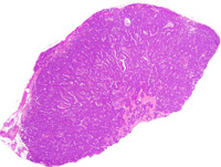
|
|
Image ID:2010 |
|
Source of Image:Ward JM |
|
Pathologist:Mikaelian I |
|
Method / Stain:H&E |
|
|
Image Caption:Mammary gland: the mammary gland is effaced by a nodular, unencapsulated, densely cellular, well-delineated neoplasm. The neoplasm is composed of solid areas with central necrosis (comedo pattern) at the center of the neoplasm, with the gradual transition to small glandular structures are the periphery of the neoplasm. The solid areas often contain small slit-like lumens with angular contours that can be identified at this low magnification.
|
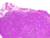
|
|
Image ID:2011 |
|
Source of Image:Ward JM |
|
Pathologist:Mikaelian I |
|
Method / Stain:H&E |
|
|
Image Caption:Mammary gland: this photomicrograph focuses on the periphery of the neoplasm where glandular differentiation is prominent. A few adipocytes are entrapped within the neoplasm. The central portions of the neoplasm are composed of large coalescing trabeculae with a few slit-like secondary lumens. The glands are lined by a one cell-thick cuboidal epithelium.
|
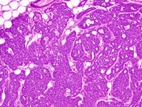
|
|
Image ID:2013 |
|
Source of Image:Ward JM |
|
Pathologist:Mikaelian I |
|
Method / Stain:H&E |
|
|
Image Caption:Mammary gland: this photomicrograph focuses on the periphery of the neoplasm where glandular differentiation is prominent. A few adipocytes are entrapped within the neoplasm. The glands are lined by a one cell-thick cuboidal epithelium where neoplastic cells often pile-up disorderly. Neoplastic cells have ill-defined cell borders and a moderate amount of acidophilic cytoplasm. The nucleus is central, oval, slightly hypochromatic, with a coarsely clumped chromatin and 1-3 small basophilic nucleoli. Anisokaryosis and anisocytosis are mild. Mitoses are numerous.
|
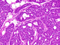
|
|
Image ID:2014 |
|
Source of Image:Ward JM |
|
Pathologist:Mikaelian I |
|
Method / Stain:H&E |
|
|
Image Caption:Mammary gland: this photomicrograph focuses on the center of the neoplasm where neoplastic cells form thick interconnected trabeculae that often show central liquefactive necrosis (comedo pattern). Although the pattern of the neoplasm is solid, there are numerous slit-like secondary lumens. There is a small to moderate amount of hyalinized fibrovascular stroma. Neoplastic cells are cuboidal to polygonal, have ill-defined cell borders and a moderate amount of acidophilic cytoplasm. The nucleus is central, oval, slightly hypochromatic, with a coarsely clumped chromatin and 1-3 small basophilic nucleoli. Anisokaryosis and anisocytosis are mild. Mitoses are numerous.
|
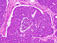
|
|
Image ID:2017 |
|
Source of Image:Ward JM |
|
Pathologist:Mikaelian I |
|
Method / Stain:H&E |
|
|
Image Caption:Mammary gland: this photomicrograph focuses on the periphery of the neoplasm where glandular differentiation is prominent. A few adipocytes are entrapped within the neoplasm. The central portions of the neoplasm are composed of large coalescing trabeculae with a few slit-like secondary lumens. The glands are lined by a one cell-thick cuboidal epithelium.
|
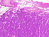
|
|
Image ID:2012 |
|
Source of Image:Ward JM |
|
Pathologist:Mikaelian I |
|
Method / Stain:H&E |
|
|
Image Caption:Mammary gland: this photomicrograph focuses on the center of the neoplasm where neoplastic cells form thick interconnected trabeculae that often show central liquefactive necrosis (comedo pattern). Although the pattern of the neoplasm is solid, there are numerous slit-like secondary lumens. There is a small to moderate amount of hyalinized fibrovascular stroma.
|
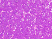
|
|
Image ID:2015 |
|
Source of Image:Ward JM |
|
Pathologist:Mikaelian I |
|
Method / Stain:H&E |
|
|
|
| MTB ID |
Tumor Name |
Organ(s) Affected |
Treatment Type |
Agents |
Strain Name |
Strain Sex |
Reproductive Status |
Tumor Frequency |
Age at Necropsy |
Description |
Reference |
| MTB:29363 |
Mammary gland adenocarcinoma - solid |
Mammary gland |
None (spontaneous) |
|
|
Female |
reproductive status not specified |
observed |
330 days |
Adenocarcinoma, solid, with secondary lumens, mammary gland |
J:94320 |
|
Image Caption:Mammary gland: the mammary gland is effaced and expanded by a large, nodular, encapsulated, well-delineated, densely cellular neoplasm. This neoplasm is composed of large (at the center of the neoplasm) to thin coalescing trabeculae supported by a moderate amount of hyalinized fibrovascular stroma.
|
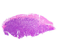
|
|
Image ID:2018 |
|
Source of Image:Ward JM |
|
Pathologist:Mikaelian I |
|
Method / Stain:H&E |
|
|
Image Caption:Mammary gland: the mammary gland is effaced and expanded by a large, nodular, encapsulated, well-delineated, densely cellular neoplasm. This neoplasm is composed of large (at the center of the neoplasm) to thin coalescing trabeculae supported by a moderate amount of hyalinized fibrovascular stroma. Neoplastic cells form a few mildly ectatic glands at the periphery of the neoplasm.
|
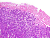
|
|
Image ID:2019 |
|
Source of Image:Ward JM |
|
Pathologist:Mikaelian I |
|
Method / Stain:H&E |
|
|
Image Caption:Mammary gland: a few secondary lumens are present in the trabeculae located at the periphery of the neoplasm.
|
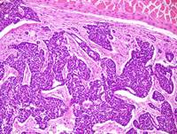
|
|
Image ID:2021 |
|
Source of Image:Ward JM |
|
Pathologist:Mikaelian I |
|
Method / Stain:H&E |
|
|
Image Caption:Mammary gland: the mammary gland is effaced and expanded by a large, nodular, encapsulated, well-delineated, densely cellular neoplasm. This neoplasm is composed of coalescing trabeculae supported by a moderate amount of hyalinized fibrovascular stroma. This photomicrograph illustrates a portion of the neoplasm where a few glands are formed. Some glands are mildly ectatic. A few adipocytes have been entrapped in the neoplasm.
|
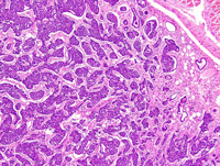
|
|
Image ID:2020 |
|
Source of Image:Ward JM |
|
Pathologist:Mikaelian I |
|
Method / Stain:H&E |
|
|
Image Caption:Mammary gland: this photomicrograph illustrates the morphology of the center of the neoplasm. Neoplastic cells form thick trabeculae with occasional central liquefactive necrosis (comedo pattern). Neoplastic cells are medium-size, cuboidal to polygonal, with distinct cell borders, and a moderate amount of acidophilic cytoplasm. The nucleus is central, round to oval, and hyperchromatic. Anisokaryosis and anisocytosis are minimal.
|
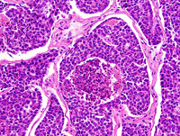
|
|
Image ID:2025 |
|
Source of Image:Ward JM |
|
Pathologist:Mikaelian I |
|
Method / Stain:H&E |
|
|
Image Caption:Mammary gland: this photomicrograph illustrates the morphology of the center of the neoplasm. Neoplastic cells form thick trabeculae with occasional central liquefactive necrosis (comedo pattern) and a few small secondary lumens.
|
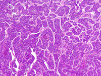
|
|
Image ID:2023 |
|
Source of Image:Ward JM |
|
Pathologist:Mikaelian I |
|
Method / Stain:H&E |
|
|
Image Caption:Mammary gland: this photomicrograph illustrates the morphology of the center of the neoplasm. Neoplastic cells form thick trabeculae with occasional central liquefactive necrosis (comedo pattern) and a few small secondary lumens. Neoplastic cells are medium-size, cuboidal to polygonal, with distinct cell borders, and a moderate amount of acidophilic cytoplasm. The nucleus is central, round to oval, and hyperchromatic. Anisokaryosis and anisocytosis are minimal.
|
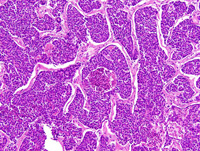
|
|
Image ID:2024 |
|
Source of Image:Ward JM |
|
Pathologist:Mikaelian I |
|
Method / Stain:H&E |
|
|
Image Caption:Mammary gland: a few secondary lumens are present in the trabeculae located at the periphery of the neoplasm. Neoplastic cells are medium-size, cuboidal to polygonal, with distinct cell borders, and a moderate amount of acidophilic cytoplasm. The nucleus is central, round to oval, and hyperchromatic. Anisokaryosis and anisocytosis are minimal.
|
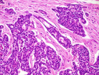
|
|
Image ID:2022 |
|
Source of Image:Ward JM |
|
Pathologist:Mikaelian I |
|
Method / Stain:H&E |
|
|
|
| MTB ID |
Tumor Name |
Organ(s) Affected |
Treatment Type |
Agents |
Strain Name |
Strain Sex |
Reproductive Status |
Tumor Frequency |
Age at Necropsy |
Description |
Reference |
| MTB:29364 |
Mammary gland adenocarcinoma - solid |
Mammary gland |
None (spontaneous) |
|
|
Female |
reproductive status not specified |
observed |
unknown |
Adenocarcinoma, solid, with secondary lumens, mamamry gland |
J:94320 |
|
Image Caption:Solid mammary adenocarcinoma: this photomicrograph illustrates an area of invasion in the mammary fat pad. The invasive portions of the neoplasm form small contoured and anastomosed glands with an inconspicuous lumen and they are surrounded by a mild scirrhous reaction. The center of some of the solid areas has undergone liquefactive necrosis (comedo pattern). Neoplastic cells are low columnar to polygonal, with indistinct cell borders and a moderate amout of acidophilic cytoplasm. The nucleus is basal, oval, medium-size, hypochromatic, and generally oriented in the long axis of the cell (at right angle with the basement membrane). Anisokaryosis and anisocytosis are mild. A few mitoses are present.
|
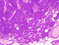
|
|
Image ID:2031 |
|
Source of Image:Ward JM |
|
Pathologist:Mikaelian I |
|
Method / Stain:H&E |
|
|
Image Caption:Solid mammary adenocarcinoma: this photomicrograph illustrates an area of invasion in the mammary fat pad. The invasive portions of the neoplasm form small contoured and anastomosed glands with an inconspicuous lumen and they are surrounded by a mild scirrhous reaction. The center of some of the solid areas has undergone liquefactive necrosis (comedo pattern). Neoplastic cells are low columnar to polygonal, with indistinct cell borders and a moderate amout of acidophilic cytoplasm. The nucleus is basal, oval, medium-size, hypochromatic, and generally oriented in the long axis of the cell (at right angle with the basement membrane). Anisokaryosis and anisocytosis are mild. A few mitoses are present.
|
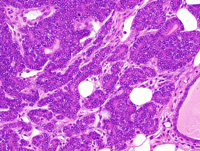
|
|
Image ID:2032 |
|
Source of Image:Ward JM |
|
Pathologist:Mikaelian I |
|
Method / Stain:H&E |
|
|
Image Caption:Solid mammary adenocarcinoma: a solid mass of neoplastic cells is protruding in the lumen of an interlobular mammary duct.
|
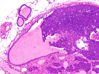
|
|
Image ID:2028 |
|
Source of Image:Ward JM |
|
Pathologist:Mikaelian I |
|
Method / Stain:H&E |
|
|
Image Caption:Solid mammary adenocarcinoma: this photomicrograph illustrates a solid area of the neoplasm with central liquefactive necrosis (comedo pattern). The arrowheads point at secondary lumens.
|
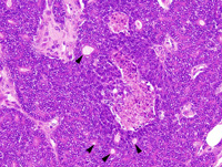
|
|
Image ID:2034 |
|
Source of Image:Ward JM |
|
Pathologist:Mikaelian I |
|
Method / Stain:H&E |
|
|
Image Caption:Mammary gland: the mammary gland is expanded by a multilobular, partially encapsulated, locally invasive, densely cellular neoplasm which multifocally grows into the lumen of mammary ducts (arrowheads). The rest of the mammary gland is lactating.
|
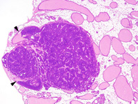
|
|
Image ID:2026 |
|
Source of Image:Ward JM |
|
Pathologist:Mikaelian I |
|
Method / Stain:H&E |
|
|
Image Caption:Solid mammary adenocarcinoma: this photomicrograph illustrates an area of invasion in the mammary fat pad. The invasive portions of the neoplasm form small contoured and anastomosed glands with an inconspicuous lumen and they are surrounded by a mild scirrhous reaction.
|
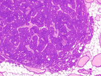
|
|
Image ID:2030 |
|
Source of Image:Ward JM |
|
Pathologist:Mikaelian I |
|
Method / Stain:H&E |
|
|
Image Caption:Solid mammary adenocarcinoma: this photomicrograph illustrates a solid area of the neoplasm with central liquefactive necrosis (comedo pattern).
|
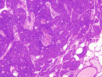
|
|
Image ID:2033 |
|
Source of Image:Ward JM |
|
Pathologist:Mikaelian I |
|
Method / Stain:H&E |
|
|
Image Caption:Mammary gland: the mammary gland is expanded by a multilobular, partially encapsulated, locally invasive, densely cellular neoplasm which multifocally grows in the lumen of a duct. The neoplasm is composed of coalescing solid areas and trabeculae supported by a small to moderate amount of fibrovascular stroma. Secondary lumens are formed in some solid areas.
|
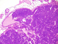
|
|
Image ID:2027 |
|
Source of Image:Ward JM |
|
Pathologist:Mikaelian I |
|
Method / Stain:H&E |
|
|
Image Caption:Solid mammary adenocarcinoma: a solid mass of neoplastic cells is protruding in the lumen of an interlobular mammary duct.
|
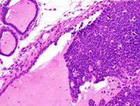
|
|
Image ID:2029 |
|
Source of Image:Ward JM |
|
Pathologist:Mikaelian I |
|
Method / Stain:H&E |
|
|
|
| MTB ID |
Tumor Name |
Organ(s) Affected |
Treatment Type |
Agents |
Strain Name |
Strain Sex |
Reproductive Status |
Tumor Frequency |
Age at Necropsy |
Description |
Reference |
| MTB:29369 |
Mammary gland hyperplasia - alveolar |
Mammary gland |
None (spontaneous) |
|
|
Female |
reproductive status not specified |
observed |
210 days |
Hyperplastic alveolar nodule, with squamous metaplasia, mammary gland |
J:94320 |
|
Image Caption:Mammary gland: a mammary lobule is greatly expanded as a result of marked proliferation of its' epithelium and ductal and alveolar ectasia. There is also mild to moderate interstitial fibrosis and lymphocytic, plasmacytic and granulocytic inflammation. There is a focal area of squamous metaplasia with the formation of a keratin pearl (arrowhead).
|
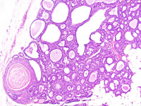
|
|
Image ID:2037 |
|
Source of Image:Ward JM |
|
Pathologist:Mikaelian I |
|
Method / Stain:H&E |
|
|
Image Caption:Hyperplastic alveolar nodule: this photomicrograph illustrates an area of prominent secretory activity. There is no evidence of atypia. There is mild interstitial lymphocytic, plasmacytic and granulocytic inflammation. A few pigment-ladden macrophages are scattered in the stroma of the hyperplastic alveolar nodule.
|
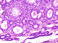
|
|
Image ID:2039 |
|
Source of Image:Ward JM |
|
Pathologist:Mikaelian I |
|
Method / Stain:H&E |
|
|
Image Caption:Mammary gland: a mammary lobule is greatly expanded as a result of marked proliferation of its' epithelium and ductal and alveolar ectasia.
|
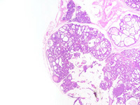
|
|
Image ID:2035 |
|
Source of Image:Ward JM |
|
Pathologist:Mikaelian I |
|
Method / Stain:H&E |
|
|
Image Caption:Mammary gland: a mammary lobule is greatly expanded as a result of marked proliferation of its' epithelium and ductal and alveolar ectasia. There is also mild to moderate interstitial fibrosis and lymphocytic and plasmacytic inflammation. There is a focal area of squamous metaplasia with the formation of a keratin pearl (arrowhead).
|
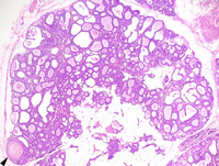
|
|
Image ID:2036 |
|
Source of Image:Ward JM |
|
Pathologist:Mikaelian I |
|
Method / Stain:H&E |
|
|
Image Caption:Hyperplastic alveolar nodule: this photomicrograph illustrates an area of squamous metaplasia. There is no evidence of atypia. Many luminal cells of the mammary gland contain lipid vacuoles. There is mild interstitial lymphocytic, plasmacytic and granulocytic inflammation.
|
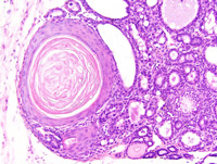
|
|
Image ID:2038 |
|
Source of Image:Ward JM |
|
Pathologist:Mikaelian I |
|
Method / Stain:H&E |
|
|
|
| MTB ID |
Tumor Name |
Organ(s) Affected |
Treatment Type |
Agents |
Strain Name |
Strain Sex |
Reproductive Status |
Tumor Frequency |
Age at Necropsy |
Description |
Reference |
| MTB:29370 |
Mammary gland metaplasia - squamous |
Mammary gland |
None (spontaneous) |
|
|
Female |
reproductive status not specified |
observed |
210 days |
Hyperplastic alveolar nodule, with squamous metaplasia, mammary gland |
J:94320 |
|
Image Caption:Mammary gland: a mammary lobule is greatly expanded as a result of marked proliferation of its' epithelium and ductal and alveolar ectasia. There is also mild to moderate interstitial fibrosis and lymphocytic, plasmacytic and granulocytic inflammation. There is a focal area of squamous metaplasia with the formation of a keratin pearl (arrowhead).
|
|
|
|
Image ID:2037 |
|
Source of Image:Ward JM |
|
Pathologist:Mikaelian I |
|
Method / Stain:H&E |
|
|
Image Caption:Mammary gland: a mammary lobule is greatly expanded as a result of marked proliferation of its' epithelium and ductal and alveolar ectasia.
|
|
|
|
Image ID:2035 |
|
Source of Image:Ward JM |
|
Pathologist:Mikaelian I |
|
Method / Stain:H&E |
|
|
Image Caption:Mammary gland: a mammary lobule is greatly expanded as a result of marked proliferation of its' epithelium and ductal and alveolar ectasia. There is also mild to moderate interstitial fibrosis and lymphocytic and plasmacytic inflammation. There is a focal area of squamous metaplasia with the formation of a keratin pearl (arrowhead).
|
|
|
|
Image ID:2036 |
|
Source of Image:Ward JM |
|
Pathologist:Mikaelian I |
|
Method / Stain:H&E |
|
|
Image Caption:Hyperplastic alveolar nodule: this photomicrograph illustrates an area of prominent secretory activity. There is no evidence of atypia. There is mild interstitial lymphocytic, plasmacytic and granulocytic inflammation. A few pigment-ladden macrophages are scattered in the stroma of the hyperplastic alveolar nodule.
|
|
|
|
Image ID:2039 |
|
Source of Image:Ward JM |
|
Pathologist:Mikaelian I |
|
Method / Stain:H&E |
|
|
Image Caption:Hyperplastic alveolar nodule: this photomicrograph illustrates an area of squamous metaplasia. There is no evidence of atypia. Many luminal cells of the mammary gland contain lipid vacuoles. There is mild interstitial lymphocytic, plasmacytic and granulocytic inflammation.
|
|
|
|
Image ID:2038 |
|
Source of Image:Ward JM |
|
Pathologist:Mikaelian I |
|
Method / Stain:H&E |
|
|
|
| MTB ID |
Tumor Name |
Organ(s) Affected |
Treatment Type |
Agents |
Strain Name |
Strain Sex |
Reproductive Status |
Tumor Frequency |
Age at Necropsy |
Description |
Reference |
| MTB:29371 |
Mammary gland adenoma - tubular |
Mammary gland |
None (spontaneous) |
|
|
Female |
reproductive status not specified |
observed |
210 days |
Adenoma, tubular, arising in an hyperplastic alveolar nodule, mammary gland |
J:94320 |
|
Image Caption:Mammary gland: there is, within an hyperplastic alveolar nodule, a small, densely cellular, unencapsulated, expensile, neoplasm (arrowheads). This neoplasm is composed of closely-packed tubules and solid areas supported by a scant amount of fibrovascular stroma.
|
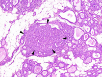
|
|
Image ID:2040 |
|
Source of Image:Ward JM |
|
Pathologist:Mikaelian I |
|
Method / Stain:H&E |
|
|
Image Caption:Tubular adenoma of the mammary gland: the neoplasm is composed of closely packed tubules and solid areas supported by a scant amount of fibrovascular stroma. Neoplastic cells are cuboidal to polygonal, with indistinct cell borders and a moderate amount of homogeneous acidophilic cytoplasm. The nucleus is central, round, normochromatic, and with a small basophilic nucleolus. Anisokaryosis and anisocytosis are mild. A few mitoses are present.
|
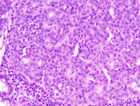
|
|
Image ID:2042 |
|
Source of Image:Ward JM |
|
Pathologist:Mikaelian I |
|
Method / Stain:H&E |
|
|
Image Caption:Mammary gland: there is, within an hyperplastic alveolar nodule, a small, densely cellular, unencapsulated, expensile, neoplasm (arrowheads). This neoplasm is composed of closely packed tubules and solid areas supported by a scant amount of fibrovascular stroma. Neoplastic cells are cuboidal to polygonal, with indistinct cell borders and a moderate amount of homogeneous acidophilic cytoplasm. The nucleus is central, round, normochromatic, and with a small basophilic nucleolus. Anisokaryosis and anisocytosis are mild. A few mitoses are present (arrowheads). A small to moderate number of inflammatory cells, including pigment-laden macrophages, infiltrates the connective tissue in the hyperplastic alveolar nodule.
|
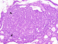
|
|
Image ID:2041 |
|
Source of Image:Ward JM |
|
Pathologist:Mikaelian I |
|
Method / Stain:H&E |
|
|
|
| MTB ID |
Tumor Name |
Organ(s) Affected |
Treatment Type |
Agents |
Strain Name |
Strain Sex |
Reproductive Status |
Tumor Frequency |
Age at Necropsy |
Description |
Reference |
| MTB:29372 |
Mammary gland hyperplasia - alveolar |
Mammary gland |
None (spontaneous) |
|
|
Female |
reproductive status not specified |
observed |
210 days |
Adenoma, tubular, arising in an hyperplastic alveolar nodule, mammary gland |
J:94320 |
|
Image Caption:Mammary gland: there is, within an hyperplastic alveolar nodule, a small, densely cellular, unencapsulated, expensile, neoplasm (arrowheads). This neoplasm is composed of closely-packed tubules and solid areas supported by a scant amount of fibrovascular stroma.
|
|
|
|
Image ID:2040 |
|
Source of Image:Ward JM |
|
Pathologist:Mikaelian I |
|
Method / Stain:H&E |
|
|
Image Caption:Tubular adenoma of the mammary gland: the neoplasm is composed of closely packed tubules and solid areas supported by a scant amount of fibrovascular stroma. Neoplastic cells are cuboidal to polygonal, with indistinct cell borders and a moderate amount of homogeneous acidophilic cytoplasm. The nucleus is central, round, normochromatic, and with a small basophilic nucleolus. Anisokaryosis and anisocytosis are mild. A few mitoses are present.
|
|
|
|
Image ID:2042 |
|
Source of Image:Ward JM |
|
Pathologist:Mikaelian I |
|
Method / Stain:H&E |
|
|
Image Caption:Mammary gland: there is, within an hyperplastic alveolar nodule, a small, densely cellular, unencapsulated, expensile, neoplasm (arrowheads). This neoplasm is composed of closely packed tubules and solid areas supported by a scant amount of fibrovascular stroma. Neoplastic cells are cuboidal to polygonal, with indistinct cell borders and a moderate amount of homogeneous acidophilic cytoplasm. The nucleus is central, round, normochromatic, and with a small basophilic nucleolus. Anisokaryosis and anisocytosis are mild. A few mitoses are present (arrowheads). A small to moderate number of inflammatory cells, including pigment-laden macrophages, infiltrates the connective tissue in the hyperplastic alveolar nodule.
|
|
|
|
Image ID:2041 |
|
Source of Image:Ward JM |
|
Pathologist:Mikaelian I |
|
Method / Stain:H&E |
|
|
|
| MTB ID |
Tumor Name |
Organ(s) Affected |
Treatment Type |
Agents |
Strain Name |
Strain Sex |
Reproductive Status |
Tumor Frequency |
Age at Necropsy |
Description |
Reference |
| MTB:29373 |
Mammary gland hyperplasia - alveolar |
Mammary gland |
None (spontaneous) |
|
|
Female |
reproductive status not specified |
observed |
210 days |
Hyperplastic alveolar nodule, mammary gland |
J:94320 |
|
Image Caption:Mammary gland: a mammary lobule is markedly hyperplastic. All alveoli are ectatic and contain a moderate amount of proteinaceous material. They are lined by a prominent cuboidal epithelium. There is mild to moderate interstitial fibrosis with moderate pleocellular inflammation.
|
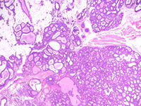
|
|
Image ID:2043 |
|
Source of Image:Ward JM |
|
Pathologist:Mikaelian I |
|
Method / Stain:H&E |
|
|
Image Caption:Mammary gland, hyperplastic alveolar nodule: all alveoli are ectatic and contain a moderate amount of proteinaceous material. They are lined by a prominent one cell-thick cuboidal epithelium. These cells often contain lipid vacuoles of various sizes. There is no evidence of atypia. There is mild interstitial fibrosis with moderate pleocellular inflammation.
|
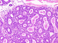
|
|
Image ID:2046 |
|
Source of Image:Ward JM |
|
Pathologist:Mikaelian I |
|
Method / Stain:H&E |
|
|
Image Caption:Mammary gland, hyperplastic alveolar nodule: all alveoli are ectatic and contain a moderate amount of proteinaceous material. They are lined by a prominent one cell-thick cuboidal epithelium. These cells often contain lipid vacuoles of various sizes. There is no evidence of atypia. There is mild interstitial fibrosis with moderate pleocellular inflammation.
|
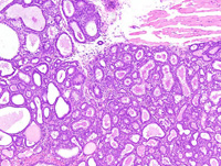
|
|
Image ID:2045 |
|
Source of Image:Ward JM |
|
Pathologist:Mikaelian I |
|
Method / Stain:H&E |
|
|
Image Caption:Mammary gland, hyperplastic alveolar nodule: all acini are ectatic and contain a moderate amount of proteinaceous material. They are lined by a prominent one cell-thick cuboidal epithelium. These cells often contain lipid vacuoles of various sizes and also a fw proteinaceous droplets. There is no evidence of atypia. There is mild interstitial fibrosis with moderate pleocellular inflammation.
|
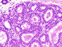
|
|
Image ID:2047 |
|
Source of Image:Ward JM |
|
Pathologist:Mikaelian I |
|
Method / Stain:H&E |
|
|
Image Caption:Mammary gland: a mammary lobule is markedly hyperplastic. All alveoli are ectatic and contain a moderate amount of proteinaceous material. They are lined by a prominent cuboidal epithelium. There is mild to moderate interstitial fibrosis with moderate pleocellular inflammation. In addition, a small adenoma (*) is present.
|
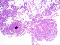
|
|
Image ID:2044 |
|
Source of Image:Ward JM |
|
Pathologist:Mikaelian I |
|
Method / Stain:H&E |
|
|
|
| MTB ID |
Tumor Name |
Organ(s) Affected |
Treatment Type |
Agents |
Strain Name |
Strain Sex |
Reproductive Status |
Tumor Frequency |
Age at Necropsy |
Description |
Reference |
| MTB:29374 |
Mammary gland adenoma - mixed |
Mammary gland |
None (spontaneous) |
|
|
Female |
reproductive status not specified |
observed |
210 days |
Adenoma, solid and tubular, arising in an hyperplastic alveolar nodule, mammary gland |
J:94320 |
|
Image Caption:Solid and tubular adenoma of the mammary gland: this photomicrograph illustrates a peripheral portion of the neoplasm where there is a small group of large cells that are columnar and have a more abundant and more acidophilic cytoplasm than most neoplastic cells. The other neoplastic cells are polygonal, with indistinct cell borders and a moderate amount of amphophilic cytoplasm. The nucleus is central, round, normochromatic, round, and with a small basophilic nucleolus. Anisokaryosis and anisocytosis overall are mild. Mitoses are numerous.
|
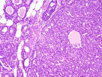
|
|
Image ID:2050 |
|
Source of Image:Ward JM |
|
Pathologist:Mikaelian I |
|
Method / Stain:H&E |
|
|
Image Caption:Solid and tubular adenoma of the mammary gland: this photomicrograph illustrates a peripheral portion of the neoplasm where there is a small group of large cells that are columnar and have a more abundant and more acidophilic cytoplasm than most neoplastic cells. These cells undergo squamous metaplasia (*) that is not associated with the presence of trichohyalin or keratohyalin granules. The other neoplastic cells are polygonal, with indistinct cell borders and a moderate amount of amphophilic cytoplasm. The nucleus is central, round, normochromatic, round, and with a small basophilic nucleolus. Anisokaryosis and anisocytosis overall are mild. Mitoses are numerous.
|
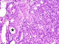
|
|
Image ID:2051 |
|
Source of Image:Ward JM |
|
Pathologist:Mikaelian I |
|
Method / Stain:H&E |
|
|
Image Caption:Mammary gland: the mammary gland is expanded by a small, unenencapsulated, densely cellular, expensile neoplasm arising in an hyperplastic alveolar nodule. The neoplasm is composed of solid areas with secondary lumen and a few closely-packed glands that are lined by a one cell-thick cuboidal epithelium and are supported by a scant amount of fibrovascular stroma. Epithelial cells in the hyperplastic alveolar nodule (in the right and lower right portions of the photomicrograph) show more atypia than generally observed in typical hyperplastic alveolar nodules. Also, the contours of the alveoli in the hyperplastic alveolar nodule are more irregular than typically observed in an hyperplastic alveolar nodule. However, cells do not pile-up disorderly and the overall pattern of the normal mammary gland can still be identified.
|

|
|
Image ID:2049 |
|
Source of Image:Ward JM |
|
Pathologist:Mikaelian I |
|
Method / Stain:H&E |
|
|
Image Caption:Solid and tubular adenoma of the mammary gland: this photomicrograph illustrates an area of transition between the hyperplastic alveolar nodule (to the far right) and the adenoma (to the left).
|
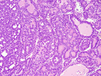
|
|
Image ID:2052 |
|
Source of Image:Ward JM |
|
Pathologist:Mikaelian I |
|
Method / Stain:H&E |
|
|
Image Caption:Mammary gland: the mammary gland is expanded by a small, unenencapsulated, densely cellular, expensile neoplasm arising in an hyperplastic alveolar nodule.
|
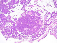
|
|
Image ID:2048 |
|
Source of Image:Ward JM |
|
Pathologist:Mikaelian I |
|
Method / Stain:H&E |
|
|
Image Caption:Solid and tubular adenoma of the mammary gland: this photomicrograph illustrates an area of transition between the hyperplastic alveolar nodule (to the far right) and the adenoma (to the left). In the tumor, cells form solid areas with secondary lumens while the epithelium is one cell-thick in the hyperplastic alveolar nodule.
|
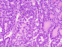
|
|
Image ID:2053 |
|
Source of Image:Ward JM |
|
Pathologist:Mikaelian I |
|
Method / Stain:H&E |
|
|
|
| MTB ID |
Tumor Name |
Organ(s) Affected |
Treatment Type |
Agents |
Strain Name |
Strain Sex |
Reproductive Status |
Tumor Frequency |
Age at Necropsy |
Description |
Reference |
| MTB:29375 |
Mammary gland hyperplasia - alveolar |
Mammary gland |
None (spontaneous) |
|
|
Female |
reproductive status not specified |
observed |
210 days |
Adenoma, solid and tubular, arising in an hyperplastic alveolar nodule, mammary gland |
J:94320 |
|
Image Caption:Solid and tubular adenoma of the mammary gland: this photomicrograph illustrates a peripheral portion of the neoplasm where there is a small group of large cells that are columnar and have a more abundant and more acidophilic cytoplasm than most neoplastic cells. These cells undergo squamous metaplasia (*) that is not associated with the presence of trichohyalin or keratohyalin granules. The other neoplastic cells are polygonal, with indistinct cell borders and a moderate amount of amphophilic cytoplasm. The nucleus is central, round, normochromatic, round, and with a small basophilic nucleolus. Anisokaryosis and anisocytosis overall are mild. Mitoses are numerous.
|
|
|
|
Image ID:2051 |
|
Source of Image:Ward JM |
|
Pathologist:Mikaelian I |
|
Method / Stain:H&E |
|
|
Image Caption:Solid and tubular adenoma of the mammary gland: this photomicrograph illustrates an area of transition between the hyperplastic alveolar nodule (to the far right) and the adenoma (to the left).
|
|
|
|
Image ID:2052 |
|
Source of Image:Ward JM |
|
Pathologist:Mikaelian I |
|
Method / Stain:H&E |
|
|
Image Caption:Solid and tubular adenoma of the mammary gland: this photomicrograph illustrates an area of transition between the hyperplastic alveolar nodule (to the far right) and the adenoma (to the left). In the tumor, cells form solid areas with secondary lumens while the epithelium is one cell-thick in the hyperplastic alveolar nodule.
|
|
|
|
Image ID:2053 |
|
Source of Image:Ward JM |
|
Pathologist:Mikaelian I |
|
Method / Stain:H&E |
|
|
Image Caption:Solid and tubular adenoma of the mammary gland: this photomicrograph illustrates a peripheral portion of the neoplasm where there is a small group of large cells that are columnar and have a more abundant and more acidophilic cytoplasm than most neoplastic cells. The other neoplastic cells are polygonal, with indistinct cell borders and a moderate amount of amphophilic cytoplasm. The nucleus is central, round, normochromatic, round, and with a small basophilic nucleolus. Anisokaryosis and anisocytosis overall are mild. Mitoses are numerous.
|
|
|
|
Image ID:2050 |
|
Source of Image:Ward JM |
|
Pathologist:Mikaelian I |
|
Method / Stain:H&E |
|
|
Image Caption:Mammary gland: the mammary gland is expanded by a small, unenencapsulated, densely cellular, expensile neoplasm arising in an hyperplastic alveolar nodule.
|
|
|
|
Image ID:2048 |
|
Source of Image:Ward JM |
|
Pathologist:Mikaelian I |
|
Method / Stain:H&E |
|
|
Image Caption:Mammary gland: the mammary gland is expanded by a small, unenencapsulated, densely cellular, expensile neoplasm arising in an hyperplastic alveolar nodule. The neoplasm is composed of solid areas with secondary lumen and a few closely-packed glands that are lined by a one cell-thick cuboidal epithelium and are supported by a scant amount of fibrovascular stroma. Epithelial cells in the hyperplastic alveolar nodule (in the right and lower right portions of the photomicrograph) show more atypia than generally observed in typical hyperplastic alveolar nodules. Also, the contours of the alveoli in the hyperplastic alveolar nodule are more irregular than typically observed in an hyperplastic alveolar nodule. However, cells do not pile-up disorderly and the overall pattern of the normal mammary gland can still be identified.
|
|
|
|
Image ID:2049 |
|
Source of Image:Ward JM |
|
Pathologist:Mikaelian I |
|
Method / Stain:H&E |
|
|
|
| MTB ID |
Tumor Name |
Organ(s) Affected |
Treatment Type |
Agents |
Strain Name |
Strain Sex |
Reproductive Status |
Tumor Frequency |
Age at Necropsy |
Description |
Reference |
| MTB:29376 |
Mammary gland adenoma - papillary |
Mammary gland |
None (spontaneous) |
|
|
Female |
reproductive status not specified |
observed |
210 days |
Papilloma, intraductal, with early transformation into a carcinoma, mammary gland |
J:94320 |
|
Image Caption:Mammary gland: the mammary gland is expanded by a nodular, unencapsulated, densely cellular neoplasm that arises within a duct (the duct was cut open during the preparation of the specimen). The lumen of the duct is filled with multiple papillary projections supported by delicate fibrovascular projections. Numerous hyperplastic alveolar nodules are present in the nearby mammary parenchyma.
|
|
|
Image ID:2055 |
|
Source of Image:Ward JM |
|
Pathologist:Mikaelian I |
|
Method / Stain:H&E |
|
|
Image Caption:Papillary adenoma, area of early transformation into a carcinoma: neoplastic cells at the center of the photomicrograph are larger, have a paler cytoplasm, a larger and paler nucleus, and a higher mitotic rate than in the rest of the neoplasm.
|
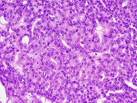
|
|
Image ID:2058 |
|
Source of Image:Ward JM |
|
Pathologist:Mikaelian I |
|
Method / Stain:H&E |
|
|
Image Caption:Papillary adenoma: the neoplasm is composed of papillary projections lined by a one cell-thick cuboidal epithelium that also forms small glands and solid areas. Neoplastic cells are cuboidal to polygonal, with indistinct cell borders and a small to moderate amount of acidophilic cytoplasm. The nucleus tends to be basal, oval, and medium-sized.
|
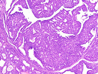
|
|
Image ID:2056 |
|
Source of Image:Ward JM |
|
Pathologist:Mikaelian I |
|
Method / Stain:H&E |
|
|
Image Caption:Papillary adenoma: the neoplasm in this area is composed of glands supported by a scant amount . Neoplastic cells are cuboidal to polygonal, with indistinct cell borders and a small to moderate amount of acidophilic cytoplasm. The nucleus tends to be basal, oval, and medium-sized. There is, at the center of the photomicrograph, an area where neoplastic cells are larger, have a paler cytoplasm, a larger and paler nucleus, and a higher mitotic rate than in the rest of the neoplasm (early transition into a carcinoma).
|
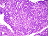
|
|
Image ID:2057 |
|
Source of Image:Ward JM |
|
Pathologist:Mikaelian I |
|
Method / Stain:H&E |
|
|
Image Caption:Mammary gland: the mammary gland is expanded by a nodular, unencapsulated, densely cellular neoplasm that arises within a duct (the duct was cut open during the preparation of the specimen). The lumen of the duct is filled with multiple papillary projections supported by delicate fibrovascular projections. Numerous hyperplastic alveolar nodules are present in the nearby mammary parenchyma.
|
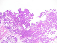
|
|
Image ID:2054 |
|
Source of Image:Ward JM |
|
Pathologist:Mikaelian I |
|
Method / Stain:H&E |
|
|
|
| MTB ID |
Tumor Name |
Organ(s) Affected |
Treatment Type |
Agents |
Strain Name |
Strain Sex |
Reproductive Status |
Tumor Frequency |
Age at Necropsy |
Description |
Reference |
| MTB:29377 |
Mammary gland carcinoma |
Mammary gland |
None (spontaneous) |
|
|
Female |
reproductive status not specified |
observed |
210 days |
Papilloma, intraductal, with early transformation into a carcinoma, mammary gland |
J:94320 |
|
Image Caption:Mammary gland: the mammary gland is expanded by a nodular, unencapsulated, densely cellular neoplasm that arises within a duct (the duct was cut open during the preparation of the specimen). The lumen of the duct is filled with multiple papillary projections supported by delicate fibrovascular projections. Numerous hyperplastic alveolar nodules are present in the nearby mammary parenchyma.
|
|
|
|
Image ID:2054 |
|
Source of Image:Ward JM |
|
Pathologist:Mikaelian I |
|
Method / Stain:H&E |
|
|
Image Caption:Papillary adenoma, area of early transformation into a carcinoma: neoplastic cells at the center of the photomicrograph are larger, have a paler cytoplasm, a larger and paler nucleus, and a higher mitotic rate than in the rest of the neoplasm.
|
|
|
|
Image ID:2058 |
|
Source of Image:Ward JM |
|
Pathologist:Mikaelian I |
|
Method / Stain:H&E |
|
|
Image Caption:Papillary adenoma: the neoplasm in this area is composed of glands supported by a scant amount . Neoplastic cells are cuboidal to polygonal, with indistinct cell borders and a small to moderate amount of acidophilic cytoplasm. The nucleus tends to be basal, oval, and medium-sized. There is, at the center of the photomicrograph, an area where neoplastic cells are larger, have a paler cytoplasm, a larger and paler nucleus, and a higher mitotic rate than in the rest of the neoplasm (early transition into a carcinoma).
|
|
|
|
Image ID:2057 |
|
Source of Image:Ward JM |
|
Pathologist:Mikaelian I |
|
Method / Stain:H&E |
|
|
Image Caption:Mammary gland: the mammary gland is expanded by a nodular, unencapsulated, densely cellular neoplasm that arises within a duct (the duct was cut open during the preparation of the specimen). The lumen of the duct is filled with multiple papillary projections supported by delicate fibrovascular projections. Numerous hyperplastic alveolar nodules are present in the nearby mammary parenchyma.
|
|
|
|
Image ID:2055 |
|
Source of Image:Ward JM |
|
Pathologist:Mikaelian I |
|
Method / Stain:H&E |
|
|
Image Caption:Papillary adenoma: the neoplasm is composed of papillary projections lined by a one cell-thick cuboidal epithelium that also forms small glands and solid areas. Neoplastic cells are cuboidal to polygonal, with indistinct cell borders and a small to moderate amount of acidophilic cytoplasm. The nucleus tends to be basal, oval, and medium-sized.
|
|
|
|
Image ID:2056 |
|
Source of Image:Ward JM |
|
Pathologist:Mikaelian I |
|
Method / Stain:H&E |
|
|
|
| MTB ID |
Tumor Name |
Organ(s) Affected |
Treatment Type |
Agents |
Strain Name |
Strain Sex |
Reproductive Status |
Tumor Frequency |
Age at Necropsy |
Description |
Reference |
| MTB:29432 |
Mammary gland hyperplasia - atypical |
Mammary gland |
None (spontaneous) |
|
|
Female |
reproductive status not specified |
observed |
84 days |
Atypical hyperplasia, mild, mammary gland |
J:94320 |
|
Image Caption:Mammary gland: the mammary ducts and alveoli are slightly more prominent than normal for a resting mammary gland.
|
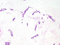
|
|
Image ID:2222 |
|
Source of Image:Ward JM |
|
Pathologist:Mikaelian I |
|
Method / Stain:H&E |
|
|
Image Caption:Mammary gland: the mammary ducts and alveoli are slightly more prominent than normal for a resting mammary gland.
|
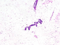
|
|
Image ID:2223 |
|
Source of Image:Ward JM |
|
Pathologist:Mikaelian I |
|
Method / Stain:H&E |
|
|
Image Caption:Mammary gland: the mammary ducts and alveoli are slightly more prominent than normal for a resting mammary gland.
|
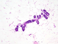
|
|
Image ID:2224 |
|
Source of Image:Ward JM |
|
Pathologist:Mikaelian I |
|
Method / Stain:H&E |
|
|
Image Caption:Mammary gland: the mammary ducts and alveoli are slightly more prominent than normal for a resting mammary gland. The ducts and the acini are lined by a cuboidal epithelium with an abnormally high mitotic activity. Cells with a morphology consistent with that of "large clear cells" (arrowheads) are more numerous than for a normal 84 days old mouse.
|
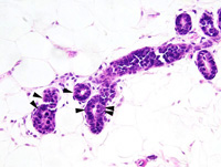
|
|
Image ID:2225 |
|
Source of Image:Ward JM |
|
Pathologist:Mikaelian I |
|
Method / Stain:H&E |
|
|
|
| MTB ID |
Tumor Name |
Organ(s) Affected |
Treatment Type |
Agents |
Strain Name |
Strain Sex |
Reproductive Status |
Tumor Frequency |
Age at Necropsy |
Description |
Reference |
| MTB:29434 |
Mammary gland adenocarcinoma |
Lung |
None (spontaneous) |
|
|
Female |
reproductive status not specified |
observed |
168 days |
Mammary adenocarcinoma, metastatic to the lungs |
J:94320 |
|
Image Caption:Lung: three branches of a large pulomonary artery are filled with large neoplastic emboli. There is moderate necrosis at the center of the neoplastic emboli.
|
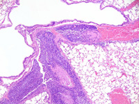
|
|
Image ID:2227 |
|
Source of Image:Ward JM |
|
Pathologist:Mikaelian I |
|
Method / Stain:H&E |
|
|
Image Caption:Lung: neoplastic cells have invaded the pulmonary parenchyma. They form anastomosed trabeculae and a few glands supported by a scant amount of fibrovascular stroma. Neoplastic cells at the periphery of neoplastic trabeculae and glands display prominent nuclear palisading, are low columnar, and have an elongated nucleus. Neoplastic cells at the center of neoplastic trabeculae are polygonal with a rounder nucleus than neoplastic cells at the periphery of neoplastic trabeculae.
|
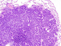
|
|
Image ID:2232 |
|
Source of Image:Ward JM |
|
Pathologist:Mikaelian I |
|
Method / Stain:H&E |
|
|
Image Caption:Lung: three branches of a large pulomonary artery are filled with large neoplastic emboli. There is moderate necrosis at the center of the neoplastic emboli.
|
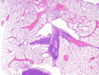
|
|
Image ID:2226 |
|
Source of Image:Ward JM |
|
Pathologist:Mikaelian I |
|
Method / Stain:H&E |
|
|
Image Caption:Lung: a neoplastic embolus (*) fills the lumen of the distal portions of a pulmonary artery.
|
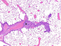
|
|
Image ID:2229 |
|
Source of Image:Ward JM |
|
Pathologist:Mikaelian I |
|
Method / Stain:H&E |
|
|
Image Caption:Lung: large neoplastic emboli are present in several pulmonary arteries (*). Neoplastic cells have invaded through the wall of the arteries into the pulmonary parenchyma. Invasion of neoplastic cells into the pulmonary parenchyma defines the metastatic stage of cancer progression.
|
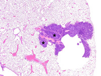
|
|
Image ID:2230 |
|
Source of Image:Ward JM |
|
Pathologist:Mikaelian I |
|
Method / Stain:H&E |
|
|
Image Caption:Lung: three branches of a large pulomonary artery are filled with large neoplastic emboli. The pulmonary artery is partly thrombosed (T) and neoplastic cells invade the thrombus. Some portions of the surface of the neoplastic embolus are covered by endothelial cells (arrowhead; re-endothelialization).
|
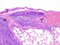
|
|
Image ID:2228 |
|
Source of Image:Ward JM |
|
Pathologist:Mikaelian I |
|
Method / Stain:H&E |
|
|
Image Caption:Lung: neoplastic cells have invaded the pulmonary parenchyma. They form anastomosed trabeculae and a few glands supported by a scant amount of fibrovascular stroma. Neoplastic cells at the periphery of neoplastic trabeculae and glands display prominent nuclear palisading, are low columnar, and have an elongated nucleus. Neoplastic cells at the center of neoplastic trabeculae are polygonal with a rounder nucleus than neoplastic cells at the periphery of neoplastic trabeculae.
|
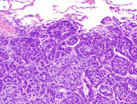
|
|
Image ID:2233 |
|
Source of Image:Ward JM |
|
Pathologist:Mikaelian I |
|
Method / Stain:H&E |
|
|
Image Caption:Lung: neoplastic cells have invaded the pulmonary parenchyma. They form anastomosed trabeculae and a few glands supported by a scant amount of fibrovascular stroma. Neoplastic cells at the periphery of neoplastic trabeculae and glands display prominent nuclear palisading, are low columnar, and have an elongated nucleus. Neoplastic cells at the center of neoplastic trabeculae are polygonal with a rounder nucleus than neoplastic cells at the periphery of neoplastic trabeculae.
|
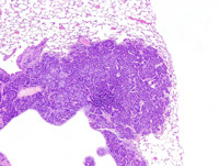
|
|
Image ID:2231 |
|
Source of Image:Ward JM |
|
Pathologist:Mikaelian I |
|
Method / Stain:H&E |
|
|
|
| MTB ID |
Tumor Name |
Organ(s) Affected |
Treatment Type |
Agents |
Strain Name |
Strain Sex |
Reproductive Status |
Tumor Frequency |
Age at Necropsy |
Description |
Reference |
| MTB:29435 |
Mammary gland adenocarcinoma - solid |
Mammary gland |
None (spontaneous) |
|
|
Female |
reproductive status not specified |
observed |
168 days |
Adenocarcinoma, solid, with epithelial to mesenchymal transition, mammary gland |
J:94320 |
|
Image Caption:Mammary gland: the mammary gland is effaced by a large, multilobular, encapsulated, densely cellular, mostly expansile, locally invasive, neoplasm with a large central area of cavitation (*).
|
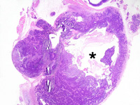
|
|
Image ID:2234 |
|
Source of Image:Ward JM |
|
Pathologist:Mikaelian I |
|
Method / Stain:H&E |
|
|
Image Caption:Solid mammary carcinoma with epithelial to mesenchymal transition: this photomicrograph illustrates a portion of the neoplasm where neoplastic cells gradually become spindloid out of a portion of the neoplasm that is solid (on the right of the photomicrograph). Cells that have undergone epithelial to mesenchymal transition are difficult to differentiate from stromal cells.
|
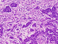
|
|
Image ID:2244 |
|
Source of Image:Ward JM |
|
Pathologist:Mikaelian I |
|
Method / Stain:H&E |
|
|
Image Caption:Solid mammary carcinoma: this photomicrograph illustrates a portion of the neoplasm that has a trabecular/solid pattern with the presence of numerous secondary lumens. Cords and trabeculae of neoplastic cells invade the capsule.
|
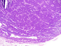
|
|
Image ID:2236 |
|
Source of Image:Ward JM |
|
Pathologist:Mikaelian I |
|
Method / Stain:H&E |
|
|
Image Caption:Solid mammary carcinoma with epithelial to mesenchymal transition: this photomicrograph illustrates a portion of the neoplasm where neoplastic cells gradually become spindloid out of a portion of the neoplasm that is solid (on the right of the photomicrograph). Cells that have undergone epithelial to mesenchymal transition are difficult to differentiate from stromal cells.
|
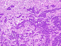
|
|
Image ID:2243 |
|
Source of Image:Ward JM |
|
Pathologist:Mikaelian I |
|
Method / Stain:H&E |
|
|
Image Caption:Mammary gland: the mammary gland is effaced by a large, multilobular, encapsulated, densely cellular, mostly expansile, locally invasive, neoplasm with a large central area of cavitation. The neoplasm is predominantly composed of solid areas with secondary lumens that can be discerned even at this low magnification. Areas indicative of epithelial to mesenchymal transition and composed of bundles and fascicles are most prominent at the periphery of the area of cavitation.
|
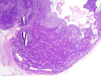
|
|
Image ID:2235 |
|
Source of Image:Ward JM |
|
Pathologist:Mikaelian I |
|
Method / Stain:H&E |
|
|
Image Caption:Solid mammary carcinoma: this photomicrograph illustrates a portion of the neoplasm that has a trabecular/solid pattern separated by a small to moderate amount of moderately dense fibrovascular stroma. Neoplastic cell are polygonal, medium-size, with distinct cell borders and a moderate amount of strongly amphophilic cytoplasm. The nucleus is central, oval, medium-size and hyperchromatic.
|
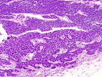
|
|
Image ID:2238 |
|
Source of Image:Ward JM |
|
Pathologist:Mikaelian I |
|
Method / Stain:H&E |
|
|
Image Caption:Solid mammary carcinoma with epithelial to mesenchymal transition: this photomicrograph illustrates a portion of the neoplasm where there is gradual transition between a solid pattern (left side of the photomicrograph) to a spindloid pattern (right side of the photomicrograph) characterized by the formation of interlacing fascicles. The stroma in the solid areas is more abundant and more fibrous than at the periphery of the neoplasm. The stroma is more edematous and less fibrous in the portions of the neoplasm composed of spindloid cells. Neoplastic cells still form a few glands with prominent lumens (arrowheads).
|
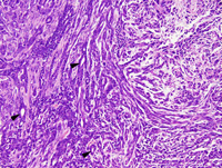
|
|
Image ID:2241 |
|
Source of Image:Ward JM |
|
Pathologist:Mikaelian I |
|
Method / Stain:H&E |
|
|
Image Caption:Solid mammary carcinoma with epithelial to mesenchymal transition: this photomicrograph illustrates a portion of the neoplasm where there is gradual transition between a solid pattern (left side of the photomicrograph) to a spindloid pattern (right side of the photomicrograph) characterized by the formation of interlacing fascicles. The stroma is more abundant and more fibrous than at the periphery of the neoplasm.
|
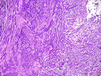
|
|
Image ID:2240 |
|
Source of Image:Ward JM |
|
Pathologist:Mikaelian I |
|
Method / Stain:H&E |
|
|
Image Caption:Solid mammary carcinoma: this photomicrograph illustrates a portion of the neoplasm that has a trabecular/solid pattern separated by a small to moderate amount of moderately dense fibrovascular stroma. Neoplastic cell are polygonal, medium-size, with distinct cell borders and a moderate amount of strongly amphophilic cytoplasm that occasionally contains a large lipid vacuole. The nucleus is central, oval, medium-size and hyperchromatic. Small clusters of neoplastic cells invade the capsule.
|
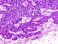
|
|
Image ID:2239 |
|
Source of Image:Ward JM |
|
Pathologist:Mikaelian I |
|
Method / Stain:H&E |
|
|
Image Caption:Solid mammary carcinoma with epithelial to mesenchymal transition: this photomicrograph illustrates a portion of the neoplasm where neoplastic cells gradually become spindloid although most of them have retained many features of epithelial cells, including angular cell contours, occasionally organization of neoplastic cells in glands, and an oval nucleus. Some neoplastic cells are fusiform with and elongated nucleus.
|
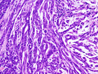
|
|
Image ID:2242 |
|
Source of Image:Ward JM |
|
Pathologist:Mikaelian I |
|
Method / Stain:H&E |
|
|
Image Caption:Solid mammary carcinoma: this photomicrograph illustrates a portion of the neoplasm that has a trabecular/solid pattern separated by a small to moderate amount of moderately dense fibrovascular stroma. Secondary lumens are prominent in the upper left portions of the photomicrograph. Neoplastic cell are polygonal, medium-size, with distinct cell borders and a moderate amount of strongly amphophilic cytoplasm.
|
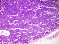
|
|
Image ID:2237 |
|
Source of Image:Ward JM |
|
Pathologist:Mikaelian I |
|
Method / Stain:H&E |
|
|
|
| MTB ID |
Tumor Name |
Organ(s) Affected |
Treatment Type |
Agents |
Strain Name |
Strain Sex |
Reproductive Status |
Tumor Frequency |
Age at Necropsy |
Description |
Reference |
| MTB:29436 |
Mammary gland adenocarcinoma - mixed |
Mammary gland |
None (spontaneous) |
|
|
Female |
reproductive status not specified |
observed |
140 days |
Adenocarcinoma, solid, comedo, mammary gland |
J:94320 |
|
Image Caption:Solid and comedo mammary carcinoma: neoplastic cells are closely packed, medium-size, polygonal, with indistinct cell borders and a small to moderate amount of amphophilic cytoplasm. The nucleus is central, oval, medium-size, with a slightly contoured nuclear membrane, normochromatic, a clumped chromatin and 1-3 small nucleoli. Anisokaryosis and anisocytosis are mild. Mitotic rate is elevated. Neoplastic cells in some places seem to be located in the stroma of the neoplasm and are not separated from the stroma by a discrete basement membrane.
|
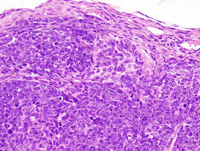
|
|
Image ID:2250 |
|
Source of Image:Ward JM |
|
Pathologist:Mikaelian I |
|
Method / Stain:H&E |
|
|
Image Caption:Mammary gland: multiple neoplasms are arising in this mammary gland. The largest neoplasm is nodular, expansile, and densely cellular. It is composed of solid areas with central necrosis.
|
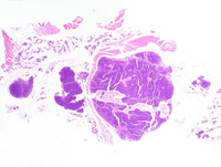
|
|
Image ID:2245 |
|
Source of Image:Ward JM |
|
Pathologist:Mikaelian I |
|
Method / Stain:H&E |
|
|
Image Caption:Mammary gland: the mammary gland is expanded by a nodular, expansile, and densely cellular neoplasm. It is composed of solid areas with central necrosis.
|
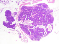
|
|
Image ID:2246 |
|
Source of Image:Ward JM |
|
Pathologist:Mikaelian I |
|
Method / Stain:H&E |
|
|
Image Caption:Mammary gland: the mammary gland is expanded by a nodular, expansile, and densely cellular neoplasm. It is composed of solid areas with central necrosis. Solid areas are separated by a small amount of moderately dense fibrovascular stroma.
|
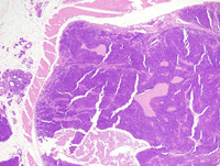
|
|
Image ID:2247 |
|
Source of Image:Ward JM |
|
Pathologist:Mikaelian I |
|
Method / Stain:H&E |
|
|
Image Caption:Mammary gland: the mammary gland is expanded by a nodular, expansile, and densely cellular neoplasm. It is composed of solid areas with central necrosis. Solid areas are separated by a small amount of moderately dense fibrovascular stroma. Neoplastic cells form a few trabeculae and nests at the periphery of the neoplasm.
|
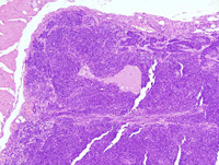
|
|
Image ID:2248 |
|
Source of Image:Ward JM |
|
Pathologist:Mikaelian I |
|
Method / Stain:H&E |
|
|
Image Caption:Solid and comedo mammary carcinoma: neoplastic cells are closely packed, medium-size, polygonal, with indistinct cell borders and a small to moderate amount of amphophilic cytoplasm. The nucleus is central, oval, medium-size, with a slightly contoured nuclear membrane, normochromatic, a clumped chromatin and 1-3 small nucleoli. Anisokaryosis and anisocytosis are mild. Mitotic rate is elevated.
|
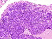
|
|
Image ID:2249 |
|
Source of Image:Ward JM |
|
Pathologist:Mikaelian I |
|
Method / Stain:H&E |
|
|
|
| MTB ID |
Tumor Name |
Organ(s) Affected |
Treatment Type |
Agents |
Strain Name |
Strain Sex |
Reproductive Status |
Tumor Frequency |
Age at Necropsy |
Description |
Reference |
| MTB:29437 |
Mammary gland carcinoma in situ - ductal |
Mammary gland |
None (spontaneous) |
|
|
Female |
reproductive status not specified |
observed |
140 days |
Ductal carcinoma in situ, mammary gland |
J:94320 |
|
Image Caption:Mammary gland: the architecture of a mammary lobule is maintained but the ducts of the mammary glands are obliterated by closely-packed neoplastic cells. Neoplastic cells are composed of several populations of cells. Some of these cells are pale, large, polygonal, with a large hypochromatic and round nucleus. Some other cells are columnar with a slightly elongated hyperchromatic nucleus. Many cells have a phenotype that is intermediate between the two types described above. Cells with a myoepithelial phenotype seem to be partly lining these ducts. The adjacent ducts and alveoli are lined by a cuboidal epithelium that has a "crowded" appearance, although there is no evidence of atypia. Mild to moderate interstitial fibrosis and pleocellular inflammation are presnt at the periphery of the ducts packed with neoplastic cells.
|
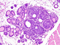
|
|
Image ID:2251 |
|
Source of Image:Ward JM |
|
Pathologist:Mikaelian I |
|
Method / Stain:H&E |
|
|
Image Caption:Mammary gland: the architecture of a mammary lobule is maintained but the ducts of the mammary glands are obliterated by closely-packed neoplastic cells. Neoplastic cells are composed of several populations of cells. Some of these cells are pale, large, polygonal, with a large hypochromatic and round nucleus. Some other cells are columnar with a slightly elongated hyperchromatic nucleus. Many cells have a phenotype that is intermediate between the two types described above. Cells with a myoepithelial phenotype seem to be partly lining these ducts. Mild to moderate interstitial fibrosis and pleocellular inflammation are presnt at the periphery of the ducts packed with neoplastic cells.
|
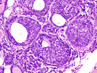
|
|
Image ID:2252 |
|
Source of Image:Ward JM |
|
Pathologist:Mikaelian I |
|
Method / Stain:H&E |
|
|
|
| MTB ID |
Tumor Name |
Organ(s) Affected |
Treatment Type |
Agents |
Strain Name |
Strain Sex |
Reproductive Status |
Tumor Frequency |
Age at Necropsy |
Description |
Reference |
| MTB:29438 |
Mammary gland hyperplasia - atypical |
Mammary gland |
None (spontaneous) |
|
|
Female |
reproductive status not specified |
observed |
140 days |
Hyperplasia, atypical, mammary gland |
J:94320 |
|
Image Caption:Mammary gland: the architecture of mammary lobules is preserved. Ducts and acini are mildly distended by a proteinaceous fluid, lipid vacuoles, and desquamated dysplastic cells. They are lined by a cuboidal epithelium where cells generally do not pile-up disordely. There is mild fibrosis and interstitial pleocellular inflammation. The mammary fat pad contains brown fat pad.
|
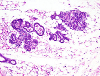
|
|
Image ID:2253 |
|
Source of Image:Ward JM |
|
Pathologist:Mikaelian I |
|
Method / Stain:H&E |
|
|
Image Caption:Mammary gland: the architecture of mammary lobules is preserved. Ducts and acini are mildly distended by a proteinaceous fluid, lipid vacuoles, and desquamated dysplastic cells. They are lined by a cuboidal epithelium where cells generally do not pile-up disordely. Some cells (arrowheads) lining these ducts resemble the "large clear cells", a type of cell that is less differentiated than most of the other luminal cells. There is mild fibrosis and interstitial pleocellular inflammation. The mammary fat pad contains brown fat pad (left portions of the photomicrograph).
|
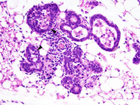
|
|
Image ID:2254 |
|
Source of Image:Ward JM |
|
Pathologist:Mikaelian I |
|
Method / Stain:H&E |
|
|
|
| MTB ID |
Tumor Name |
Organ(s) Affected |
Treatment Type |
Agents |
Strain Name |
Strain Sex |
Reproductive Status |
Tumor Frequency |
Age at Necropsy |
Description |
Reference |
| MTB:29439 |
Mammary gland carcinoma in situ - ductal |
Mammary gland |
None (spontaneous) |
|
|
Female |
reproductive status not specified |
observed |
140 days |
Ductal carcinoma in situ, mammary gland |
J:94320 |
|
Image Caption:Mammary gland: the architecture of the mammary gland is preserved. The lumen of the ducts and alveoli of a few acini is moderately distended by closely-packed neoplastic cells.
|
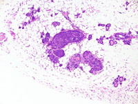
|
|
Image ID:2255 |
|
Source of Image:Ward JM |
|
Pathologist:Mikaelian I |
|
Method / Stain:H&E |
|
|
Image Caption:Mammary gland: the architecture of the mammary gland is preserved. The lumen of the ducts and alveoli of a few acini is moderately distended by closely-packed neoplastic cells that form solid areas with a few linear clefts. These cells are medium-size, polygonal, with indistinct cell borders and a small amount of amphophilic cytoplasm. The nucleus is central, oval to slightly elongated, medium-size, oval to elongated, normochromatic, with a coarsely clumped chromatin and 1-2 inconspicuous basophilic nucleoli. Anisokaryosis and anisocytosis are mild. A few mitoses are present. There is mild fibrosis and pleocllular inflammation at the periphery of the affected ducts and acini.
|
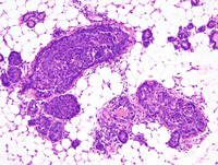
|
|
Image ID:2256 |
|
Source of Image:Ward JM |
|
Pathologist:Mikaelian I |
|
Method / Stain:H&E |
|
|
Image Caption:Mammary gland: the architecture of the mammary gland is preserved. The lumen of the ducts and alveoli of a few acini is moderately distended by closely-packed neoplastic cells. These cells are medium-size, polygonal, with indistinct cell borders and a small amount of amphophilic cytoplasm. The nucleus is central, oval, medium-size, oval, normochromatic, with a finely stippled chromatin and 1-2 inconspicuous basophilic nucleoli. Anisokaryosis and anisocytosis are mild. A few mitoses are present. A few neoplastic cells undergo single cell necrosis. There is mild fibrosis and pleocllular inflammation at the periphery of the affected ducts and acini.
|
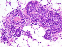
|
|
Image ID:2257 |
|
Source of Image:Ward JM |
|
Pathologist:Mikaelian I |
|
Method / Stain:H&E |
|
|
Image Caption:Mammary gland: the architecture of the mammary gland is preserved. The lumen of the ducts and alveoli of a few acini is moderately distended by closely-packed neoplastic cells that form solid areas with a few linear clefts. These cells are medium-size, polygonal, with indistinct cell borders and a small amount of amphophilic cytoplasm. The nucleus is central, oval to slightly elongated, medium-size, oval to elongated, normochromatic, with a coarsely clumped chromatin and 1-2 inconspicuous basophilic nucleoli. Anisokaryosis and anisocytosis are mild. A few mitoses are present. Myoepithelial differentiation seems to be present at this stage of the disease. There is mild fibrosis and pleocllular inflammation at the periphery of the affected ducts and acini.
|
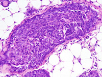
|
|
Image ID:2258 |
|
Source of Image:Ward JM |
|
Pathologist:Mikaelian I |
|
Method / Stain:H&E |
|
|
|
| MTB ID |
Tumor Name |
Organ(s) Affected |
Treatment Type |
Agents |
Strain Name |
Strain Sex |
Reproductive Status |
Tumor Frequency |
Age at Necropsy |
Description |
Reference |
| MTB:29440 |
Mammary gland carcinoma - mixed |
Mammary gland |
None (spontaneous) |
|
|
Female |
reproductive status not specified |
observed |
140 days |
Carcinoma, solid, with spindloid differentiation, mammary gland |
J:94320 |
|
Image Caption:Mammary gland: the mammary gland is effaced by a multilobular, ill-defined, densely cellular neoplasm. This neoplasm is predominantly composed of solid areas that gradually blend into the adjacent fibrovascular stroma. During this process, some neoplastic cells acquired an spindloid phenotype.
|
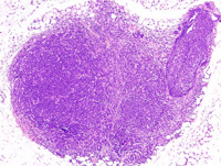
|
|
Image ID:2259 |
|
Source of Image:Ward JM |
|
Pathologist:Mikaelian I |
|
Method / Stain:H&E |
|
|
Image Caption:Mammary gland: the mammary gland is effaced by a multilobular, ill-defined, densely cellular neoplasm. This neoplasm is predominantly composed of solid areas that gradually blend into the adjacent fibrovascular stroma. During this process, some neoplastic cells acquired an spindloid phenotype. Neoplastic cells are oat-shaped to spindloid, with indistinct cell borders and a small amount of amphophilic cytoplasm. The nucleus is oval to elongated, normochromatic to slightly hypochromatic, with a clumped to stippled chromatin, and 1-3 small basophilic nucleoli. Anisokaryosis and anisocytosis are mild. Mitoses are numerous. Numerous neoplastic cells undergo single cell necrosis. There is a mild lymphocytic infiltration at the periphery of the neoplasm.
|
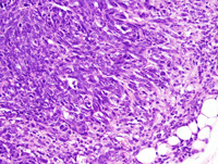
|
|
Image ID:2261 |
|
Source of Image:Ward JM |
|
Pathologist:Mikaelian I |
|
Method / Stain:H&E |
|
|
Image Caption:Mammary gland: the mammary gland is effaced by a multilobular, ill-defined, densely cellular neoplasm. This neoplasm is predominantly composed of solid areas that gradually blend into the adjacent fibrovascular stroma. During this process, some neoplastic cells acquired an spindloid phenotype. Neoplastic cells are oat-shaped to spindloid, with indistinct cell borders and a small amount of amphophilic cytoplasm. The nucleus is oval to elongated, normochromatic to slightly hypochromatic, with a clumped to stippled chromatin, and 1-3 small basophilic nucleoli. Anisokaryosis and anisocytosis are mild. Mitoses are numerous. Numerous neoplastic cells undergo single cell necrosis. There is a mild lymphocytic infiltration at the periphery of the neoplasm.
|
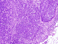
|
|
Image ID:2260 |
|
Source of Image:Ward JM |
|
Pathologist:Mikaelian I |
|
Method / Stain:H&E |
|
|
|
| MTB ID |
Tumor Name |
Organ(s) Affected |
Treatment Type |
Agents |
Strain Name |
Strain Sex |
Reproductive Status |
Tumor Frequency |
Age at Necropsy |
Description |
Reference |
| MTB:29441 |
Mammary gland adenocarcinoma |
Mammary gland |
None (spontaneous) |
|
|
Female |
reproductive status not specified |
observed |
98 days |
Adenocarcinoma, glandular, mammary gland |
J:94320 |
|
Image Caption:Mammary gland: the mammary gland is effaced by a nodular, unencapsulated, well-delineated, densely cellular, invasive neoplasm. This neoplasm is composed of closely-packed glands supported by a small to moderate amount of fibrovascular stroma.
|

|
|
Image ID:2264 |
|
Source of Image:Ward JM |
|
Pathologist:Mikaelian I |
|
Method / Stain:H&E |
|
|
Image Caption:Mammary gland: the mammary gland is effaced by a nodular, unencapsulated, well-delineated, densely cellular, invasive neoplasm. This neoplasm is composed of closely-packed glands supported by a small to moderate amount of fibrovascular stroma.
|
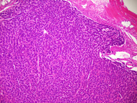
|
|
Image ID:2263 |
|
Source of Image:Ward JM |
|
Pathologist:Mikaelian I |
|
Method / Stain:H&E |
|
|
Image Caption:Glandular mammary adenocarcinoma: this photomicrograph illustrates a portion of the neoplasm where neoplastic cells had a more abundant and more acidophilic cytoplasm. The pattern of the neoplasm in this area was glandular to solid and nuclear palissading is lost.
|
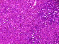
|
|
Image ID:2270 |
|
Source of Image:Ward JM |
|
Pathologist:Mikaelian I |
|
Method / Stain:H&E |
|
|
Image Caption:Mammary gland: the neoplasm is composed of closely-packed glands supported by a moderate amount of fibrovascular stroma (the stroma was less abundant in some fields). Neoplastic cells are cuboidal, medium-sized, with indistinct cell borders, and a moderate amount of strongly acidophilic cytoplasm. The nucleus is basal, oval to round, and hyperchromatic. Anisokaryosis sand anisocytosis are mild. Inflammation is minimal.
|
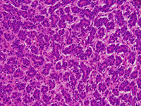
|
|
Image ID:2266 |
|
Source of Image:Ward JM |
|
Pathologist:Mikaelian I |
|
Method / Stain:H&E |
|
|
Image Caption:Glandular mammary adenocarcinoma: this photomicrograph illustrates a portion of the neoplasm where neoplastic cells had a more abundant and more acidophilic cytoplasm. The pattern of the neoplasm in this area was glandular to solid.
|
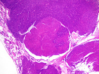
|
|
Image ID:2268 |
|
Source of Image:Ward JM |
|
Pathologist:Mikaelian I |
|
Method / Stain:H&E |
|
|
Image Caption:Mammary gland: the neoplasm is composed of closely-packed glands supported by a small to moderate amount of fibrovascular stroma. Neoplastic cells are cuboidal, medium-sized, with indistinct cell borders, and a moderate amount of strongly acidophilic cytoplasm. The nucleus is basal, oval to round, and hyperchromatic. Anisokaryosis sand anisocytosis are mild. Inflammation is minimal.
|
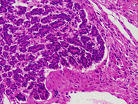
|
|
Image ID:2265 |
|
Source of Image:Ward JM |
|
Pathologist:Mikaelian I |
|
Method / Stain:H&E |
|
|
Image Caption:Glandular mammary adenocarcinoma: this photomicrograph illustrates a portion of the neoplasm where neoplastic cells had a more abundant and more acidophilic cytoplasm. The pattern of the neoplasm in this area was glandular to solid and nuclear palissading is lost.
|
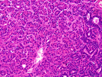
|
|
Image ID:2271 |
|
Source of Image:Ward JM |
|
Pathologist:Mikaelian I |
|
Method / Stain:H&E |
|
|
Image Caption:Glandular mammary adenocarcinoma: this photomicrograph illustrates an area of invasion of the panniculus at the periphery of the neoplasm.
|
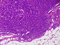
|
|
Image ID:2267 |
|
Source of Image:Ward JM |
|
Pathologist:Mikaelian I |
|
Method / Stain:H&E |
|
|
Image Caption:Glandular mammary adenocarcinoma: this photomicrograph illustrates a portion of the neoplasm where neoplastic cells had a more abundant and more acidophilic cytoplasm. The pattern of the neoplasm in this area was glandular to solid and nuclear palissading is lost.
|
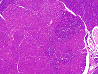
|
|
Image ID:2269 |
|
Source of Image:Ward JM |
|
Pathologist:Mikaelian I |
|
Method / Stain:H&E |
|
|
Image Caption:Mammary gland: the mammary gland is effaced by a nodular, unencapsulated, well-delineated, densely cellular, invasive neoplasm. This neoplasm is composed of closely-packed glands supported by a small to moderate amount of fibrovascular stroma.
|
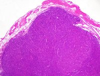
|
|
Image ID:2262 |
|
Source of Image:Ward JM |
|
Pathologist:Mikaelian I |
|
Method / Stain:H&E |
|
|
|
| MTB ID |
Tumor Name |
Organ(s) Affected |
Treatment Type |
Agents |
Strain Name |
Strain Sex |
Reproductive Status |
Tumor Frequency |
Age at Necropsy |
Description |
Reference |
| MTB:29442 |
Mammary gland hyperplasia - atypical - ductal |
Mammary gland |
None (spontaneous) |
|
|
Female |
reproductive status not specified |
observed |
98 days |
Atypical ductal hyperplasia, mammary gland |
J:94320 |
|
Image Caption:Mammary gland: an intralobular duct and most acini or a mammary lobule are lined by a prominent epithelium. Cells forming this epithelium are cuboidal to polygonal, with a moderate amount of pale basophilic cytoplasm and a medium-size hypochromatic nucleus.
|
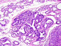
|
|
Image ID:2272 |
|
Source of Image:Ward JM |
|
Pathologist:Mikaelian I |
|
Method / Stain:H&E |
|
|
Image Caption:Mammary gland: an intralobular duct and most acini or a mammary lobule are lined by a prominent epithelium. Cells forming this epithelium are cuboidal to polygonal, with a moderate amount of pale basophilic cytoplasm and a medium-size hypochromatic nucleus. Some of these clear cells are surrounded by 2 cells with a small elongated hyperchromatic nucleus oriented at right angle with the basement membrane (arrowheads). Myoepithelial differentiation can still be recognized (arrow).
|
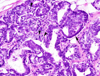
|
|
Image ID:2273 |
|
Source of Image:Ward JM |
|
Pathologist:Mikaelian I |
|
Method / Stain:H&E |
|
|
|
| MTB ID |
Tumor Name |
Organ(s) Affected |
Treatment Type |
Agents |
Strain Name |
Strain Sex |
Reproductive Status |
Tumor Frequency |
Age at Necropsy |
Description |
Reference |
| MTB:29443 |
Mammary gland carcinoma in situ - ductal |
Mammary gland |
None (spontaneous) |
|
|
Female |
reproductive status not specified |
observed |
98 days |
Ductal carcinoma in situ, mammary gland |
J:94320 |
|
Image Caption:Mammary gland: a mammary lobule is more prominent than nearby lobules. The intralobular ducts and the acini of this lobule are mildly distended by epithelial cells. The center of some solid areas is often necrotic. Cells at the periphery of the lobules are smaller, often slightly attenuated, with a nucleus oriented parallel to the basement membrane, and with a small amount of strongly amphophilic cytoplasm. These features are consistent with myoepithelial differentiation. Cells at the center of the lobules are larger, polygonal, with a moderate amount of pale basophilic cytoplasm and a medium-size hypochromatic nucleus. Mitotic figures are numerous.
|
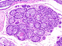
|
|
Image ID:2275 |
|
Source of Image:Ward JM |
|
Pathologist:Mikaelian I |
|
Method / Stain:H&E |
|
|
Image Caption:Mammary gland: a mammary lobule is more prominent than nearby lobules. The intralobular ducts and the acini of this lobule are mildly distended by epithelial cells. The center of some solid areas is often necrotic. Cells at the periphery of the lobules are smaller, often slightly attenuated, with a nucleus oriented parallel to the basement membrane, and with a small amount of strongly amphophilic cytoplasm. These features are consistent with myoepithelial differentiation. Cells at the center of the lobules are larger, polygonal, with a moderate amount of pale basophilic cytoplasm and a medium-size hypochromatic nucleus. Mitotic figures are numerous.There is mild interstitial fibrosis.
|
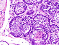
|
|
Image ID:2276 |
|
Source of Image:Ward JM |
|
Pathologist:Mikaelian I |
|
Method / Stain:H&E |
|
|
Image Caption:Mammary gland: a mammary lobule is more prominent than nearby lobules. The intralobular ducts and the acini of this lobule are mildly distended by epithelial cells.
|
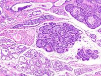
|
|
Image ID:2274 |
|
Source of Image:Ward JM |
|
Pathologist:Mikaelian I |
|
Method / Stain:H&E |
|
|
|
| MTB ID |
Tumor Name |
Organ(s) Affected |
Treatment Type |
Agents |
Strain Name |
Strain Sex |
Reproductive Status |
Tumor Frequency |
Age at Necropsy |
Description |
Reference |
| MTB:29462 |
Mammary gland adenocarcinoma - papillary |
Mammary gland |
None (spontaneous) |
|
|
Female |
reproductive status not specified |
observed |
unknown |
Adenocarcinoma, papillary, mammary gland |
J:94320 |
|
Image Caption:Mammary gland: the neoplasm is composed of ectatic glands which are partially filled with papillary projections. These epithelial structures are lined by a one cell-thick cuboidal epithelium supported by a small amount of dense fibrovascular stroma. The lumen of some glands contains clumps of degenerated neoplastic cells. Neoplastic cells are cuboidal and have a moderate amount of acidophilic cytoplasm. The nucleus is medium-sized, central and oval. Fixation artifacts prevent further interpretation of these tissues.
|
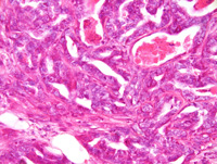
|
|
Image ID:2409 |
|
Source of Image:Ward JM |
|
Pathologist:Mikaelian I |
|
Method / Stain:H&E |
|
|
Image Caption:Mammary gland: the neoplasm is composed of ectatic glands which are partially filled with papillary projections. These epithelial structures are lined by a one cell-thick cuboidal epithelium supported by a small amount of dense fibrovascular stroma.
|
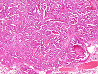
|
|
Image ID:2407 |
|
Source of Image:Ward JM |
|
Pathologist:Mikaelian I |
|
Method / Stain:H&E |
|
|
Image Caption:Mammary gland: the neoplasm is composed of ectatic glands which are partially filled with slender papillary projections. These epithelial structures are lined by a one cell-thick cuboidal epithelium supported by a small amount of dense fibrovascular stroma. The lumen of some glands contains clumps of degenerated neoplastic cells. Neoplastic cells are cuboidal and have a moderate amount of acidophilic cytoplasm. The nucleus is medium-sized, central and oval. Fixation artifacts prevent further interpretation of these tissues.
|
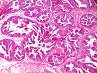
|
|
Image ID:2410 |
|
Source of Image:Ward JM |
|
Pathologist:Mikaelian I |
|
Method / Stain:H&E |
|
|
Image Caption:Mammary gland: the neoplasm is composed of ectatic glands which are partially filled with papillary projections. These epithelial structures are lined by a one cell-thick cuboidal epithelium supported by a small amount of dense fibrovascular stroma.
|
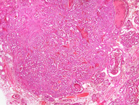
|
|
Image ID:2406 |
|
Source of Image:Ward JM |
|
Pathologist:Mikaelian I |
|
Method / Stain:H&E |
|
|
Image Caption:Mammary gland: the mammary gland is effaced by a multilobular, densely cellular, unencapsulated neoplastic mass.
|
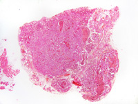
|
|
Image ID:2405 |
|
Source of Image:Ward JM |
|
Pathologist:Mikaelian I |
|
Method / Stain:H&E |
|
|
Image Caption:Mammary gland: the neoplasm is composed of ectatic glands which are partially filled with papillary projections. These epithelial structures are lined by a one cell-thick cuboidal epithelium supported by a small amount of dense fibrovascular stroma. The lumen of some glands contains clumps of degenerated neoplastic cells. Neoplastic cells are cuboidal and have a moderate amount of acidophilic cytoplasm. The nucleus is medium-sized, central and oval. Fixation artifacts prevent further interpretation of these tissues.
|
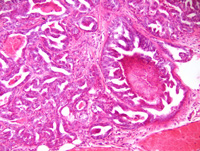
|
|
Image ID:2408 |
|
Source of Image:Ward JM |
|
Pathologist:Mikaelian I |
|
Method / Stain:H&E |
|
|
Image Caption:Mammary gland: the neoplasm is composed of ectatic glands which are partially filled with slender papillary projections. These epithelial structures are lined by a one cell-thick cuboidal epithelium supported by a small amount of dense fibrovascular stroma. The lumen of some glands contains clumps of degenerated neoplastic cells. Neoplastic cells are cuboidal and have a moderate amount of acidophilic cytoplasm. The nucleus is medium-sized, central and oval. Fixation artifacts prevent further interpretation of these tissues.
|
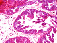
|
|
Image ID:2411 |
|
Source of Image:Ward JM |
|
Pathologist:Mikaelian I |
|
Method / Stain:H&E |
|
|
|
| MTB ID |
Tumor Name |
Organ(s) Affected |
Treatment Type |
Agents |
Strain Name |
Strain Sex |
Reproductive Status |
Tumor Frequency |
Age at Necropsy |
Description |
Reference |
| MTB:29463 |
Mammary gland hyperplasia - atypical |
Mammary gland |
None (spontaneous) |
|
|
Female |
reproductive status not specified |
observed |
unknown |
Atypical hyperplasia and interstitial fibrosis, mammary gland |
J:94320 |
|
Image Caption:Dysplasia, hyperplasia and interstitial fibrosis of the mammary gland: most intralobular ducts and most acini are filled by an amorphous acidophilic material. There is moderate to locally marked periacinar and periductular fibrosis. Cells lining mammary acini and ducts do not pile-up but they have more cytoplasm and a larger nucleus than normally. Immunohistochemistry for myoepithelial markers would be needed to determine whether myoepithelial differentiation is maintained in these lesions.
|
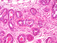
|
|
Image ID:2414 |
|
Source of Image:Ward JM |
|
Pathologist:Mikaelian I |
|
Method / Stain:H&E |
|
|
Image Caption:Mammary gland: the lobular architecture of the mammary gland is prominent. Most intralobular ducts and most acini are filled by an amorphous acidophilic material. There is moderate to locally marked periacinar and periductular fibrosis. Cells lining mammary acini and ducts do not pile-up. Immunohistochemistry for myoepithelial markers would be needed to determine whether myoepithelial differentiation is maintained in these lesions.
|
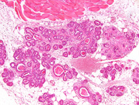
|
|
Image ID:2412 |
|
Source of Image:Ward JM |
|
Pathologist:Mikaelian I |
|
Method / Stain:H&E |
|
|
Image Caption:Mammary gland: the lobular architecture of the mammary gland is prominent. Most intralobular ducts and most acini are filled by an amorphous acidophilic material. There is moderate to locally marked periacinar and periductular fibrosis. Cells lining mammary acini and ducts do not pile-up. Immunohistochemistry for myoepithelial markers would be needed to determine whether myoepithelial differentiation is maintained in these lesions.
|
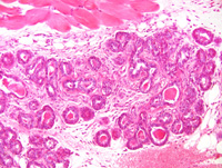
|
|
Image ID:2413 |
|
Source of Image:Ward JM |
|
Pathologist:Mikaelian I |
|
Method / Stain:H&E |
|
|
|
| MTB ID |
Tumor Name |
Organ(s) Affected |
Treatment Type |
Agents |
Strain Name |
Strain Sex |
Reproductive Status |
Tumor Frequency |
Age at Necropsy |
Description |
Reference |
| MTB:29465 |
Mammary gland carcinoma - papillary |
Mammary gland |
None (spontaneous) |
|
|
Female |
reproductive status not specified |
observed |
unknown |
Intraductal papillary carcinoma, mammary gland |
J:94320 |
|
Image Caption:Mammary gland: the epithelium of several interlobular ducts forms small papillae. The lumen of these papillae contains necrotic epithelial cells. There is moderate periductal fibrosis. Adjacent mamamry acini are moderately ectatic, contain necrotic epithelial cells, and are surrounded by moderate fibrosis with mild to moderate pleocellular inflammation.
|
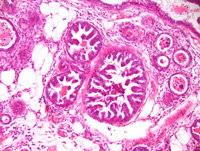
|
|
Image ID:2415 |
|
Source of Image:Ward JM |
|
Pathologist:Mikaelian I |
|
Method / Stain:H&E |
|
|
|
| MTB ID |
Tumor Name |
Organ(s) Affected |
Treatment Type |
Agents |
Strain Name |
Strain Sex |
Reproductive Status |
Tumor Frequency |
Age at Necropsy |
Description |
Reference |
| MTB:29471 |
Mammary gland adenoacanthoma |
Mammary gland |
None (spontaneous) |
|
|
Female |
reproductive status not specified |
observed |
unknown |
Adenocarcinoma, glandular, with squamous metaplasia (adenoacanthoma), mammary gland |
J:94320 |
|
Image Caption:Mammary gland: this is a nodular, well-delineated, unencapsulated, densely cellular neoplasm. Cytological details are poor because the tissue was fixed in ethanol.
|
|
|
Image ID:2416 |
|
Source of Image:Ward JM |
|
Pathologist:Mikaelian I |
|
Method / Stain:H&E |
|
|
Image Caption:Mammary gland: this is a nodular, well-delineated, unencapsulated, densely cellular neoplasm. The neoplasm is composed of interconnected ducts separated by a moderate amount of dense fibrovascular stroma. Cytological details are poor because the tissue was fixed in ethanol.
|
|
|
Image ID:2417 |
|
Source of Image:Ward JM |
|
Pathologist:Mikaelian I |
|
Method / Stain:H&E |
|
|
Image Caption:Mammary gland: this is a nodular, well-delineated, unencapsulated, invasive, densely cellular neoplasm. The neoplasm is composed of interconnected ducts separated by a moderate amount of dense fibrovascular stroma. There are numerous areas of cornification which appear as concentric whirls of a strongly acidophilic material. There is a mild to moderate lymphocytic and plasmacytic infiltrate at the periphery of the tumor. Cytological details are poor because the tissue was fixed in ethanol.
|
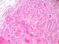
|
|
Image ID:2419 |
|
Source of Image:Ward JM |
|
Pathologist:Mikaelian I |
|
Method / Stain:H&E |
|
|
Image Caption:Mammary gland: there are numerous areas of cornification which appear as concentric whirls of a strongly acidophilic material. The cytoplasm of a few cells in these areas of cornification contain keratohyalin granules (arrowhead). There is a mild to moderate lymphocytic and plasmacytic infiltrate (L) at the periphery of the tumor. Cytological details are poor because the tissue was fixed in ethanol.
|
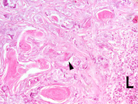
|
|
Image ID:2421 |
|
Source of Image:Ward JM |
|
Pathologist:Mikaelian I |
|
Method / Stain:H&E |
|
|
Image Caption:Mammary gland: this is a nodular, well-delineated, unencapsulated, invasive, densely cellular neoplasm. The neoplasm is composed of interconnected ducts separated by a moderate amount of dense fibrovascular stroma. There are numerous areas of cornification which appear as concentric whirls of a strongly acidophilic material. There is a mild to moderate lymphocytic and plasmacytic infiltrate at the periphery of the tumor. Cytological details are poor because the tissue was fixed in ethanol.
|
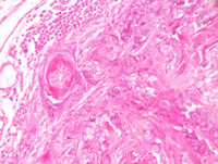
|
|
Image ID:2420 |
|
Source of Image:Ward JM |
|
Pathologist:Mikaelian I |
|
Method / Stain:H&E |
|
|
Image Caption:Mammary gland: this is a nodular, well-delineated, unencapsulated, invasive, densely cellular neoplasm. The neoplasm is composed of interconnected ducts separated by a moderate amount of dense fibrovascular stroma. There are numerous areas of cornification which appear as concentric whirls of a strongly acidophilic material. There is a mild to moderate lymphocytic and plasmacytic infiltrate at the periphery of the tumor. Cytological details are poor because the tissue was fixed in ethanol.
|
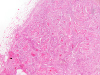
|
|
Image ID:2418 |
|
Source of Image:Ward JM |
|
Pathologist:Mikaelian I |
|
Method / Stain:H&E |
|
|
|
| MTB ID |
Tumor Name |
Organ(s) Affected |
Treatment Type |
Agents |
Strain Name |
Strain Sex |
Reproductive Status |
Tumor Frequency |
Age at Necropsy |
Description |
Reference |
| MTB:29472 |
Mammary gland adenocarcinoma - papillary |
Mammary gland |
None (spontaneous) |
|
|
Female |
reproductive status not specified |
observed |
unknown |
Adenocarcinoma, papillary, mammary gland |
J:94320 |
|
Image Caption:Mammary gland: the neoplasm is composed of interconnected ducts lined by a one cell-thick epithelium and supported by a moderate amount of fibrovascular stroma with a moderate pleocellular inflammation. The epithelium of numerous ducts forms small papillae supported by a scant amount of fibrovascular stroma. Neoplastic cells are cuboidal. Cytological details are poor because the tissue was fixed in ethanol.
|
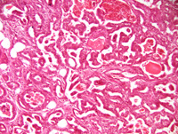
|
|
Image ID:2425 |
|
Source of Image:Ward JM |
|
Pathologist:Mikaelian I |
|
Method / Stain:H&E |
|
|
Image Caption:Mammary gland: there is a nodular, unencapsulated, densely cellular, invasive neoplasm in the right portion of the specimen. The left portion of the specimen contains multiple small areas of intraductal carcinoma. Cytological details are poor because the tissue was fixed in ethanol.
|
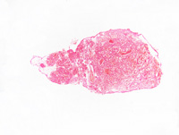
|
|
Image ID:2422 |
|
Source of Image:Ward JM |
|
Pathologist:Mikaelian I |
|
Method / Stain:H&E |
|
|
Image Caption:Mammary gland: the mammary parenchyma is effaced by a nodular, unencapsulated, densely cellular, invasive neoplasm. This neoplasm is composed of interconnected ducts lined by a one cell-thick epithelium and supported by a moderate amount of fibrovascular stroma with a moderate pleocellular inflammation. The epithelium of numerous ducts forms small papillae supported by a scant amount of fibrovascular stroma. Neoplastic cells are cuboidal. Cytological details are poor because the tissue was fixed in ethanol.
|
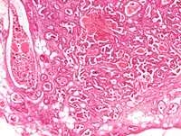
|
|
Image ID:2424 |
|
Source of Image:Ward JM |
|
Pathologist:Mikaelian I |
|
Method / Stain:H&E |
|
|
Image Caption:Mammary gland: the mammary parenchyma is effaced by a nodular, unencapsulated, densely cellular, invasive neoplasm. This neoplasm is composed of interconnected ducts lined by a one cell-thick epithelium and supported by a moderate amount of fibrovascular stroma with a moderate pleocellular inflammation. Cytological details are poor because the tissue was fixed in ethanol.
|
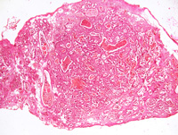
|
|
Image ID:2423 |
|
Source of Image:Ward JM |
|
Pathologist:Mikaelian I |
|
Method / Stain:H&E |
|
|
Image Caption:Mammary gland: the neoplasm is composed of interconnected ducts lined by a one cell-thick epithelium and supported by a moderate amount of fibrovascular stroma with a moderate pleocellular inflammation. The epithelium of numerous ducts forms small papillae supported by a scant amount of fibrovascular stroma. Neoplastic cells are cuboidal. Cytological details are poor because the tissue was fixed in ethanol.
|
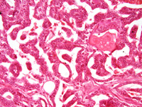
|
|
Image ID:2426 |
|
Source of Image:Ward JM |
|
Pathologist:Mikaelian I |
|
Method / Stain:H&E |
|
|
|
| MTB ID |
Tumor Name |
Organ(s) Affected |
Treatment Type |
Agents |
Strain Name |
Strain Sex |
Reproductive Status |
Tumor Frequency |
Age at Necropsy |
Description |
Reference |
| MTB:29473 |
Mammary gland carcinoma - papillary |
Mammary gland |
None (spontaneous) |
|
|
Female |
reproductive status not specified |
observed |
unknown |
Intraductal papillary carcinoma, mammary gland |
J:94320 |
|
Image Caption:Mammary gland: all mammary ducts and alveoli are moderately ectatic and are lined by a one cell-thick epithelium that forms numerous small papillary projections. There is moderate interstitial fibrosis and pleocellular inflammation at the periphery of these ducts and alveoli. Many ducts and alveoli contain a moderate number of desquamated and degenerated neoplastic epithelial cells. Cytological details are poor because the tissue was fixed in ethanol.
|
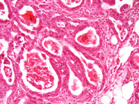
|
|
Image ID:2429 |
|
Source of Image:Ward JM |
|
Pathologist:Mikaelian I |
|
Method / Stain:H&E |
|
|
Image Caption:Mammary gland: all mammary ducts and alveoli are moderately ectatic and are lined by a one cell-thick epithelium that forms numerous small papillary projections. There is moderate interstitial fibrosis and pleocellular inflammation at the periphery of these ducts and alveoli. Cytological details are poor because the tissue was fixed in ethanol.
|
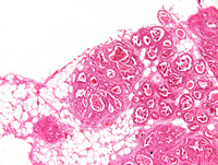
|
|
Image ID:2427 |
|
Source of Image:Ward JM |
|
Pathologist:Mikaelian I |
|
Method / Stain:H&E |
|
|
Image Caption:Mammary gland: all mammary ducts and alveoli are moderately ectatic and are lined by a one cell-thick epithelium that forms numerous small papillary projections. There is moderate interstitial fibrosis and pleocellular inflammation at the periphery of these ducts and alveoli. Many ducts and alveoli contain a moderate number of desquamated and degenerated neoplastic epithelial cells. Cytological details are poor because the tissue was fixed in ethanol.
|
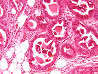
|
|
Image ID:2430 |
|
Source of Image:Ward JM |
|
Pathologist:Mikaelian I |
|
Method / Stain:H&E |
|
|
Image Caption:Mammary gland: all mammary ducts and alveoli are moderately ectatic and are lined by a one cell-thick epithelium that forms numerous small papillary projections. There is moderate interstitial fibrosis and pleocellular inflammation at the periphery of these ducts and alveoli. Many ducts and alveoli contain a moderate number of desquamated and degenerated neoplastic epithelial cells. Cytological details are poor because the tissue was fixed in ethanol.
|
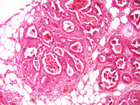
|
|
Image ID:2428 |
|
Source of Image:Ward JM |
|
Pathologist:Mikaelian I |
|
Method / Stain:H&E |
|
|
|
| MTB ID |
Tumor Name |
Organ(s) Affected |
Treatment Type |
Agents |
Strain Name |
Strain Sex |
Reproductive Status |
Tumor Frequency |
Age at Necropsy |
Description |
Reference |
| MTB:69591 |
Spleen hyperplasia - splenic marginal zone |
Spleen |
None (spontaneous) |
|
|
Unspecified |
reproductive status not specified |
observed |
|
splenic marginal zone hyperplasia |
J:107304 |
|
Image Caption:This image was submitted by JM Ward. Image is from study set created by Herbert C. Morse III (NIAID, NIH), Torgny Fredrickson (NIAID, NIH), and Jerrold M. Ward (NCI, NIH) in 2001. A whole-slide scan image cane be viewed at https://images.jax.org/webclient/img_detail/15602/.
|
|
|
Image ID:5752 |
|
Source of Image:Ward JM |
|
Pathologist:Ward JM |
|
Method / Stain:H&E |
|
|
|
| MTB ID |
Tumor Name |
Organ(s) Affected |
Treatment Type |
Agents |
Strain Name |
Strain Sex |
Reproductive Status |
Tumor Frequency |
Age at Necropsy |
Description |
Reference |
| MTB:100592 |
(Unspecified organ) normal tissue (control) |
(Unspecified organ) |
None (spontaneous) |
|
|
Unspecified |
reproductive status not specified |
not applicable |
|
Normal Control |
J:107304 |
|
|