|
| MTB ID |
Tumor Name |
Organ(s) Affected |
Treatment Type |
Agents |
Strain Name |
Strain Sex |
Reproductive Status |
Tumor Frequency |
Age at Necropsy |
Description |
Reference |
| MTB:27655 |
Intestine - Small Intestine normal tissue (control) |
Intestine - Small Intestine |
None (spontaneous) |
|
|
Unspecified |
reproductive status not specified |
not applicable |
35 days |
Small intestine - normal tissue. CDK4 immunohistochemistry |
J:94320 |
|
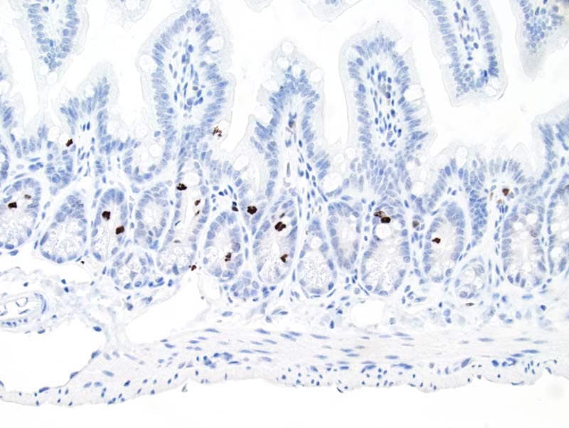
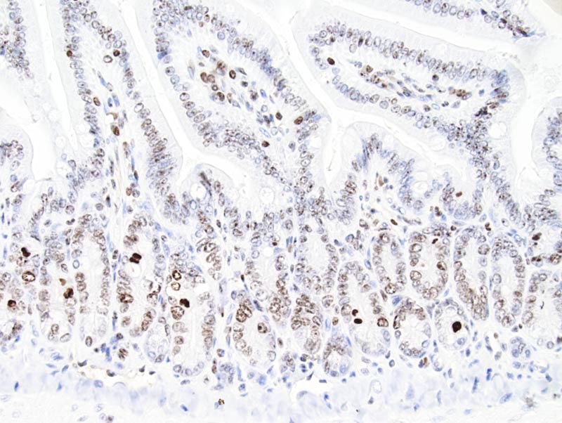
Image Caption:Small intestine - normal tissue. There is strong labeling of all mitotic figures for cdk4 (antibody diluted at 1:50, tissue fixed in 10% neutral buffered formalin, heat-mediated antigen retrieval in citrate buffer at pH=6.0).
|
|
|
Image ID:1336 |
|
Source of Image:Mikaelian I |
|
Pathologist:Mikaelian I |
|
Method / Stain:IHC for CDK4 |
|
|
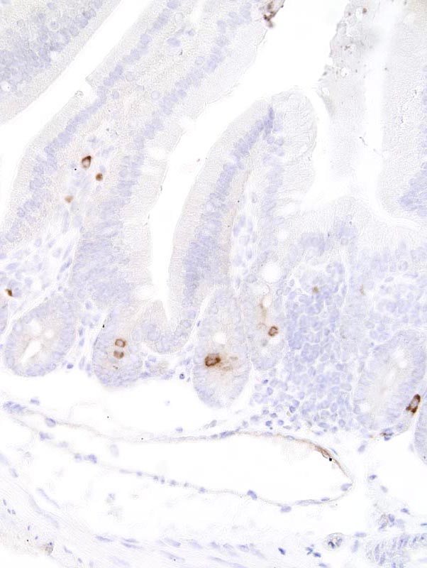
Image Caption:Small intestine - normal tissue. There is strong labeling of all mitotic figures for cdk4 (antibody diluted at 1:50, tissue fixed in Fekete's solution, heat-mediated antigen retrieval in 0.1mM EDTA at pH=7.5). Cytological details are obscured by the antigen retrieval method.
|
|
|
Image ID:1335 |
|
Source of Image:Mikaelian I |
|
Pathologist:Mikaelian I |
|
Method / Stain:IHC for CDK4 |
|
|
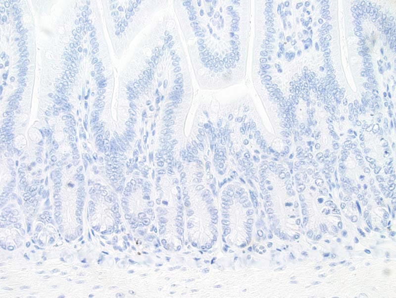
Image Caption:Small intestine - normal tissue. Labeling of mitotic figures and enterocytes was lost at this dilution (antibody diluted at 1:50, tissue fixed in Bouin's solution, heat-mediated antigen retrieval in citrate buffer at pH=6.0).
|
|
|
Image ID:1334 |
|
Source of Image:Mikaelian I |
|
Pathologist:Mikaelian I |
|
Method / Stain:IHC for CDK4 |
|
|
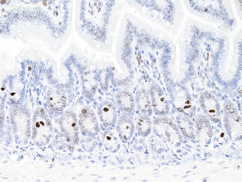
Image Caption:Small intestine - normal tissue. There is strong labeling of all mitotic figures for cdk4 (antibody diluted at 1:50, tissue fixed in Bouin's solution, heat-mediated antigen retrieval in citrate buffer at pH=6.0). There is also moderate nuclear labeling of most enterocytes and scattered cells (plasma cells?) in the lamina propria of the small intestine. Enterocytes and cells in the lamina propria did not label for cdk4 for tissues fixed in fixatives other than Bouin's solution. Hence labeling of the nucleus of plasma cells and enterocytes was interpreted as an artifact.
|
|
|
Image ID:1333 |
|
Source of Image:Mikaelian I |
|
Pathologist:Mikaelian I |
|
Method / Stain:IHC for CDK4 |
|
|
Image Caption:Small intestine - normal tissue. There is strong labeling of all mitotic figures for cdk4 (antibody diluted at 1:25, tissue fixed in Bouin's solution, heat-mediated antigen retrieval in citrate buffer at pH=6.0). There is also moderate labeling of the nucleus of most enterocytes and scattered cells (plasma cells?) in the lamina propria of the small intestine. Enterocytes and cells in the lamina propria did not label for cdk4 for tissues fixed in fixatives other than Bouin's solution. Hence labeling of the nucleus of plasma cells and enterocytes was interpreted as an artifact.
|
|
|
Image ID:1332 |
|
Source of Image:Mikaelian I |
|
Pathologist:Mikaelian I |
|
Method / Stain:IHC for CDK4 |
|
|
|
| MTB ID |
Tumor Name |
Organ(s) Affected |
Treatment Type |
Agents |
Strain Name |
Strain Sex |
Reproductive Status |
Tumor Frequency |
Age at Necropsy |
Description |
Reference |
| MTB:27683 |
Peyer's patch normal tissue (control) |
Peyer's patch |
None (spontaneous) |
|
|
Unspecified |
reproductive status not specified |
not applicable |
35 days |
Peyer's patch - Normal tissue. CDK4 immunohistochemistry |
J:94320 |
|
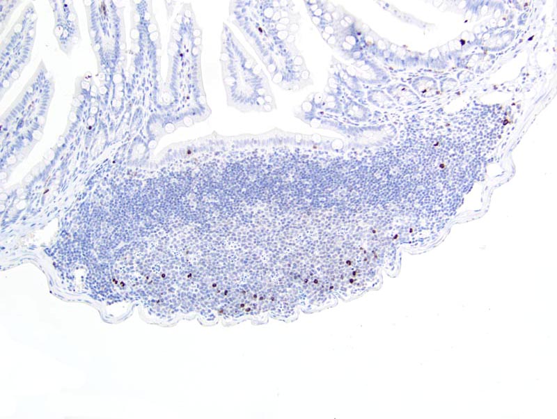
Image Caption:Small intestine, Peyer's patch - normal tissue. There is strong labeling of all mitotic figures for cdk4 in the germinative center of a Peyer's patch (antibody diluted at 1:50, tissue fixed in 10% neutral buffered formalin, heat-mediated antigen retrieval in citrate buffer at pH=6.0). There is also strong labeling of scattered mitotic figures in intestinal crypts.
|
|
|
Image ID:1341 |
|
Source of Image:Mikaelian I |
|
Pathologist:Mikaelian I |
|
Method / Stain:IHC for CDK4 |
|
|
|
| MTB ID |
Tumor Name |
Organ(s) Affected |
Treatment Type |
Agents |
Strain Name |
Strain Sex |
Reproductive Status |
Tumor Frequency |
Age at Necropsy |
Description |
Reference |
| MTB:27704 |
Thymus normal tissue (control) |
Thymus |
None (spontaneous) |
|
|
Unspecified |
reproductive status not specified |
not applicable |
35 days |
Thymus - Normal tissue. CDK4 immunohistochemistry |
J:94320 |
|
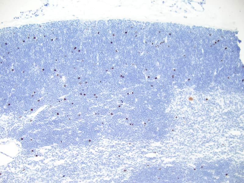
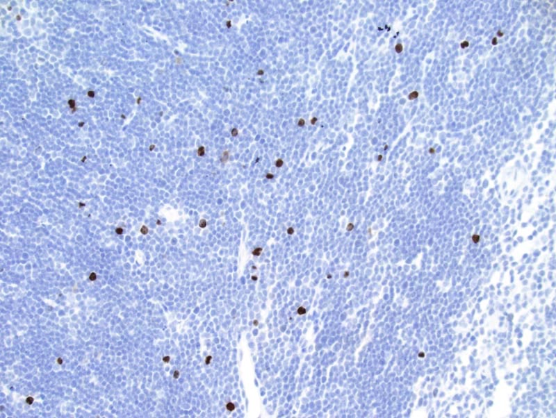
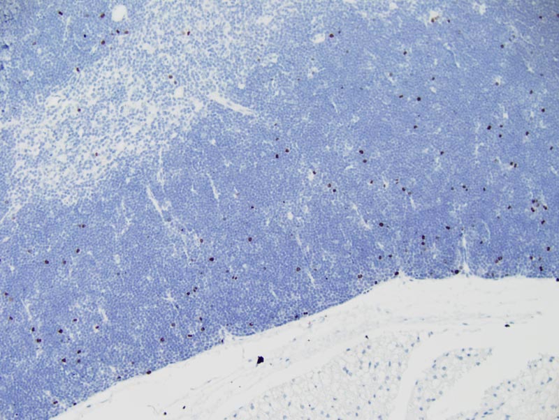
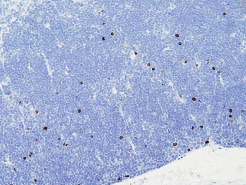
Image Caption:Thymus - normal tissue. There is strong nuclear labeling of scattered cortical thymocytes (antibody diluted at 1:50, tissue fixed in 4% paraformaldehyde, heat-mediated antigen retrieval in citrate buffer at pH=6.0).
|
|
|
Image ID:1346 |
|
Source of Image:Mikaelian I |
|
Pathologist:Mikaelian I |
|
Method / Stain:IHC for CDK4 |
|
|
Image Caption:Thymus - normal tissue. There is strong nuclear labeling of scattered cortical thymocytes (antibody diluted at 1:50, tissue fixed in 4% paraformaldehyde, heat-mediated antigen retrieval in citrate buffer at pH=6.0).
|
|
|
Image ID:1345 |
|
Source of Image:Mikaelian I |
|
Pathologist:Mikaelian I |
|
Method / Stain:IHC for CDK4 |
|
|
Image Caption:Thymus - normal tissue. There is strong nuclear labeling of scattered cortical thymocytes (antibody diluted at 1:50, tissue fixed in 10% neutral buffered formalin, heat-mediated antigen retrieval in citrate buffer at pH=6.0).
|
|
|
Image ID:1344 |
|
Source of Image:Mikaelian I |
|
Pathologist:Mikaelian I |
|
Method / Stain:IHC for CDK4 |
|
|
Image Caption:Thymus - normal tissue. There is strong nuclear labeling of scattered cortical thymocytes (antibody diluted at 1:50, tissue fixed in 10% neutral buffered formalin, heat-mediated antigen retrieval in citrate buffer at pH=6.0).
|
|
|
Image ID:1343 |
|
Source of Image:Mikaelian I |
|
Pathologist:Mikaelian I |
|
Method / Stain:IHC for CDK4 |
|
|