|
| MTB ID |
Tumor Name |
Organ(s) Affected |
Treatment Type |
Agents |
Strain Name |
Strain Sex |
Reproductive Status |
Tumor Frequency |
Age at Necropsy |
Description |
Reference |
| MTB:18036 |
Mammary gland adenocarcinoma |
Mammary gland |
None (spontaneous) |
|
|
Female |
reproductive status not specified |
observed |
186 days |
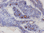
Mammary gland, adenocarcinoma. |
J:94310 |
|
Image Caption:Mammary gland, adenocarcinoma. This is a high magnification picture of an area of a mammary adenocarcinoma which displays acinar and solid patterns concurrently. Immunolabeling is restricted to the areas of acinar differentiation and generally is restricted to the apical cellular membrane of neoplastic cells. In addition, the cytoplasm of a few neoplastic cells is intensely labeled for MMTV nucleoid protein p27.
|
|
|
Image ID:223 |
|
Source of Image:Mikaelian I |
|
Pathologist:Mikaelian I |
|
Method / Stain:IHC for MMTV nucleoid protein p27 |
|
|
|
| MTB ID |
Tumor Name |
Organ(s) Affected |
Treatment Type |
Agents |
Strain Name |
Strain Sex |
Reproductive Status |
Tumor Frequency |
Age at Necropsy |
Description |
Reference |
| MTB:18075 |
Mammary gland adenocarcinoma |
Mammary gland |
None (spontaneous) |
|
|
Female |
reproductive status not specified |
observed |
165 days |
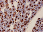
Mammary gland, adenocarcinoma. |
J:94310 |
|
Image Caption:Mammary gland, adenocarcinoma. This is a high magnification image of an acinar portion of the neoplasm. Neoplastic cells show intense apical labeling for MMTV nucleoid protein p27.
|
|
|
Image ID:226 |
|
Source of Image:Mikaelian I |
|
Pathologist:Mikaelian I |
|
Method / Stain:IHC for MMTV nucleoid protein p27 |
|
|
Image Caption:Mammary gland, adenocarcinoma. This is a high magnification image of an acinar portion of the neoplasm. Neoplastic cells show intense apical labeling for MMTV nucleoid protein p27.
|
|
|
Image ID:225 |
|
Source of Image:Mikaelian I |
|
Pathologist:Mikaelian I |
|
Method / Stain:IHC for MMTV nucleoid protein p27 |
|
|
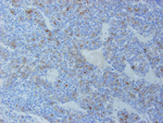
Image Caption:Mammary gland, adenocarcinoma. This low magnification picture of a mammary adenocarcinoma in a MMTV-infected mouse shows intense labeling of neoplastic cells in the portions of the neoplasm with an acinar pattern (lower left corner). Minimal or no labeling is observed in the solid portions of the tumor (upper right corner).
|
|
|
Image ID:224 |
|
Source of Image:Mikaelian I |
|
Pathologist:Mikaelian I |
|
Method / Stain:IHC for MMTV nucleoid protein p27 |
|
|
|
| MTB ID |
Tumor Name |
Organ(s) Affected |
Treatment Type |
Agents |
Strain Name |
Strain Sex |
Reproductive Status |
Tumor Frequency |
Age at Necropsy |
Description |
Reference |
| MTB:18118 |
Mammary gland adenocarcinoma |
Mammary gland |
None (spontaneous) |
|
|
Female |
reproductive status not specified |
observed |
249 days |
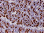
Mammary gland, adenocarcinoma. |
J:94313 |
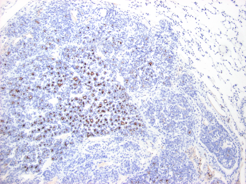
Image Caption:Mammary gland, adenocarcinoma.
There is moderate to strong labeling for MMTV nucleoid protein p27 of the proteinaceous material within the lumen of neoplastic acini. There is mild labeling of the cytoplasm of neoplastic cells.
|
|
|
Image ID:275 |
|
Source of Image:Mikaelian I |
|
Pathologist:Mikaelian I |
|
Method / Stain:IHC for MMTV nucleoid protein p27 |
|
|
|
| MTB ID |
Tumor Name |
Organ(s) Affected |
Treatment Type |
Agents |
Strain Name |
Strain Sex |
Reproductive Status |
Tumor Frequency |
Age at Necropsy |
Description |
Reference |
| MTB:25464 |
Mammary gland adenocarcinoma |
Lung |
None (spontaneous) |
|
|
Female |
reproductive status not specified |
observed |
439 days |
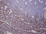
Mammary adenocarcinoma, metastatic to the lung: immunohistolabeling for MMTV gp36 |
J:94320 |
|
Image Caption:Mammary adenocarcinoma, metastatic to the lung: there is strong labeling for gp36 of the apical pole of neoplastic cells in the areas of acinar differentiation. There is no evidence of immunolabeling in the solid portions of the neoplasm and in the nearby pulmonary parenchyma.
|
|
|
Image ID:1044 |
|
Source of Image:Mikaelian I |
|
Pathologist:Mikaelian I |
|
Method / Stain:IHC for MMTV nucleoid protein p27 |
|
|
|
| MTB ID |
Tumor Name |
Organ(s) Affected |
Treatment Type |
Agents |
Strain Name |
Strain Sex |
Reproductive Status |
Tumor Frequency |
Age at Necropsy |
Description |
Reference |
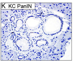
| MTB:41480 |
Pancreas - Duct pancreatic intraepithelial neoplasia (PanIN) |
Pancreas - Duct |
None (spontaneous) |
|
|
Unspecified |
reproductive status not specified |
observed |
|
PanIN, pancreatic intraepithelial neoplasia |
J:156461 |
|
Image Caption:Fig. 2K.
|
|
|
Image ID:4307 |
|
Source of Image:Morton JP |
|
Pathologist:Morton JP |
|
|