|
| MTB ID |
Tumor Name |
Organ(s) Affected |
Treatment Type |
Agents |
Strain Name |
Strain Sex |
Reproductive Status |
Tumor Frequency |
Age at Necropsy |
Description |
Reference |
| MTB:27649 |
Intestine - Large Intestine - Colon normal tissue (control) |
Intestine - Large Intestine - Colon |
None (spontaneous) |
|
|
Unspecified |
reproductive status not specified |
not applicable |
unknown |
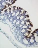
Large intestine, colon -Normal tissue. |
J:94310 |
|
Image Caption:Large intestine, colon - Normal tissue. There is strong labeling of the superficial portions of the colonic epithelium with a monoclonal antibody raised against human keratins 8 and 18.
|
|
|
Image ID:259 |
|
Source of Image:Mikaelian I |
|
Pathologist:Mikaelian I |
|
Method / Stain:IHC for keratin 8 and 18 |
|
|
|
| MTB ID |
Tumor Name |
Organ(s) Affected |
Treatment Type |
Agents |
Strain Name |
Strain Sex |
Reproductive Status |
Tumor Frequency |
Age at Necropsy |
Description |
Reference |
| MTB:27725 |
Intestine - Small Intestine - Duodenum normal tissue (control) |
Intestine - Small Intestine - Duodenum |
None (spontaneous) |
|
|
Unspecified |
reproductive status not specified |
not applicable |
unknown |
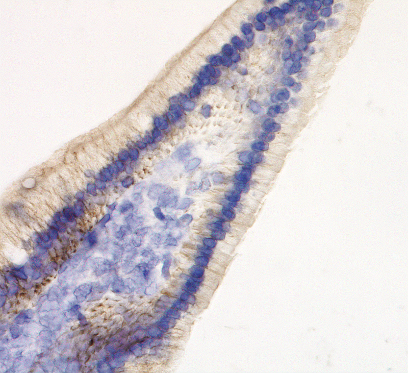
Duodenum - normal tissue. Keratins 8 and 18 immunohistochemistry |
J:94320 |
|
Image Caption:Duodenum - normal tissue. There is mild to moderate cytoplasmic labeling of enterocytes for keratins 8 and 18 (tissues fixed in Fekete's solution, 1:25 dilution, trypsin digestion).
|
|
|
Image ID:690 |
|
Source of Image:Mikaelian I |
|
Pathologist:Mikaelian I |
|
Method / Stain:IHC for keratin 8 and 18 |
|
|
|
| MTB ID |
Tumor Name |
Organ(s) Affected |
Treatment Type |
Agents |
Strain Name |
Strain Sex |
Reproductive Status |
Tumor Frequency |
Age at Necropsy |
Description |
Reference |
| MTB:25809 |
Mammary gland adenocarcinoma - type P |
Mammary gland |
None (spontaneous) |
|
|
Female |
reproductive status not specified |
observed |
256 days |
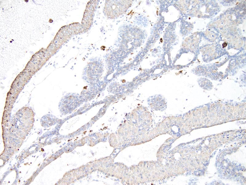
Mammary adenocarcinoma type P, immunohistochemistry for keratins 8/18 |
J:94320 |
|
Image Caption:Type P mammary adenocarcinoma, immunohistochemistry for keratins 8/18: cytoplasmic labeling for keratins 8/18 is moderate for the large acidophilic cells and weak for most of the small basophilic cells. In addition, there is a continuous row of unlabeled basal cells with a myoepithelial phenotype. Cytoplasmic staining of macrophages in the stroma and in the ducts is the result of endogenous peroxidase activity which could not be totally quenched.
|
|
|
Image ID:1191 |
|
Source of Image:Mikaelian I |
|
Pathologist:Mikaelian I |
|
Method / Stain:IHC for keratin 8 and 18 |
|
|
|
| MTB ID |
Tumor Name |
Organ(s) Affected |
Treatment Type |
Agents |
Strain Name |
Strain Sex |
Reproductive Status |
Tumor Frequency |
Age at Necropsy |
Description |
Reference |
| MTB:26706 |
Mammary gland adenocarcinoma |
Mammary gland |
None (spontaneous) |
|
|
Female |
reproductive status not specified |
observed |
unknown |
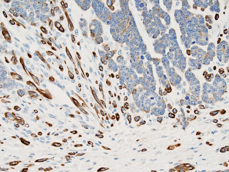
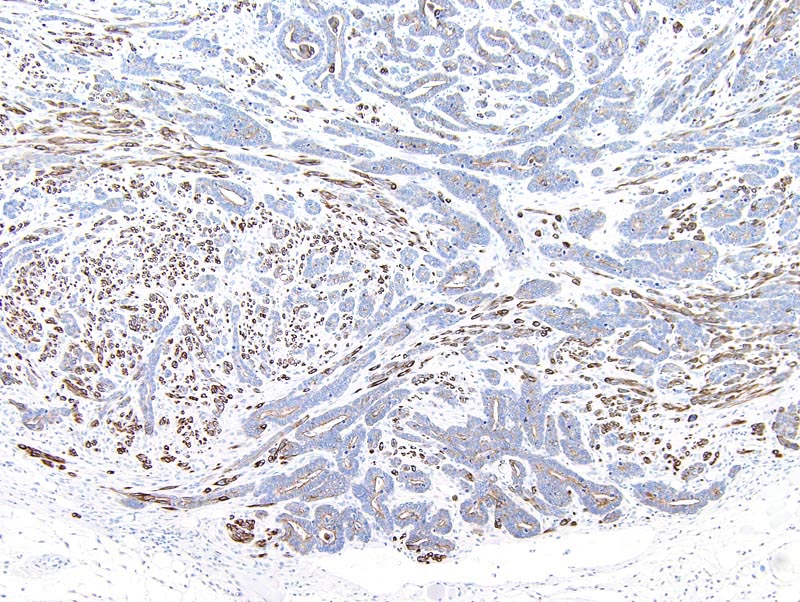
Adenocarcinoma with epithelial to mesenchymal transition, immunohistochemistry for keratins 8/18, mammary gland |
J:94320 |
|
Image Caption:Mammary adenocarcinoma with epithelial to mesenchymal transition, immunohistochemistry for keratins 8/18: there is strong cytoplasmic labeling of most spindloid cells for keratins 8/18. Cells with a luminal phenotype located in neoplastic glands show moderate apical labeling for keratins 8/18.
|
|
|
Image ID:1368 |
|
Source of Image:Fancher K |
|
Pathologist:Mikaelian I |
|
Method / Stain:IHC for keratin 8 and 18 |
|
|
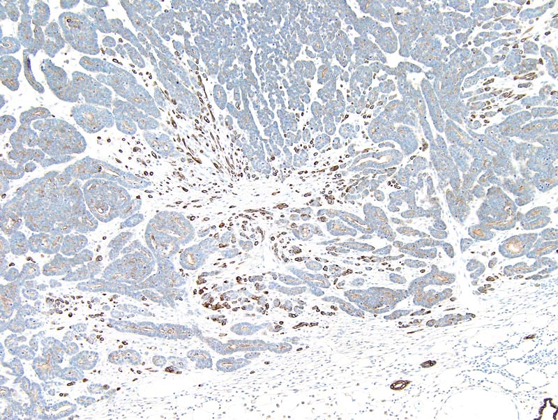
Image Caption:Mammary adenocarcinoma with epithelial to mesenchymal transition, immunohistochemistry for keratins 8/18: there is strong cytoplasmic labeling of most spindloid cells for keratins 8/18. Cells with a luminal phenotype located in neoplastic glands show moderate labeling for keratins 8/18.
|
|
|
Image ID:1363 |
|
Source of Image:Fancher K |
|
Pathologist:Mikaelian I |
|
Method / Stain:IHC for keratin 8 and 18 |
|
|
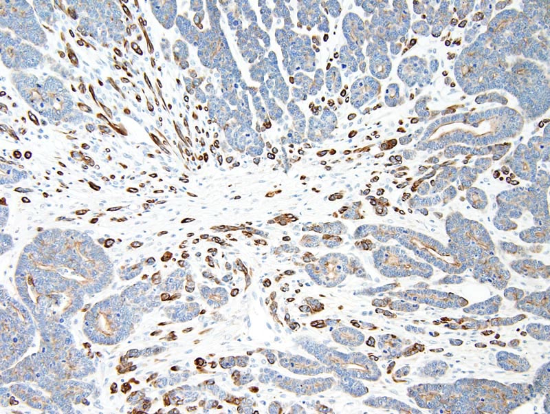
Image Caption:Mammary adenocarcinoma with epithelial to mesenchymal transition, immunohistochemistry for keratins 8/18: there is strong cytoplasmic labeling of most spindloid cells for keratins 8/18. Cells with a luminal phenotype located in neoplastic glands show moderate apical labeling for keratins 8/18.
|
|
|
Image ID:1366 |
|
Source of Image:Fancher K |
|
Pathologist:Mikaelian I |
|
Method / Stain:IHC for keratin 8 and 18 |
|
|
Image Caption:Mammary adenocarcinoma with epithelial to mesenchymal transition, immunohistochemistry for keratins 8/18: there is strong cytoplasmic labeling of most spindloid cells for keratins 8/18. Cells with a luminal phenotype located in neoplastic glands show moderate apical labeling for keratins 8/18.
|

|
|
Image ID:1365 |
|
Source of Image:Fancher K |
|
Pathologist:Mikaelian I |
|
Method / Stain:IHC for keratin 8 and 18 |
|
|
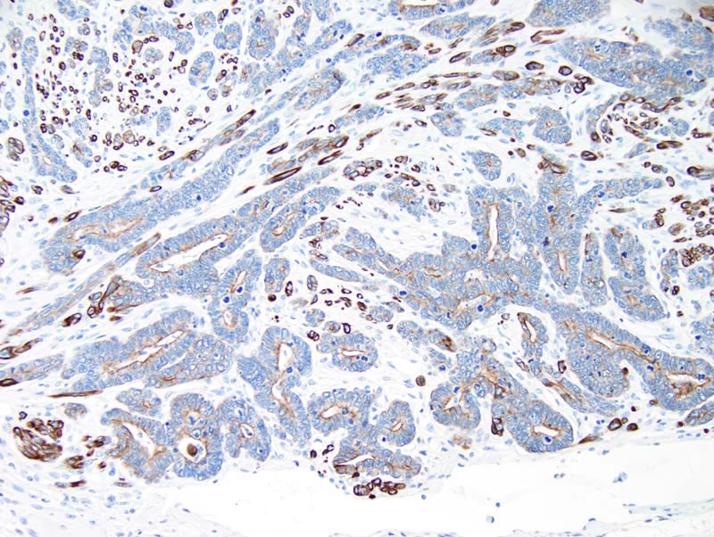
Image Caption:Mammary adenocarcinoma with epithelial to mesenchymal transition, immunohistochemistry for keratins 8/18: there is strong cytoplasmic labeling of most spindloid cells for keratins 8/18. Cells with a luminal phenotype located in neoplastic glands show moderate apical labeling for keratins 8/18. A few cells with a basal location and located in neoplastic glands also display moderate to strong cytoplasmic labeling for keratins 8/18.
|
|
|
Image ID:1364 |
|
Source of Image:Fancher K |
|
Pathologist:Mikaelian I |
|
Method / Stain:IHC for keratin 8 and 18 |
|
|
Image Caption:Mammary adenocarcinoma with epithelial to mesenchymal transition, immunohistochemistry for keratins 8/18: there is strong cytoplasmic labeling of most spindloid cells for keratins 8/18. Cells with a luminal phenotype located in neoplastic glands show moderate apical labeling for keratins 8/18.
|
|
|
Image ID:1367 |
|
Source of Image:Fancher K |
|
Pathologist:Mikaelian I |
|
Method / Stain:IHC for keratin 8 and 18 |
|
|
|
| MTB ID |
Tumor Name |
Organ(s) Affected |
Treatment Type |
Agents |
Strain Name |
Strain Sex |
Reproductive Status |
Tumor Frequency |
Age at Necropsy |
Description |
Reference |
| MTB:26716 |
Mammary gland adenocarcinoma - solid |
Mammary gland |
None (spontaneous) |
|
|
Female |
reproductive status not specified |
observed |
unknown |
Solid adenocarcinoma, immunohistochemistry for keratins 8/18, mammary gland |
J:94320 |
|
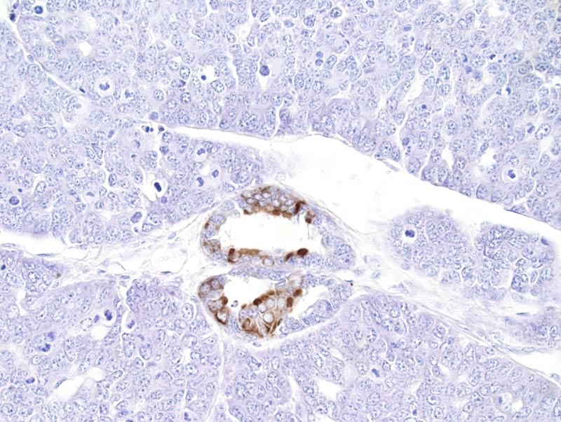
Image Caption:Solid adenocarcinoma, immunohistochemistry for keratins 8/18: a small number of neoplastic cells with a luminal phenotype (predominantly) and rare cells with a basal phenotype located in an area of glandular differentiation show moderate cytoplasmic labeling for keratins 8/18. Other neoplastic cells are not labeled for keratins 8/18.
|
|
|
Image ID:1392 |
|
Source of Image:Fancher K |
|
Pathologist:Mikaelian I |
|
Method / Stain:IHC for keratin 8 and 18 |
|
|