|
| MTB ID |
Tumor Name |
Organ(s) Affected |
Treatment Type |
Agents |
Strain Name |
Strain Sex |
Reproductive Status |
Tumor Frequency |
Age at Necropsy |
Description |
Reference |
| MTB:27721 |
Eye - Cornea normal tissue (control) |
Eye - Cornea |
None (spontaneous) |
|
|
Unspecified |
reproductive status not specified |
not applicable |
unknown |
Eye - Cornea - Normal tissue. |
J:94313 |
|
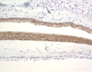
Image Caption:Eye, cornea (C) and conjunctiva of the nictitans (N) - Normal tissue: There is strong diffuse labeling of the full-thickness of the corneal and conjunctival epithelia by a monoclonal antibody raised against human keratin 5.
|
|
|
Image ID:272 |
|
Source of Image:Mikaelian I |
|
Pathologist:Mikaelian I |
|
Method / Stain:IHC for keratin 5 and 6 |
|
|
|
| MTB ID |
Tumor Name |
Organ(s) Affected |
Treatment Type |
Agents |
Strain Name |
Strain Sex |
Reproductive Status |
Tumor Frequency |
Age at Necropsy |
Description |
Reference |
| MTB:27720 |
Eye - Eyelid normal tissue (control) |
Eye - Eyelid |
None (spontaneous) |
|
|
Unspecified |
reproductive status not specified |
not applicable |
|
Eye - eyelid - Normal tissue. |
J:94313 |
|
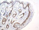
Image Caption:Eye - Normal tissue. Cross-section of the eyelid with the conjunctiva (C) and the epidermis (E): there is strong labeling of the basal layers of the epidermis and the conjunctiva by a monoclonal antibody raised against human keratin 5. The outer root sheath of hair follicles and reserve cells of sebaceous glands, as well as a few suprabasal epidermal and conjunctival cells are also labeled.
|
|
|
Image ID:273 |
|
Source of Image:Mikaelian I |
|
Pathologist:Mikaelian I |
|
Method / Stain:IHC for keratin 5 and 6 |
|
|
|
| MTB ID |
Tumor Name |
Organ(s) Affected |
Treatment Type |
Agents |
Strain Name |
Strain Sex |
Reproductive Status |
Tumor Frequency |
Age at Necropsy |
Description |
Reference |
| MTB:27687 |
Preputial/Clitoral gland (sex not specified) normal tissue (control) |
Preputial/Clitoral gland (sex not specified) |
None (spontaneous) |
|
|
Unspecified |
reproductive status not specified |
not applicable |
|
Preputial gland - . |
J:94313 |
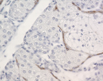
Image Caption:Preputial gland - Normal tissue:
There is moderate labeling of a single layer of cells located between reserve cells and fully differentiated sebocytes in the ducts of the preputial gland.
|
|
|
Image ID:271 |
|
Source of Image:Mikaelian I |
|
Pathologist:Mikaelian I |
|
Method / Stain:IHC for keratin 5 and 6 |
|
|