|
| MTB ID |
Tumor Name |
Organ(s) Affected |
Treatment Type |
Agents |
Strain Name |
Strain Sex |
Reproductive Status |
Tumor Frequency |
Age at Necropsy |
Description |
Reference |
| MTB:27717 |
Adrenal gland - Medulla normal tissue (control) |
Adrenal gland - Medulla |
None (spontaneous) |
|
|
Unspecified |
reproductive status not specified |
not applicable |
35 days |
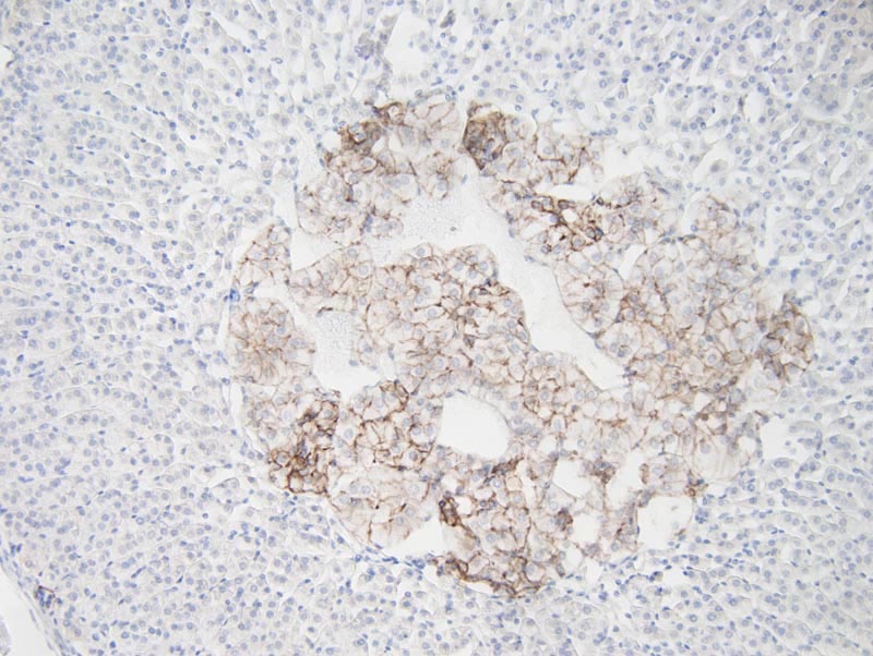
Adrenal gland - Medulla - Normal tissue. NCAM (CD56) immunohistochemistry. |
J:94320 |
|
Image Caption:Adrenal gland - Medulla - normal tissue. There is strong intercellular labeling of the cells of the adrenal medulla for NCAM (antibody diluted at 1:650, tissue fixed in Fekete's solution, heat-mediated antigen retrieval in citrate buffer at pH=6.0).
|
|
|
Image ID:542 |
|
Source of Image:Mikaelian I |
|
Pathologist:Mikaelian I |
|
Method / Stain:IHC for NCAM (CD56) |
|
|
|
| MTB ID |
Tumor Name |
Organ(s) Affected |
Treatment Type |
Agents |
Strain Name |
Strain Sex |
Reproductive Status |
Tumor Frequency |
Age at Necropsy |
Description |
Reference |
| MTB:27644 |
Eye - Retina normal tissue (control) |
Eye - Retina |
None (spontaneous) |
|
|
Unspecified |
reproductive status not specified |
not applicable |
35 days |
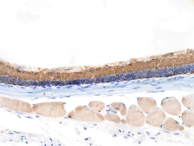
Eye - Retina - Normal tissue. NCAM (CD56) immunohistochemistry |
J:94320 |
|
Image Caption:Retina - normal tissue. There is strong labeling of ganglion cell layer and of the internal plexiform layer for NCAM (CD56) (antibody diluted at 1:650, tissue fixed in Fekete's solution, heat-mediated antigen retrieval in citrate buffer at pH=6.0). There is also severe retinal degeneration, a common lesion in C57BL6/J mice. Labeling of skeletal muscle fibers is interpreted as an artifact.
|
|
|
Image ID:600 |
|
Source of Image:Mikaelian I |
|
Pathologist:Mikaelian I |
|
Method / Stain:IHC for NCAM (CD56) |
|
|
|
| MTB ID |
Tumor Name |
Organ(s) Affected |
Treatment Type |
Agents |
Strain Name |
Strain Sex |
Reproductive Status |
Tumor Frequency |
Age at Necropsy |
Description |
Reference |
| MTB:27651 |
Intestine - Large Intestine - Colon normal tissue (control) |
Intestine - Large Intestine - Colon |
None (spontaneous) |
|
|
Unspecified |
reproductive status not specified |
not applicable |
35 days |
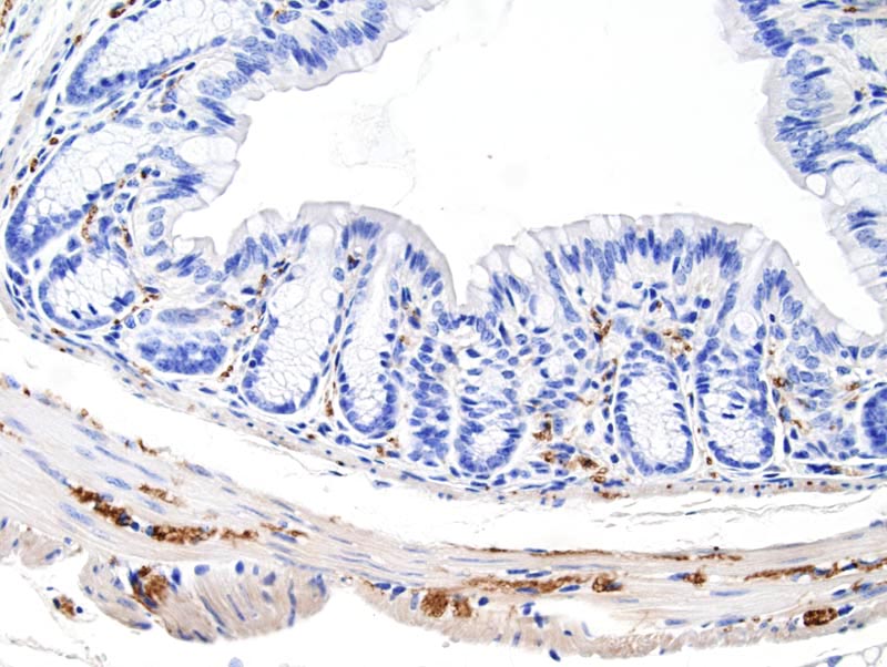
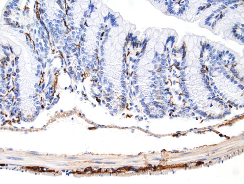
Intestine - Large Intestine - Colon -Normal tissue. NCAM (CD56) immunohistochemistry |
J:94320 |
|
Image Caption:Colon - normal tissue. There is strong labeling of myenteric plexuses and of the axones of intestinal nerves for NCAM (antibody diluted at 1:650, tissue fixed in 4% paraformaldehyde). There is mild background labeling of smooth muscle fibers of the wall of the colon. Unlike tissues undergoing heat-mediated antigen retrieval in citrate buffer, there is no artifactual staining of epithelial cells in the colon.
|
|
|
Image ID:664 |
|
Source of Image:Mikaelian I |
|
Pathologist:Mikaelian I |
|
Method / Stain:IHC for NCAM (CD56) |
|
|
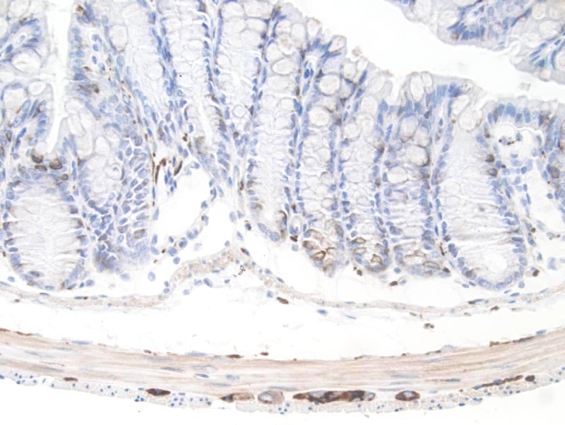
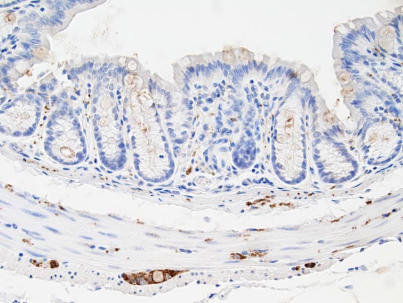
Image Caption:Colon - normal tissue. There is strong labeling of myenteric plexuses of the axones of intestinal nerves for NCAM (antibody diluted at 1:650, tissue fixed in Fekete's solution, heat-mediated antigen retrieval in citrate buffer at pH=6.0). There is mild background labeling of smooth muscle fibers of the wall of the colon as well as of scattered cells of the colon.
|
|
|
Image ID:663 |
|
Source of Image:Mikaelian I |
|
Pathologist:Mikaelian I |
|
Method / Stain:IHC for NCAM (CD56) |
|
|
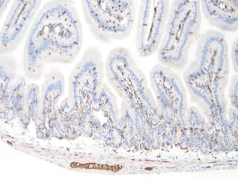
Image Caption:Colon - normal tissue. There is strong labeling of myenteric plexuses of the axones of intestinal nerves for NCAM (antibody diluted at 1:650, tissue fixed in Fekete's solution). There is mild artifactual staining of smooth muscle fibers of the colonic wall.
|
|
|
Image ID:662 |
|
Source of Image:Mikaelian I |
|
Pathologist:Mikaelian I |
|
Method / Stain:IHC for NCAM (CD56) |
|
|
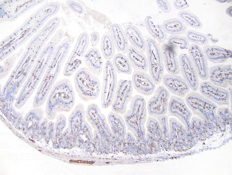
Image Caption:Colon - normal tissue. There is strong labeling of myenteric plexuses and of the axones of intestinal nerves for NCAM (antibody diluted at 1:650, tissue fixed in 4% paraformaldehyde). There is mild background staining of smooth muscle fibers of the wall of the colon and of goblet cells. Artifactual staining of goblet cells is less intense for tissues fixed in 4% paraformaldehyde than for tissues fixed in Fekete's solution.
|
|
|
Image ID:665 |
|
Source of Image:Mikaelian I |
|
Pathologist:Mikaelian I |
|
Method / Stain:IHC for NCAM (CD56) |
|
|
|
| MTB ID |
Tumor Name |
Organ(s) Affected |
Treatment Type |
Agents |
Strain Name |
Strain Sex |
Reproductive Status |
Tumor Frequency |
Age at Necropsy |
Description |
Reference |
| MTB:27657 |
Intestine - Small Intestine - Duodenum normal tissue (control) |
Intestine - Small Intestine - Duodenum |
None (spontaneous) |
|
|
Unspecified |
reproductive status not specified |
not applicable |
35 days |
Duodenum - normal tissue. NCAM (CD56) immunohistochemistry |
J:94320 |
|
Image Caption:Duodenum - normal tissue. There is strong labeling of myenteric plexuses and of the axones of intestinal nerves for NCAM (antibody diluted at 1:650, tissue fixed in Fekete's solution, heat-mediated antigen retrieval in citrate buffer at pH=6.0). In addition, there is strong artifactual staining of all goblet cells.
|
|
|
Image ID:692 |
|
Source of Image:Mikaelian I |
|
Pathologist:Mikaelian I |
|
Method / Stain:IHC for NCAM (CD56) |
|
|
Image Caption:Duodenum - normal tissue. There is strong labeling of myenteric plexuses and of the axones of intestinal nerves for NCAM (antibody diluted at 1:650, tissue fixed in Fekete's solution, heat-mediated antigen retrieval in citrate buffer at pH=6.0). In addition, there is strong artifactual staining of all goblet cells.
|
|
|
Image ID:691 |
|
Source of Image:Mikaelian I |
|
Pathologist:Mikaelian I |
|
Method / Stain:IHC for NCAM (CD56) |
|
|
|
| MTB ID |
Tumor Name |
Organ(s) Affected |
Treatment Type |
Agents |
Strain Name |
Strain Sex |
Reproductive Status |
Tumor Frequency |
Age at Necropsy |
Description |
Reference |
| MTB:27674 |
Nerve normal tissue (control) |
Nerve |
None (spontaneous) |
|
|
Unspecified |
reproductive status not specified |
not applicable |
35 days |
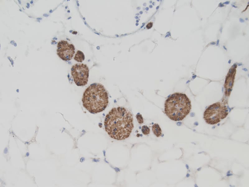
Nerve - Normal tissue. NCAM (CD56) immunohistochemistry |
J:94320 |
|
Image Caption:Subcutaneous adipose tissue - normal tissue. There is strong labeling of nerves for NCAM (antibody diluted at 1:650, tissue fixed in Fekete's solution, heat-mediated antigen retrieval in citrate buffer at pH=6.0).
|
|
|
Image ID:736 |
|
Source of Image:Mikaelian I |
|
Pathologist:Mikaelian I |
|
Method / Stain:IHC for NCAM (CD56) |
|
|