|
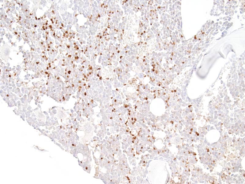
Image Caption:Bone marrow - normal tissue. There is strong nuclear immunolabeling of numerous cells scattered throughout the bone marrow (antibody diluted at 1:100, tissues fixed in 4% paraformaldehyde and decalcified for 2 hours, heat-mediated antigen retrieval in citrate buffer at pH=6.0). The tissue was decalcified, which has altered its' cytologic features.
|
|
|
Image ID:558 |
|
Source of Image:Mikaelian I |
|
Pathologist:Mikaelian I |
|
Method / Stain:IHC for p15 [Cdkn2b] [INK 4b] |
|
|
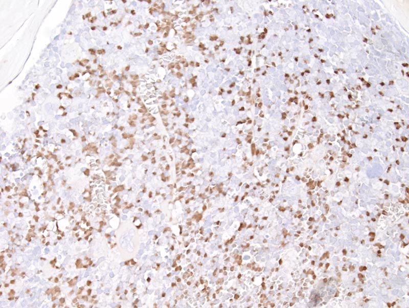
Image Caption:Bone marrow - normal tissue. There is strong nuclear immunolabeling of numerous cells scattered throughout the bone marrow (antibody diluted at 1:100, tissues fixed in 10% neutral buffered formalin and decalcified for 2 hours). The tissue was decalcified, which has altered its' cytologic features.
|
|
|
Image ID:557 |
|
Source of Image:Mikaelian I |
|
Pathologist:Mikaelian I |
|
Method / Stain:IHC for p15 [Cdkn2b] [INK 4b] |
|
|
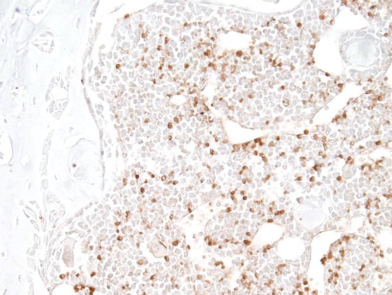
Image Caption:Bone marrow - normal tissue. There is strong nuclear immunolabeling of numerous cells scattered throughout the bone marrow (antibody diluted at 1:100, tissues fixed in Fekete's solution and decalcified for 2 hours). The tissue was decalcified which has altered its' cytologic features.
|
|
|
Image ID:556 |
|
Source of Image:Mikaelian I |
|
Pathologist:Mikaelian I |
|
Method / Stain:IHC for p15 [Cdkn2b] [INK 4b] |
|
|
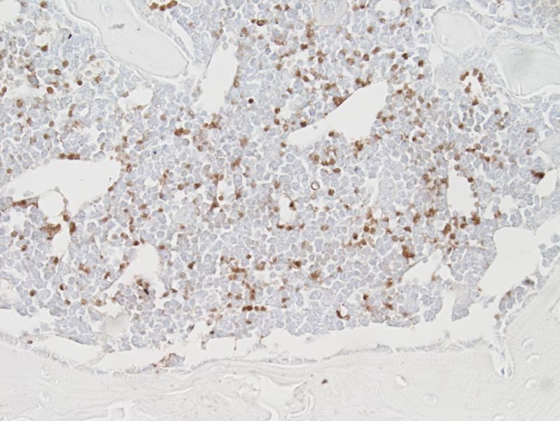
Image Caption:Bone marrow - normal tissue. There is strong nuclear immunolabeling of numerous cells scattered throughout the bone marrow (antibody diluted at 1:100, tissues fixed in Fekete's solution and decalcified for 2 hours). The tissue was decalcified which has altered its' cytologic features.
|
|
|
Image ID:555 |
|
Source of Image:Mikaelian I |
|
Pathologist:Mikaelian I |
|
Method / Stain:IHC for p15 [Cdkn2b] [INK 4b] |
|