|
| MTB ID |
Tumor Name |
Organ(s) Affected |
Treatment Type |
Agents |
Strain Name |
Strain Sex |
Reproductive Status |
Tumor Frequency |
Age at Necropsy |
Description |
Reference |
| MTB:27638 |
Epididymis normal tissue (control) |
Epididymis |
None (spontaneous) |
|
|
Male |
reproductive status not specified |
not applicable |
35 days |
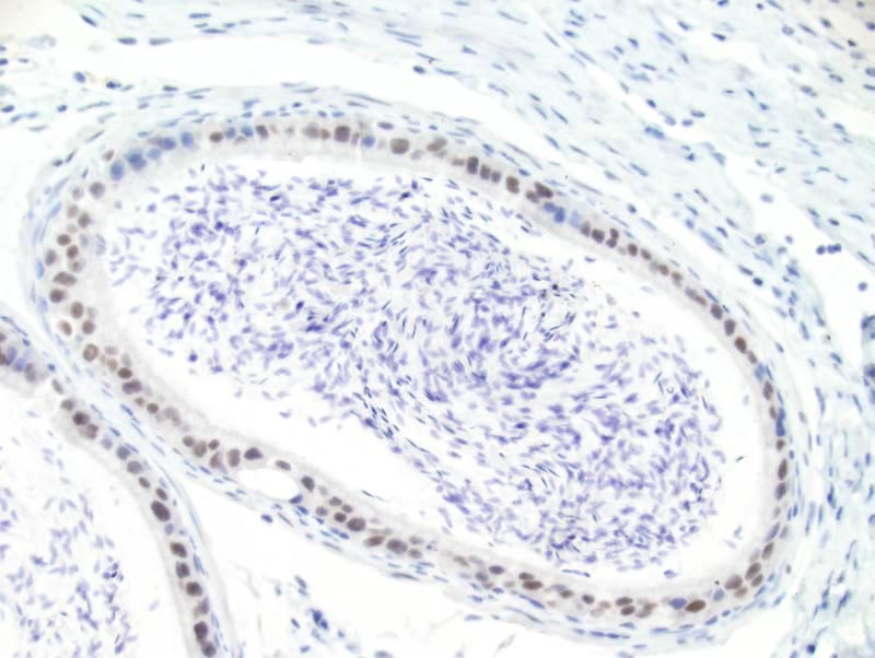
Epididymis - Normal tissue. Androgen receptor (AR) immunohistochemistry. |
J:94320 |
|
Image Caption:Epididymis - normal tissue. There is moderate to strong nuclear labeling for androgen receptor of the epithelial cells of the epididymis (antibody diluted at 1:100, tissue fixed in 4% paraformaldehyde, heat-mediated antigen retrieval in citrate buffer at pH=6.0).
|
|
|
Image ID:567 |
|
Source of Image:Mikaelian I |
|
Pathologist:Mikaelian I |
|
Method / Stain:IHC for androgen receptor (AR) |
|
|
|
| MTB ID |
Tumor Name |
Organ(s) Affected |
Treatment Type |
Agents |
Strain Name |
Strain Sex |
Reproductive Status |
Tumor Frequency |
Age at Necropsy |
Description |
Reference |
| MTB:27689 |
Prostate gland normal tissue (control) |
Prostate gland |
None (spontaneous) |
|
|
Male |
reproductive status not specified |
not applicable |
35 days |
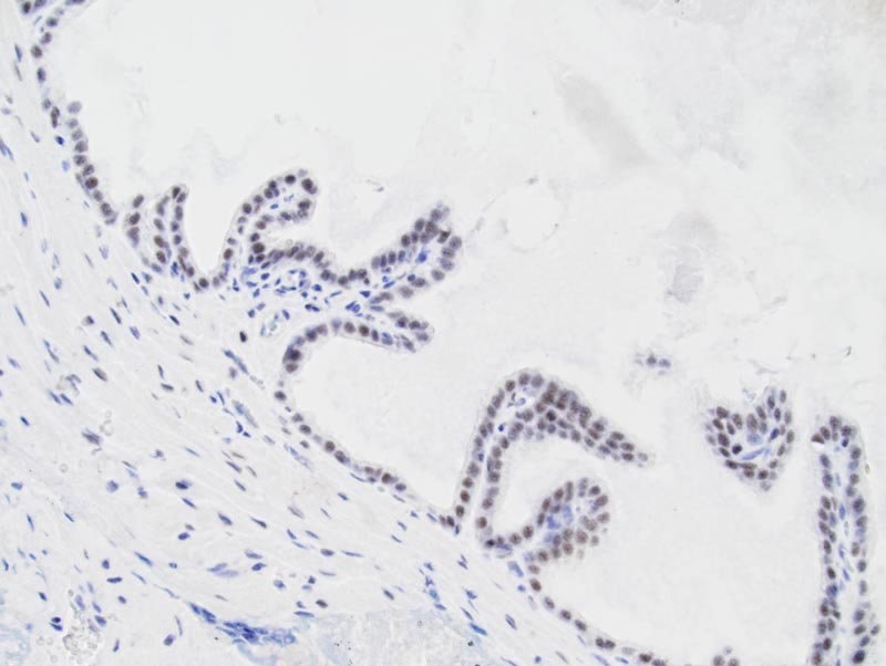
Prostate gland - Normal tissue. Androgen receptor (AR) immunohistochemistry. |
J:94320 |
|
Image Caption:Prostate - normal tissue. There is moderate nuclear labeling for androgen receptor of the epithelial cells of the prostate (antibody diluted at 1:500, tissue fixed in IHC Zinc fixative, heat-mediated antigen retrieval in citrate buffer at pH=6.0).
|
|
|
Image ID:569 |
|
Source of Image:Mikaelian I |
|
Pathologist:Mikaelian I |
|
Method / Stain:IHC for androgen receptor (AR) |
|
|
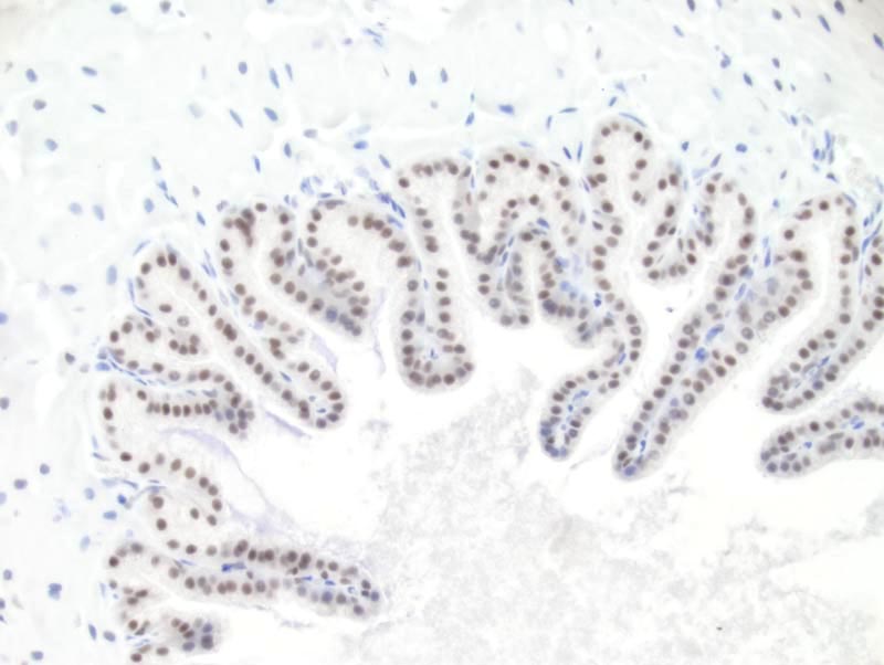
Image Caption:Prostate - normal tissue. There is moderate to strong nuclear labeling for androgen receptor of the epithelial cells of the prostate (antibody diluted at 1:100, tissue fixed in 4% paraformaldehyde, heat-mediated antigen retrieval in citrate buffer at pH=6.0).
|
|
|
Image ID:568 |
|
Source of Image:Mikaelian I |
|
Pathologist:Mikaelian I |
|
Method / Stain:IHC for androgen receptor (AR) |
|
|
|
| MTB ID |
Tumor Name |
Organ(s) Affected |
Treatment Type |
Agents |
Strain Name |
Strain Sex |
Reproductive Status |
Tumor Frequency |
Age at Necropsy |
Description |
Reference |
| MTB:60944 |
Prostate gland adenocarcinoma |
Prostate gland |
Genetic Modification |
|
|
Male |
reproductive status not specified |
observed |
|
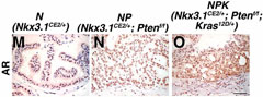
prostate tumor; comparision of tumors from "NPK" and "NP" mice and normal prostate tissue from "N" mice; MRI, H&E, immunohistochemistry, & immunofluorescence |
J:200917 |
|
Image Caption:Fig. 2M-O "Immunohistochemical staining for AR."
|
|
|
Image ID:5178 |
|
Source of Image:Aytes A |
|
Pathologist:Aytes A |
|
Method / Stain:IHC for androgen receptor |
|
|
|
| MTB ID |
Tumor Name |
Organ(s) Affected |
Treatment Type |
Agents |
Strain Name |
Strain Sex |
Reproductive Status |
Tumor Frequency |
Age at Necropsy |
Description |
Reference |
| MTB:60973 |
Prostate gland adenocarcinoma |
Prostate gland |
Genetic Modification |
tamoxifen (for inducing expression from mutant alleles) |
|
Male |
reproductive status not specified |
~72 |
|
prostate tumor; comparision of tumors from "NPK" and "NP" mice and normal prostate tissue from "N" mice; MRI, H&E, immunohistochemistry, & immunofluorescence |
J:200917 |
|
Image Caption:Fig. 2M-O "Immunohistochemical staining for AR."
|
|
|
|
Image ID:5178 |
|
Source of Image:Aytes A |
|
Pathologist:Aytes A |
|
Method / Stain:IHC for androgen receptor |
|
|
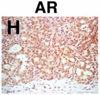
Image Caption:Fig. 4H Immunostaining for androgen receptor in a primary prostate tumor from an NPK mouse.
|
|
|
Image ID:5187 |
|
Source of Image:Aytes A |
|
Pathologist:Aytes A |
|
Method / Stain:IHC for androgen receptor |
|
|
|
| MTB ID |
Tumor Name |
Organ(s) Affected |
Treatment Type |
Agents |
Strain Name |
Strain Sex |
Reproductive Status |
Tumor Frequency |
Age at Necropsy |
Description |
Reference |
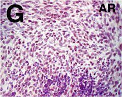
| MTB:60975 |
Prostate gland adenocarcinoma |
Lymph node |
Genetic Modification |
|
|
Male |
reproductive status not specified |
observed |
|
lymph node metastases from prostate tumor; whole slide scan, H&E, and immunohistochemistry for various markers |
J:200917 |
|
Image Caption:SupFig. S4G lumbar lymph node metastasis from an NPK tumor-bearing mouse
|
|
|
Image ID:5220 |
|
Source of Image:Aytes A |
|
Pathologist:Aytes A |
|
Method / Stain:IHC for androgen receptor |
|
|