|
| MTB ID |
Tumor Name |
Organ(s) Affected |
Treatment Type |
Agents |
Strain Name |
Strain Sex |
Reproductive Status |
Tumor Frequency |
Age at Necropsy |
Description |
Reference |
| MTB:27655 |
Intestine - Small Intestine normal tissue (control) |
Intestine - Small Intestine |
None (spontaneous) |
|
|
Unspecified |
reproductive status not specified |
not applicable |
35 days |
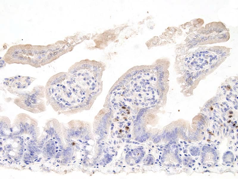
Small intestine - normal tissue. IRF-4 immunohistochemistry |
J:94320 |
|
Image Caption:Small intestine - normal tissue. There is strong nuclear labeling for IRF-4 of plasma cells in the lamina propria of the intestine (1:25 dilution, tissues fixed in 10% neutral buffered formalin, heat-mediated antigen retrieval in 0.1mM EDTA).
|
|
|
Image ID:682 |
|
Source of Image:Mikaelian I |
|
Pathologist:Mikaelian I |
|
Method / Stain:IHC for IRF-4 |
|
|
|
| MTB ID |
Tumor Name |
Organ(s) Affected |
Treatment Type |
Agents |
Strain Name |
Strain Sex |
Reproductive Status |
Tumor Frequency |
Age at Necropsy |
Description |
Reference |
| MTB:27704 |
Thymus normal tissue (control) |
Thymus |
None (spontaneous) |
|
|
Unspecified |
reproductive status not specified |
not applicable |
35 days |
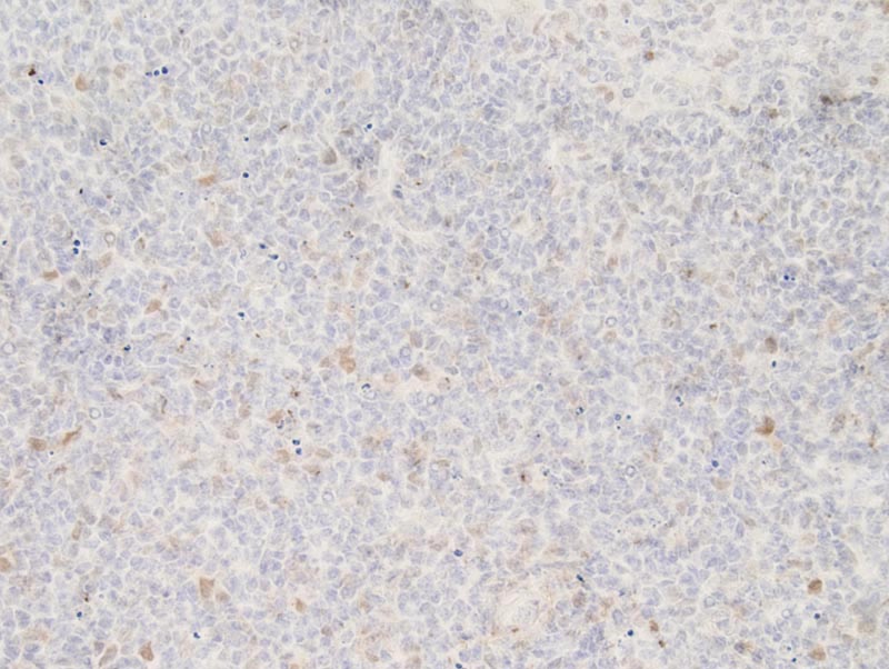
Thymus - Normal tissue. IRF-4 immunohistochemistry |
J:94320 |
|
Image Caption:Thymus - normal tissue. There is mild to moderate nuclear labeling for IRF-4 of lymphocytes scattered in the cortex of the thymus (1:25 dilution, tissues fixed in Fekete's solution, heat-mediated antigen retrieval in 0.1mM EDTA). Poor cytological detail is the result of heat-mediated antigen retrieval in EDTA.
|
|
|
Image ID:901 |
|
Source of Image:Mikaelian I |
|
Pathologist:Mikaelian I |
|
Method / Stain:IHC for IRF-4 |
|
|