|
| MTB ID |
Tumor Name |
Organ(s) Affected |
Treatment Type |
Agents |
Strain Name |
Strain Sex |
Reproductive Status |
Tumor Frequency |
Age at Necropsy |
Description |
Reference |
| MTB:27655 |
Intestine - Small Intestine normal tissue (control) |
Intestine - Small Intestine |
None (spontaneous) |
|
|
Unspecified |
reproductive status not specified |
not applicable |
35 days |
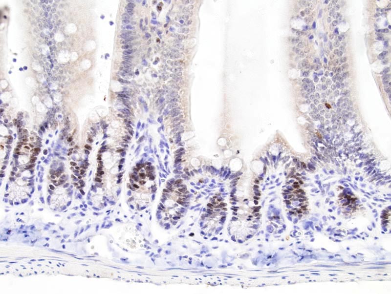
Small intestine - normal tissue. Ki67 immunohistochemistry |
J:94320 |
|
Image Caption:Small intestine - normal tissue. There is strong nuclear labeling for Ki67 of the enterocytes of the crypts (antibody diluted at 1:200, tissues fixed in IHC Zinc fixative (TM), heat-mediated antigen retrieval in citrate buffer at pH=6.0). There is weak background staining of the cytoplasm of the enterocytes of the villi.
|
|
|
Image ID:683 |
|
Source of Image:Mikaelian I |
|
Pathologist:Mikaelian I |
|
Method / Stain:IHC for Ki-67 |
|
|
|
| MTB ID |
Tumor Name |
Organ(s) Affected |
Treatment Type |
Agents |
Strain Name |
Strain Sex |
Reproductive Status |
Tumor Frequency |
Age at Necropsy |
Description |
Reference |
| MTB:48025 |
Pancreas - Islet of Langerhans tumor - neuroendocrine |
Pancreas - Islet of Langerhans |
None (spontaneous) |
|
|
Mixed Population |
reproductive status not specified |
85.71% |
10-12 months |
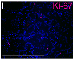
pancreas neuroendocrine tumor (PNET) |
J:176507 |
|
Image Caption:Fig. 3I shows a PNET section stained for Ki-67, pink staining.
|
|
|
Image ID:4761 |
|
Source of Image:Yu R |
|
Pathologist:Yu R |
|
|
|
| MTB ID |
Tumor Name |
Organ(s) Affected |
Treatment Type |
Agents |
Strain Name |
Strain Sex |
Reproductive Status |
Tumor Frequency |
Age at Necropsy |
Description |
Reference |
| MTB:27683 |
Peyer's patch normal tissue (control) |
Peyer's patch |
None (spontaneous) |
|
|
Unspecified |
reproductive status not specified |
not applicable |
35 days |
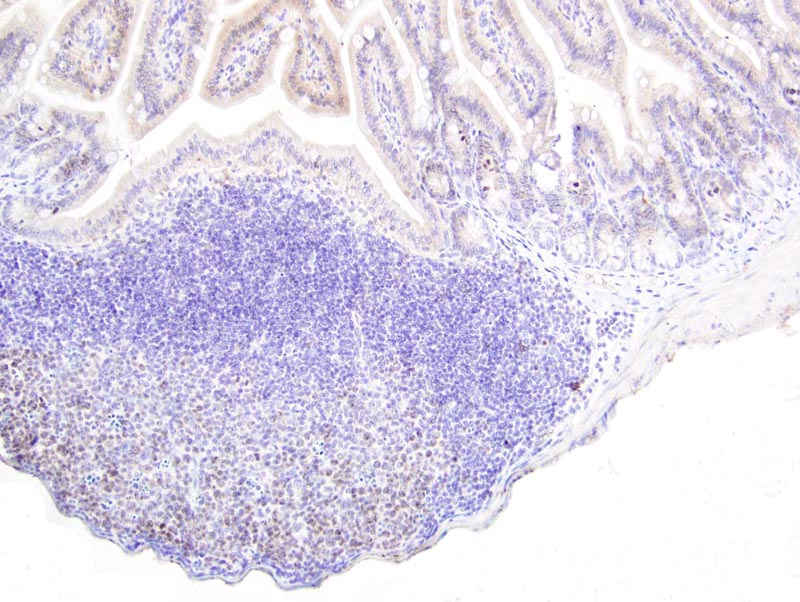
Peyer's patch - Normal tissue. Ki67 immunohistochemistry |
J:94320 |
|
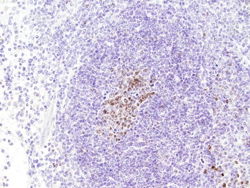
Image Caption:Peyer's patch - normal tissue. There is moderate nuclear labeling for Ki67 in the germinal center of a Peyer's patch (antibody diluted at 1:200, tissues fixed in Bouin's solution, heat-mediated antigen retrieval in citrate buffer at pH=6.0).
|
|
|
Image ID:752 |
|
Source of Image:Mikaelian I |
|
Pathologist:Mikaelian I |
|
Method / Stain:IHC for Ki-67 |
|
|
|
| MTB ID |
Tumor Name |
Organ(s) Affected |
Treatment Type |
Agents |
Strain Name |
Strain Sex |
Reproductive Status |
Tumor Frequency |
Age at Necropsy |
Description |
Reference |
| MTB:27696 |
Spleen normal tissue (control) |
Spleen |
None (spontaneous) |
|
|
Unspecified |
reproductive status not specified |
not applicable |
35 days |
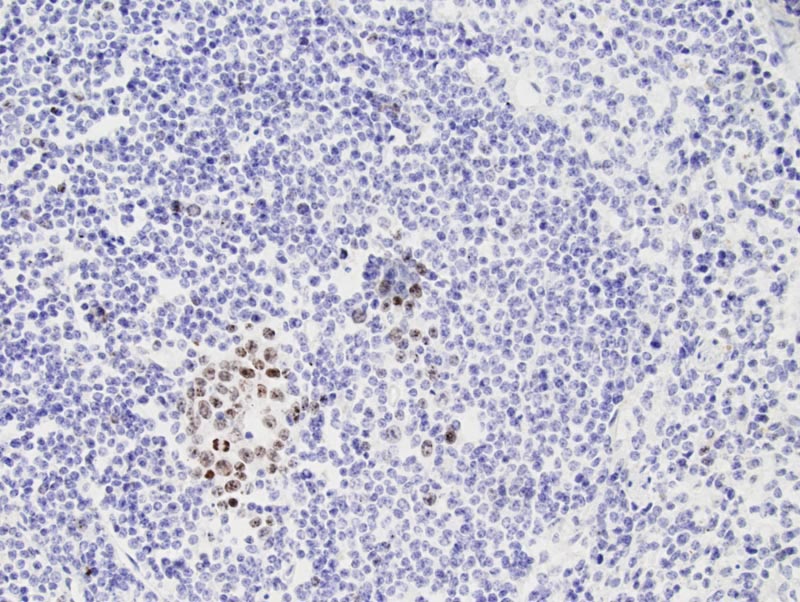
Spleen - Normal tissue. Ki67 immunohistochemistry |
J:94320 |
|
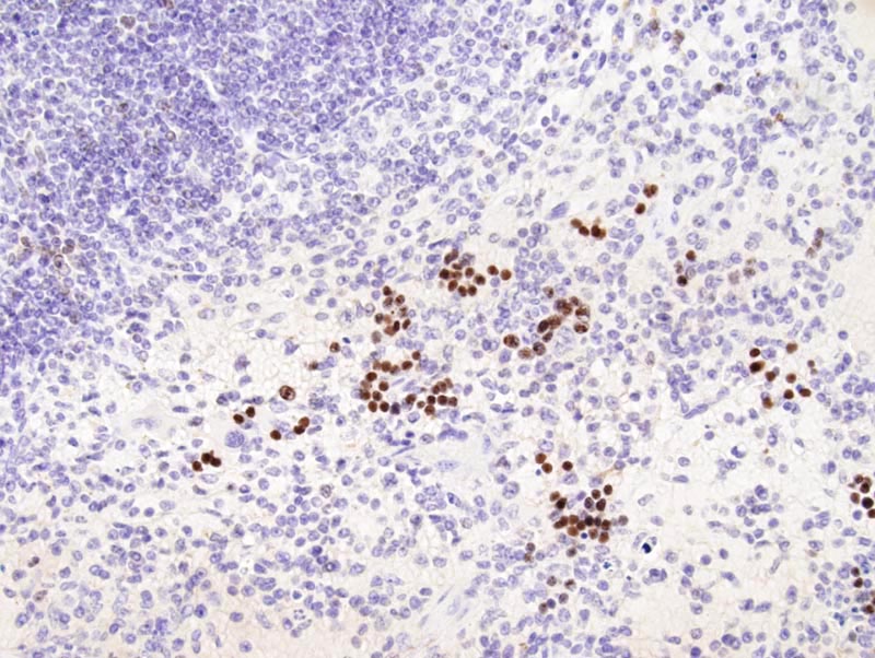
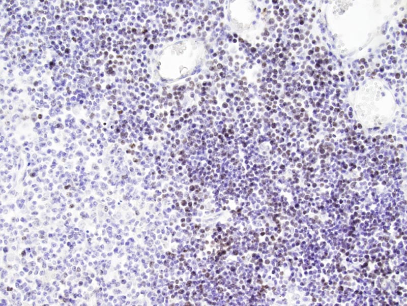
Image Caption:Spleen - normal tissue. There is strong nuclear labeling for Ki67 of lymphocytes of a germinal center of the white pulp of the spleen (antibody diluted at 1:200, tissues fixed in IHC Zinc fixative (TM), heat-mediated antigen retrieval in citrate buffer at pH=6.0).
|
|
|
Image ID:777 |
|
Source of Image:Mikaelian I |
|
Pathologist:Mikaelian I |
|
Method / Stain:IHC for Ki-67 |
|
|
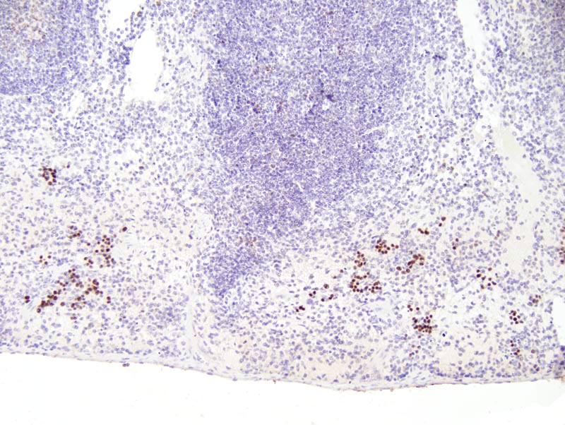
Image Caption:Spleen - normal tissue. There is strong nuclear labeling for Ki67 of lymphocytes of a germinal center of the white pulp of the spleen (antibody diluted at 1:200, tissues fixed in Bouin's solution, heat-mediated antigen retrieval in citrate buffer at pH=6.0).
|
|
|
Image ID:776 |
|
Source of Image:Mikaelian I |
|
Pathologist:Mikaelian I |
|
Method / Stain:IHC for Ki-67 |
|
|
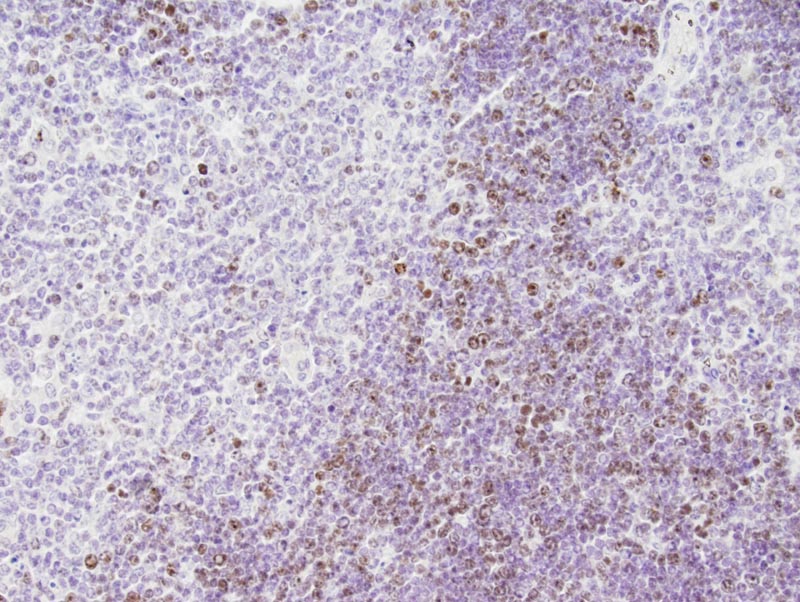
Image Caption:Spleen - normal tissue. There is strong nuclear labeling for Ki67 of periarteriolar lymphocytes (antibody diluted at 1:200, tissues fixed in Bouin's solution, heat-mediated antigen retrieval in citrate buffer at pH=6.0). Labeling is weaker for lymphocytes of the white pulp.
|
|
|
Image ID:775 |
|
Source of Image:Mikaelian I |
|
Pathologist:Mikaelian I |
|
Method / Stain:IHC for Ki-67 |
|
|
Image Caption:Spleen - normal tissue. There is strong nuclear labeling for Ki67 of periarteriolar lymphocytes (antibody diluted at 1:200, tissues fixed in Bouin's solution, heat-mediated antigen retrieval in citrate buffer at pH=6.0).
|
|
|
Image ID:774 |
|
Source of Image:Mikaelian I |
|
Pathologist:Mikaelian I |
|
Method / Stain:IHC for Ki-67 |
|
|
|
| MTB ID |
Tumor Name |
Organ(s) Affected |
Treatment Type |
Agents |
Strain Name |
Strain Sex |
Reproductive Status |
Tumor Frequency |
Age at Necropsy |
Description |
Reference |
| MTB:27704 |
Thymus normal tissue (control) |
Thymus |
None (spontaneous) |
|
|
Unspecified |
reproductive status not specified |
not applicable |
35 days |
Thymus - Normal tissue. Ki67 immunohistochemistry |
J:94320 |
|
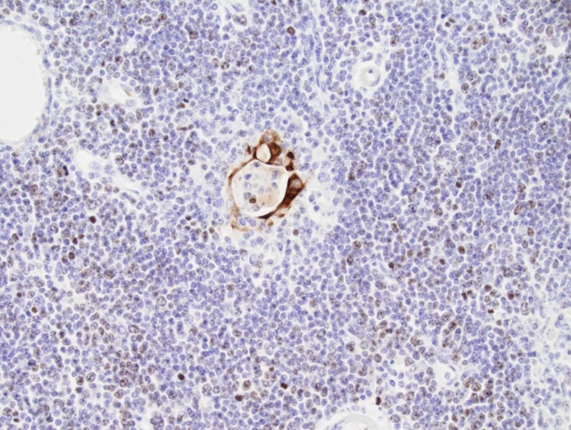
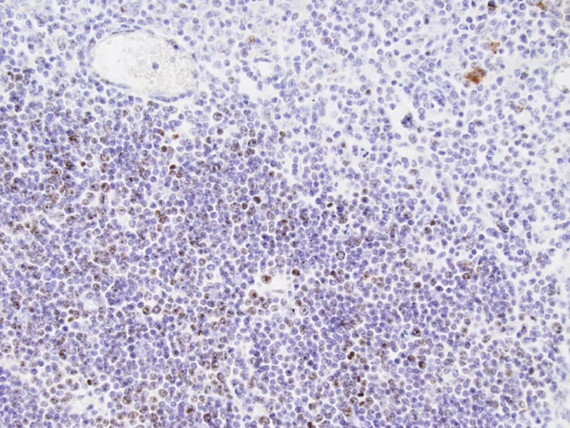
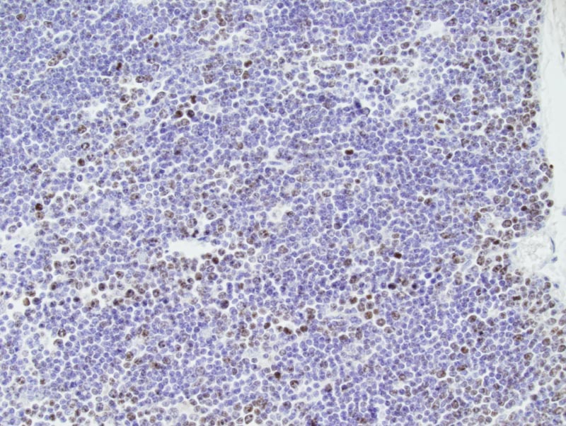
Image Caption:Thymus - normal tissue. There is strong nuclear labeling for Ki67 of most cortical lymphocytes (antibody diluted at 1:200, tissues fixed in Bouin's solution, heat-mediated antigen retrieval in citrate buffer at pH=6.0).
|
|
|
Image ID:903 |
|
Source of Image:Mikaelian I |
|
Pathologist:Mikaelian I |
|
Method / Stain:IHC for Ki-67 |
|
|
Image Caption:Thymus - normal tissue. There is moderate nuclear labeling for Ki67 of about 75% of cortical lymphocytes (antibody diluted at 1:200, tissues fixed in IHC Zinc fixative, heat-mediated antigen retrieval in citrate buffer at pH=6.0). Less nuclei are labeled for tissues fixed in IHC Zinc fixative (TM) than for tissues fixed in Bouin's solution.
|
|
|
Image ID:907 |
|
Source of Image:Mikaelian I |
|
Pathologist:Mikaelian I |
|
Method / Stain:IHC for Ki-67 |
|
|
Image Caption:Thymus - normal tissue. There is moderate nuclear labeling for Ki67 of about 50% of cortical lymphocytes (antibody diluted at 1:100, tissues fixed in Fekete's solution, heat-mediated antigen retrieval in citrate buffer at pH=6.0). Less nuclei are labeled for tissues fixed in Fekete's solution than for tissues fixed in Bouin's solution. In addition, there is strong artifactual staining of the keratinocytes of a Hassal's corpuscule. Strong artifactual staining was also present in the corneal epithelium for tissues fixed in Fekete's solution.
|
|
|
Image ID:906 |
|
Source of Image:Mikaelian I |
|
Pathologist:Mikaelian I |
|
Method / Stain:IHC for Ki-67 |
|
|
Image Caption:Thymus - normal tissue. There is moderate nuclear labeling for Ki67 of about 50% of cortical lymphocytes (antibody diluted at 1:100, tissues fixed in Fekete's solution, heat-mediated antigen retrieval in citrate buffer at pH=6.0). Less nuclei are labeled for tissues fixed in Fekete's solution than for tissues fixed in Bouin's solution.
|
|
|
Image ID:905 |
|
Source of Image:Mikaelian I |
|
Pathologist:Mikaelian I |
|
Method / Stain:IHC for Ki-67 |
|
|
Image Caption:Thymus - normal tissue. There is moderate nuclear labeling for Ki67 of about 50% of cortical lymphocytes (antibody diluted at 1:100, tissues fixed in Fekete's solution, heat-mediated antigen retrieval in citrate buffer at pH=6.0). Less nuclei are labeled for tissues fixed in Fekete's solution than for tissues fixed in Bouin's solution.
|
|
|
Image ID:904 |
|
Source of Image:Mikaelian I |
|
Pathologist:Mikaelian I |
|
Method / Stain:IHC for Ki-67 |
|
|