|
| MTB ID |
Tumor Name |
Organ(s) Affected |
Treatment Type |
Agents |
Strain Name |
Strain Sex |
Reproductive Status |
Tumor Frequency |
Age at Necropsy |
Description |
Reference |
| MTB:27660 |
Leukocyte - Lymphocyte normal tissue (control) |
Lymph node |
None (spontaneous) |
|
|
Unspecified |
reproductive status not specified |
not applicable |
35 days |
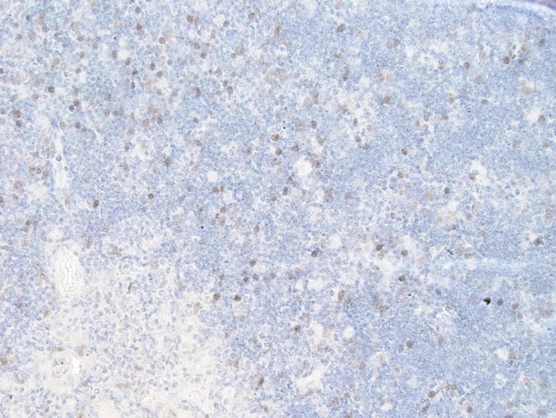
Lymph node lymphocytes - Normal tissue. Cyclin A immunohistochemistry |
J:94320 |
|
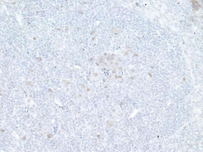
Image Caption:Lymph node - normal tissue. There is weak to moderate nuclear immunolabeling for cyclin A of lymphocytes located at the center of a germinative follicle as well as of a few lymphocytes scattered throughout the B area of this lymphoid follicle (antibody diluted at 1:10, tissue fixed in Fekete's solution, heat-mediated antigen retrieval in 0.1 mM EDTA at pH=7.5). There is a mild background staining.
|
|
|
Image ID:727 |
|
Source of Image:Mikaelian I |
|
Pathologist:Mikaelian I |
|
Method / Stain:IHC for cyclin A |
|
|
|
| MTB ID |
Tumor Name |
Organ(s) Affected |
Treatment Type |
Agents |
Strain Name |
Strain Sex |
Reproductive Status |
Tumor Frequency |
Age at Necropsy |
Description |
Reference |
| MTB:27683 |
Peyer's patch normal tissue (control) |
Peyer's patch |
None (spontaneous) |
|
|
Unspecified |
reproductive status not specified |
not applicable |
35 days |
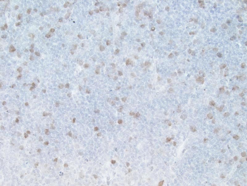
Peyer's patch - Normal tissue. Cyclin A immunohistochemistry |
J:94320 |
|
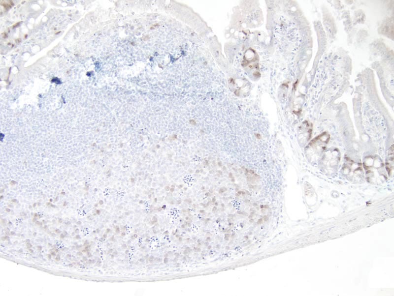
Image Caption:Peyer's patch - normal tissue. There is moderate nuclear immunolabeling for cyclin A of lymphocytes located at the center of a germinative follicle (antibody diluted at 1:10, tissue fixed in Fekete's solution, heat-mediated antigen retrieval in 0.1 mM EDTA at pH=7.5). There is also moderate nuclear immunolabeling of the enterocytes located in the intestinal crypts.
|
|
|
Image ID:751 |
|
Source of Image:Mikaelian I |
|
Pathologist:Mikaelian I |
|
Method / Stain:IHC for cyclin A |
|
|
|
| MTB ID |
Tumor Name |
Organ(s) Affected |
Treatment Type |
Agents |
Strain Name |
Strain Sex |
Reproductive Status |
Tumor Frequency |
Age at Necropsy |
Description |
Reference |
| MTB:27704 |
Thymus normal tissue (control) |
Thymus |
None (spontaneous) |
|
|
Unspecified |
reproductive status not specified |
not applicable |
35 days |
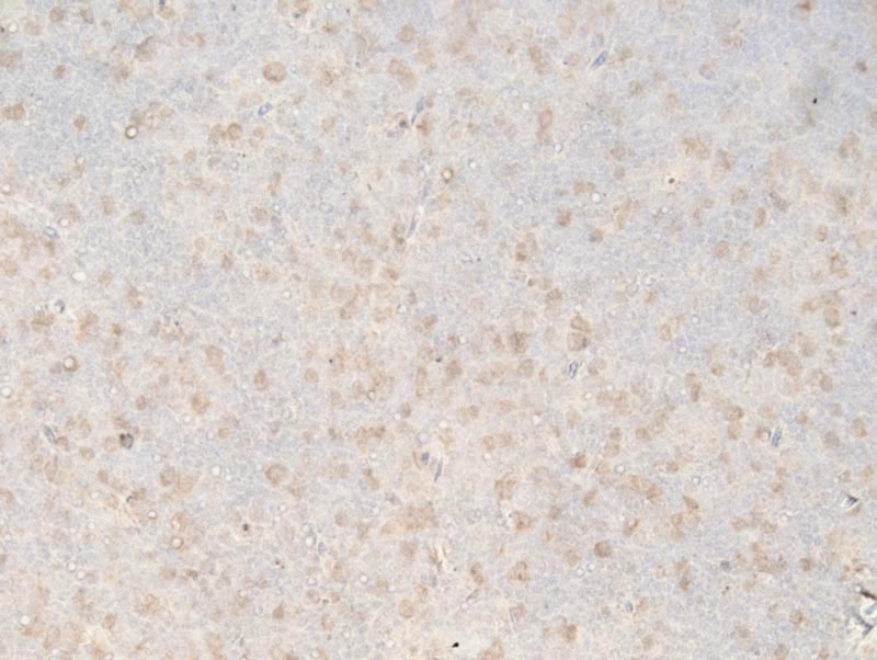
Thymus - Normal tissue. Cyclin A immunohistochemistry |
J:94320 |
|
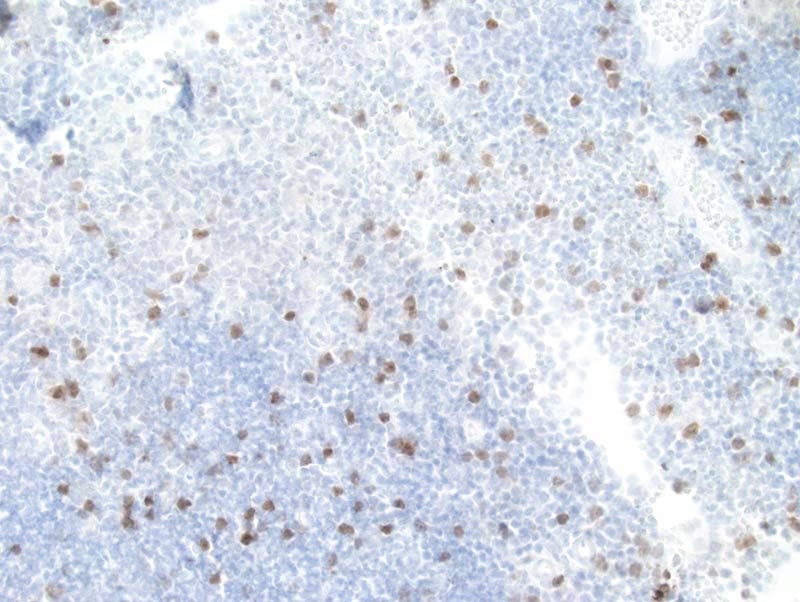
Image Caption:Thymus - normal tissue. There is strong nuclear immunolabeling for cyclin A of thymocytes scattered in the cortex of the thymus (antibody diluted at 1:50, tissue fixed in IHC Zinc Fixative (TM), heat-mediated antigen retrieval in 0.1 mM EDTA at pH=7.5).
|
|
|
Image ID:897 |
|
Source of Image:Mikaelian I |
|
Pathologist:Mikaelian I |
|
Method / Stain:IHC for cyclin A |
|
|
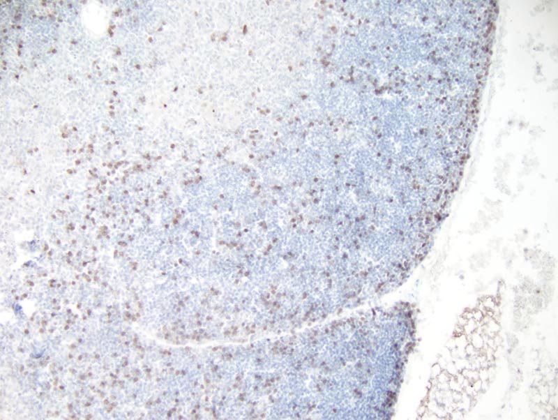
Image Caption:Thymus - normal tissue. There is strong nuclear immunolabeling for cyclin A of thymocytes scattered in the cortex of the thymus (antibody diluted at 1:50, tissue fixed in IHC Zinc Fixative (TM), heat-mediated antigen retrieval in 0.1 mM EDTA at pH=7.5).
|
|
|
Image ID:896 |
|
Source of Image:Mikaelian I |
|
Pathologist:Mikaelian I |
|
Method / Stain:IHC for cyclin A |
|
|
Image Caption:Thymus - normal tissue. There is moderate nuclear immunolabeling for cyclin A of thymocytes scattered in the cortex of the thymus (antibody diluted at 1:10, tissue fixed in 10% neutral buffered formalin, heat-mediated antigen retrieval in 0.1 mM EDTA at pH=7.5).
|
|
|
Image ID:895 |
|
Source of Image:Mikaelian I |
|
Pathologist:Mikaelian I |
|
Method / Stain:IHC for cyclin A |
|
|
Image Caption:Thymus - normal tissue. There is moderate nuclear immunolabeling for cyclin A of thymocytes scattered in the cortex of the thymus (antibody diluted at 1:25, tissue fixed in Fekete's solution, heat-mediated antigen retrieval in 0.1 mM EDTA at pH=7.5).
|
|
|
Image ID:894 |
|
Source of Image:Mikaelian I |
|
Pathologist:Mikaelian I |
|
Method / Stain:IHC for cyclin A |
|
|
Image Caption:Thymus - normal tissue. There is weak to moderate nuclear immunolabeling for cyclin A of thymocytes scattered in the cortex of the thymus (antibody diluted at 1:25, tissue fixed in Bouin's solution, heat-mediated antigen retrieval in 0.1 mM EDTA at pH=7.5). There is a mild to moderate background staining.
|
|
|
Image ID:893 |
|
Source of Image:Mikaelian I |
|
Pathologist:Mikaelian I |
|
Method / Stain:IHC for cyclin A |
|
|