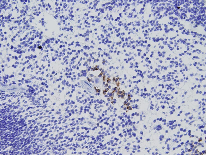
Image Caption:Spleen - normal tissue. There is strong labeling of the plasma membrane and of the cytoplasm of scattered round to dendritic cells of the red pulp (tissue fixed in 4% paraformaldehyde, antibody diluted at 1:25, heat-mediated antigen retrieval in citrate buffer).
NOTE: This pattern of labeling was interpreted as an artifact because L-selectin is normally expressed on peripheral lymphocytes, NK cells, neutrophils, eosinophils, and monocytes while only cells with a monocytic or dendritic phenotype labeled with this antibody.
|
|
|
Image ID:779 |
|
Source of Image:Mikaelian I |
|
Pathologist:Mikaelian I |
|
Method / Stain:IHC for L-selectin (CD62L) |
|
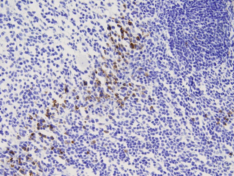
Image Caption:Spleen - normal tissue. There is strong cytoplasmic and plasma membrane labeling of scattered round to dendritic cells located in the red pulp (tissue fixed in 4% paraformaldehyde, antibody diluted at 1:100, heat-mediated antigen retrieval in citrate buffer).
NOTE: This pattern of labeling was interpreted as an artifact because L-selectin is normally expressed on peripheral lymphocytes, NK cells, neutrophils, eosinophils, and monocytes while only cells with a monocytic or dendritic phenotype labeled with this antibody.
|
|
|
Image ID:778 |
|
Source of Image:Mikaelian I |
|
Pathologist:Mikaelian I |
|
Method / Stain:IHC for L-selectin (CD62L) |
|
|