|
| MTB ID |
Tumor Name |
Organ(s) Affected |
Treatment Type |
Agents |
Strain Name |
Strain Sex |
Reproductive Status |
Tumor Frequency |
Age at Necropsy |
Description |
Reference |
| MTB:50789 |
Adrenal gland hyperplasia - spindle cell |
Adrenal gland |
None (spontaneous) |
|
|
Female |
reproductive status not specified |
observed |
877 days |
adrenal cortical spindle cell sarcoma and hyperplasia |
J:122261 |
|
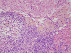
Image Caption:This is a 25x image that is a higher magnification of the right-center area of the 10x image.
|
|
|
Image ID:5022 |
|
Source of Image:Sundberg J |
|
Pathologist:Sundberg J |
|
|
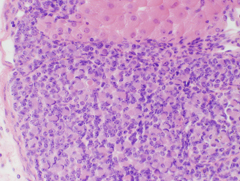
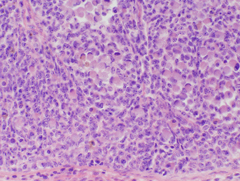
Image Caption:This is a 40x image, 40xa, that is a higher magnification of the left-center area of the 4x image.
|
|
|
Image ID:5023 |
|
Source of Image:Sundberg J |
|
Pathologist:Sundberg J |
|
|
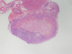
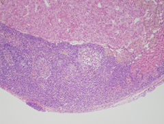
Image Caption:This is a 4x image.
|
|
|
Image ID:5020 |
|
Source of Image:Sundberg J |
|
Pathologist:Sundberg J |
|
|
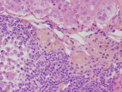
Image Caption:This is a 40x image, 40xc, that is a higher magnification of the center area of the 25x image.
|
|
|
Image ID:5025 |
|
Source of Image:Sundberg J |
|
Pathologist:Sundberg J |
|
|
Image Caption:This is a 10x image that is a higher magnification of the lower-center area of the 4x image.
|
|
|
Image ID:5021 |
|
Source of Image:Sundberg J |
|
Pathologist:Sundberg J |
|
|
Image Caption:This is a 40x image, 40xb, that is a higher magnification of the lower-left area of the 10x image.
|
|
|
Image ID:5024 |
|
Source of Image:Sundberg J |
|
Pathologist:Sundberg J |
|
|
|
| MTB ID |
Tumor Name |
Organ(s) Affected |
Treatment Type |
Agents |
Strain Name |
Strain Sex |
Reproductive Status |
Tumor Frequency |
Age at Necropsy |
Description |
Reference |
| MTB:64312 |
Adrenal gland hyperplasia - spindle cell |
Adrenal gland |
None (spontaneous) |
|
|
Female |
reproductive status not specified |
observed |
562 days |
adrenal gland spindle cell hyperplasia |
J:122261 |
|
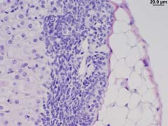
Image Caption:This is a 2.5x image.
|

|
|
Image ID:5561 |
|
Source of Image:Sundberg J |
|
Pathologist:Sundberg J |
|
|
Image Caption:This is a 40x image, 40x, that is a higher magnification of the right, middle region of the 4x image.
|
|
|
Image ID:5563 |
|
Source of Image:Sundberg J |
|
Pathologist:Sundberg J |
|
|
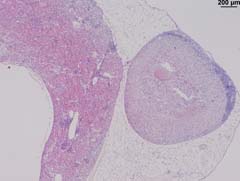
Image Caption:This is a 4x image, 4x, that is a higher magnification of the right, middle area of the 2.5x image.
|
|
|
Image ID:5562 |
|
Source of Image:Sundberg J |
|
Pathologist:Sundberg J |
|
|
|
| MTB ID |
Tumor Name |
Organ(s) Affected |
Treatment Type |
Agents |
Strain Name |
Strain Sex |
Reproductive Status |
Tumor Frequency |
Age at Necropsy |
Description |
Reference |
| MTB:120772 |
Adrenal gland tumor |
Adrenal gland |
None (spontaneous) |
|
|
Mixed Population |
reproductive status not specified |
20% |
|
adrenal tumors from various cohorts; microvessel density (CD31 IHC) |
J:295972 |
|
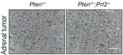
Image Caption:Suppl. Fig. S3B "(B) IHC analysis of staining with CD31 antibody to evaluate the angiogenesis response within the adrenal tumors. No significant differences in MVD between adrenal tumors in Pten+/- and Pten+/-;Prl2-/- mice."
|
|
|
Image ID:6860 |
|
Source of Image:Li Q |
|
Pathologist:Li Q |
|
Method / Stain:IHC for CH31 |
|
|
|
| MTB ID |
Tumor Name |
Organ(s) Affected |
Treatment Type |
Agents |
Strain Name |
Strain Sex |
Reproductive Status |
Tumor Frequency |
Age at Necropsy |
Description |
Reference |
| MTB:120803 |
Adrenal gland tumor |
Adrenal gland |
None (spontaneous) |
|
|
Mixed Population |
reproductive status not specified |
12.3% |
|
adrenal tumors from various cohorts; microvessel density (CD31 IHC) |
J:295972 |
|
Image Caption:Suppl. Fig. S3B "(B) IHC analysis of staining with CD31 antibody to evaluate the angiogenesis response within the adrenal tumors. No significant differences in MVD between adrenal tumors in Pten+/- and Pten+/-;Prl2-/- mice."
|
|
|
|
Image ID:6860 |
|
Source of Image:Li Q |
|
Pathologist:Li Q |
|
Method / Stain:IHC for CH31 |
|
|
|
| MTB ID |
Tumor Name |
Organ(s) Affected |
Treatment Type |
Agents |
Strain Name |
Strain Sex |
Reproductive Status |
Tumor Frequency |
Age at Necropsy |
Description |
Reference |
| MTB:11341 |
Adrenal gland - Cortex adenoma |
Adrenal gland - Cortex |
None (spontaneous) |
|
|
Male |
reproductive status not specified |
observed |
|
|
J:94307 |
|
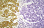
Image Caption:Adrenocortical adenoma. Chromium fixation delineates the adrenal medulla (brown staining). The blue nodule in the cortex compresses the adjacent normal cells. The tumor cells form small islands of polyhedral cells in a typical "endocrine" pattern.
|
|
|
Image ID:122 |
|
Source of Image:Sundberg J |
|
Pathologist:Sundberg J |
|
Method / Stain:chromium fixation method for adrenal medulla |
|
|
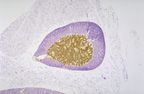
Image Caption:Adrenocortical adenoma. The classical histochemical approach to evaluating the adrenal medulla is to fix it in chromium based fixatives as shown here. The medulla stains brown while the remainder of the gland, the cortex, is blue due to the counterstain. In the upper right quadrant of the cortex is a dark blue nodule that is an adrenal cortical adenoma since it does not stain brown, is located within the adrenal cortex, and has cellular features consistent with the cortical cells.
|
|
|
Image ID:121 |
|
Source of Image:Sundberg J |
|
Pathologist:Sundberg J |
|
Method / Stain:chromium fixation method for adrenal medulla |
|
|
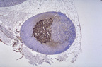
Image Caption:Adrenocortical adenoma. The kidney is in the upper left corner of this image. The adrenal gland has been subjected to immunohistochemistry for detection of expression of tyrosine hydroxylase. The center of the gland (adrenal medulla) is positive for this enzyme (dark brown). The cortex is blue due to the counterstain (hematoxylin). To the right of the medulla is an round mass that compresses the cortex. The cells resemble the cortex and do not express tyrosine hydroxylase and therefore are most likely of adrenal cortex origin.
|
|
|
Image ID:120 |
|
Source of Image:Sundberg J |
|
Pathologist:Sundberg J |
|
Method / Stain:IHC for tyrosine hydroxylase |
|
|