Caption |
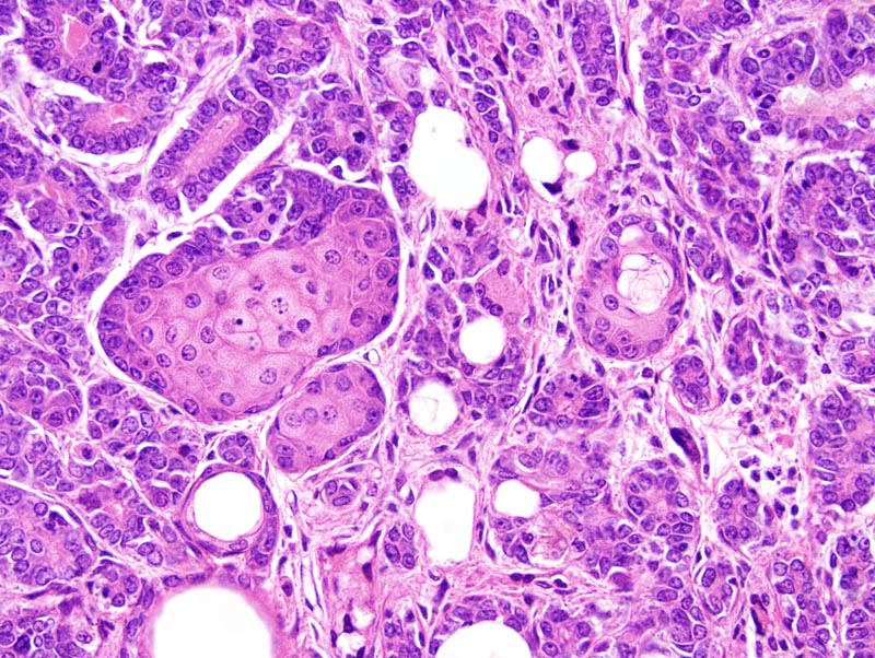
Mammary gland: focally (at the center of the photomicrograph), neoplastic cells undergo sebaceous differentiation. In this area, neoplastic cells form nests characterized by the presence of one row of attenuated cells resembling reserve cells of sebaceous glands, and multiple layers of epithelial cells with various degrees of sebaceous differentiation. Adjacent neoplastic ducts are lined by a several cell thick epithelium which shows squamous differentiation, a feature which is reminiscent of sebaceous ducts.
NOTE: Epidermal or follicular differentiation are common in transgenic mice with activated Wnt/beta catenin pathways, and also in spontaneous mammary tumors. However, sebaceous differentiation is very uncommon in mouse mammary adenocarcinomas. |