Caption |
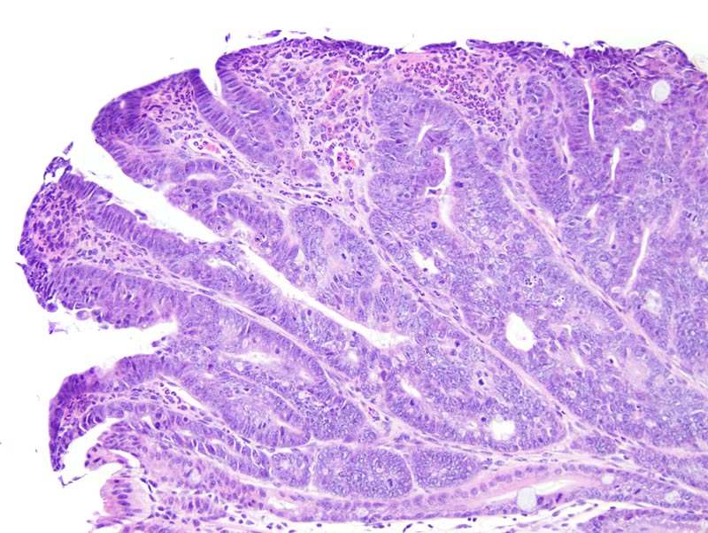
Colon: this photomicrograph illustrates the superficial portions of the adenoma. The neoplasm is characterized by the formation of slightly contoured and anastomosed glands lined by a one cell-thick columnar epithelium supported by a small to moderate amount of fibrovascular stroma with a mild lymphocytic and plasmacytic inflammation. Neoplastic cells have indistinct cell borders and a moderate amount of strongly amphophilic cytoplasm. The nucleus is central, oval, oriented in the long axis of the cell, and with a clumped chromatin. Mitoses are numerous. The epithelium at the surface of the adenoma is eroded and the underlying lamina propria is infiltrated by a moderate number of neutrophils. A moderate number of apoptotic neoplastic cells are scattered throughout the neoplasm. |