Pathology Image Detail
Caption |
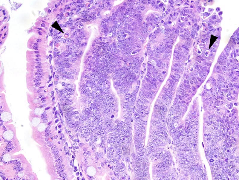
Small intestine: this high power photomicrograph illustrates a portion of the neoplasm where neoplastic cells pile-up disoderly up to 5 cells thick. Nuclear disorientation is prominent, too. A few globule leukocytes are present (arrowheads). |
Description |
Adenoma, small intestine |
Age at Necropsy |
84 days |
Contributor |
Kemp CJ (J:97126) |
Pathologist |
Mikaelian I (J:94320) |
Method |
H&E |
Model |
|
Strain |
|