Caption |
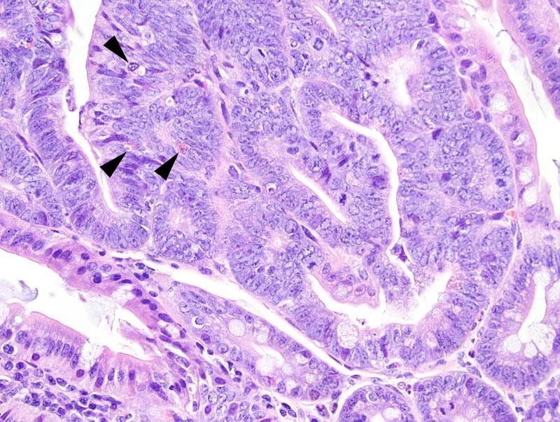
Intestinal adenoma: this high power photomicrograph illustrates portions of the neoplasm where neoplastic cells pie-up disorderly (upper left portions of the photomicrograph) and other areas where neoplastic cells are columnar and form a one cell-thick epithelium. Neoplastic cells have indistinct cell borders and a moderate amount of amphophilic cytoplasm. The nucleus is oval, oriented in the long axis of the cell, hypochromatic, with a clumped chromatin and 1-2 small basophilic nucleoli. Mitoses are present. A few globule leukocytes (arrowheads) are present in the solid portions of the neoplasm. |