Pathology Image Detail

Caption

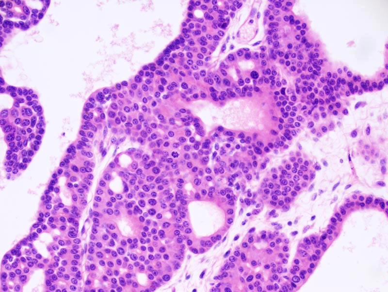
Mammary gland: the cyst is mostly lined by a several (2-4) cells-thick epithelium (earlier stages of macrocysts are lined by a one cell-thick epithelium). In some areas, the epithelium has retained the one cell-thick appearance, which is typical of early stages of macrocysts. The neoplastic epithelium forms glands which are variably ectatic. The cells are cuboidal, with indistinct cell borders, and a moderate amount of acidophilic cytoplasm. Their nucleus is basal, oval, and medium-sized. There is no evidence of atypia and the mitotic rate is low.

Description

Mammary gland, macrocyst (late stage)

Age at Necropsy

399 days

Contributor

Mikaelian I (J:94320)

Pathologist

Mikaelian I (J:94320)

Method

H&E

Model

MMHC ID
MTB:29166
Tumor Name
Mammary gland • cyst - macrocyst
Treatment Type
None (spontaneous)
Tumor Synonyms
Mammary gland, macrocyst (late stage)
Organ Affected
Mammary gland
Frequency
observed
Reference
J:94320

Strain

Strain
STOCK Tg(MMTVTGFA)29Rjc
Strain Types
transgenic
General Note
Hemizygous mice. The genetic background of these mice was a mixture of FVB/NJ, C57BL/6, and DBA/2.
Strain Synonyms
Strain Sex
Unspecified
Reproductive Status
reproductive status not specified
Age of Detection
399 days