Caption |
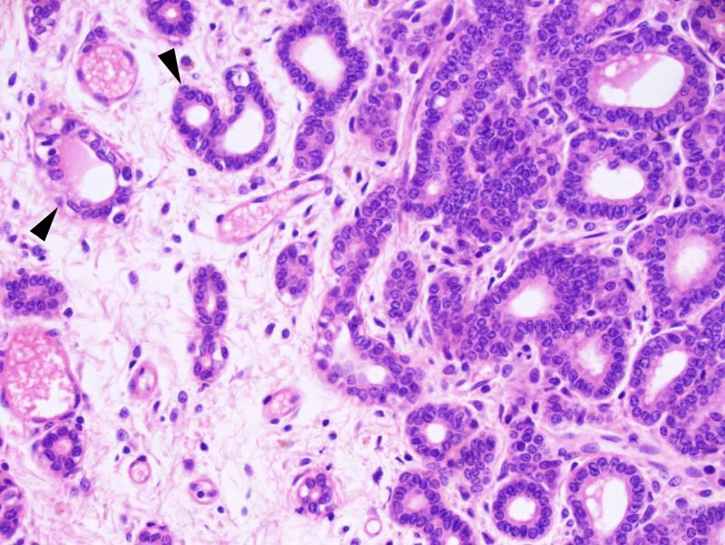
Mammary gland: this is a photomicrograph of the center of a "plaque": the tubules at the center of the plaque are lined by a one cell-thick cuboidal to low columnar epithelium. Occasionally, a discontinuous tubules are lined by two layers of cells. The basal layer is composed of attenuated cells, which were identified by immunohistochemistry as having a myoepithelial phenotype (labeling for alpha smooth muscle actin and/or keratin 5 and/or keratin 14 and/or keratin 17). The luminal have indistinct cell borders and a moderate amount of acidophilic cytoplasm. The nucleus is basal, oval, medium-sized, and hyperchromatic. There is no evidence of atypia and mitotic rate is low. The stroma is diffusely edematous. |