Pathology Image Detail

Caption

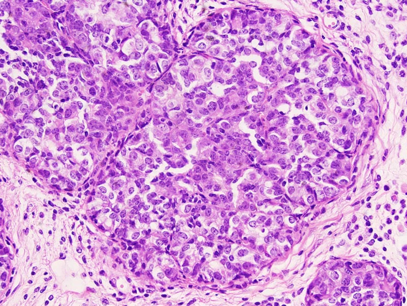
In situ mammary carcinoma: there is prominent proliferation of cells in a mammary alveolus. Thin fibrovascular projections subdivide the alveolus. Proliferating cells are large, with indistinct cell borders and a moderate amount of pale acidophilic cytoplasm. The nucleus is central, oval, medium-sized and slightly hypochromatic. Anisokaryosis and anisocytosis are mild. There is no evidence of invasion. There is mild to moderate interstitial fibrosis and lymphocytic and plasmacytic inflammation. Myoepithelial differentiation is not detected in this portion of the lesion.

Description

Carcinoma in situ, mammary gland

Age at Necropsy

unknown

Contributor

Ward JM (J:107304)

Pathologist

Mikaelian I (J:94320)

Method

H&E

Model

MMHC ID
MTB:29204
Tumor Name
Mammary gland • carcinoma in situ
Treatment Type
None (spontaneous)
Tumor Synonyms
Carcinoma in situ, mammary gland
Organ Affected
Mammary gland
Frequency
observed
Reference
J:94320

Strain

Strain
B6;C-Tg(MMTV-RET)127Ina
Strain Types
transgenic
General Note
The genetic background of these mice was a mixture of C57BL/6 and BALB/c.
Strain Synonyms
(C57BL6xBalb/c)-Tg(MMTVRet)1Ina
Strain Sex
Female
Reproductive Status
reproductive status not specified
Age of Detection
unknown