Caption |
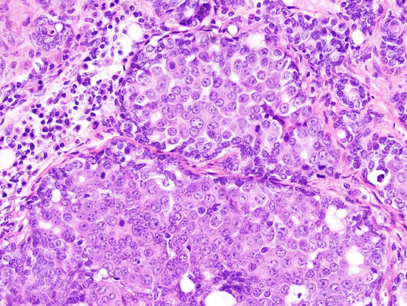
Mammary gland: there is moderate diffuse expansion of a mammary lobule. Within this lobule, a few acini are markedly expanded by closely packed epithelial cells that form solid areas and small glands with an inconspicuous lumen. The cells are polygonal, with distinct cell borders and a moderate amount of pale acidophilic cytoplasm. The nucleus is central, oval, medium-sized and hypochromatic. Anisokaryosis and anisocytosis are mild. There is no evidence of local invasion. Mitotic figures are numerous. There is moderate interstitial lymphocytic and plasmacytic inflammation and fibrosis. Myoepithelial differentiation is prominent and appears as a continuous layer of cells with a nucleus oriented parallel to the basement membrane and a scant amount of cytoplasm. |