Pathology Image Detail
Caption |
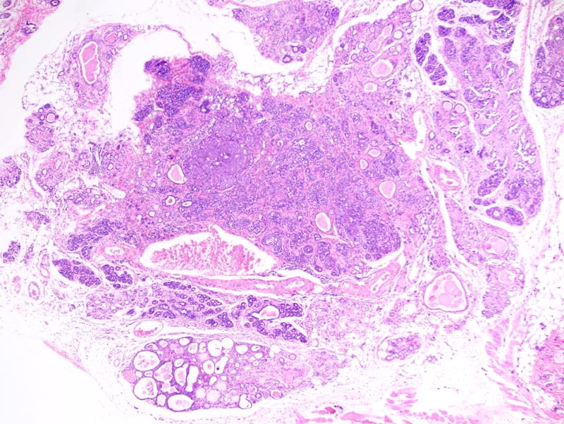
In situ mammary carcinoma: this low magnification photomicrograph represents portions of a mammary gland with areas of in situ mammary carcinoma (at the center of the photomicrographs) with multiple areas of atypical hyperplasia more at the periphery of the neoplasm. There is moderate intersitial fibrosis and lymphoplasmacytic inflammation that involves all mammary lobules. |
Description |
Carcinoma in situ, mammary gland |
Age at Necropsy |
unknown |
Contributor |
Ward JM (J:107304) |
Pathologist |
Mikaelian I (J:94320) |
Method |
H&E |
Model |
|
Strain |
|