Caption |
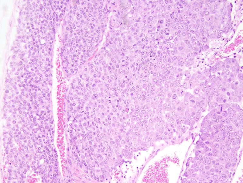
Mammary gland: the portion of the neoplasm located at the center of the photomicrograph is composed of large polygonal cells with distinct cell borders, a large amount of acidophilic cytoplasm, and medium-size hypochromatic nuclei. The portions of the neoplasm located at the periphery of this photomicrograph are composed of crowded small cells with indistinct cell borders, a small amount of acidophilic cytoplasm, and a slightly smaller and more hypochromatic nucleus. Anisokaryosis and anisocytosis are mild. Mitoses are numerous. |