Pathology Image Detail
Caption |
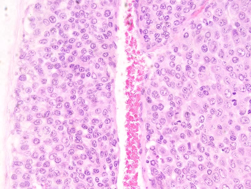
Solid mammary adenocarcinoma: this photomicrograph illustrates a portion of the neoplasm composed of large neoplastic cells (on the right of the photomicrograph) separated by a large vascular space from a portion of the neoplasm composed of smaller cells with less cytoplasm, and smaller and slightly more hyperchromatic nucleus. Mitoses are more numerous in the portion of the neoplasm composed of large neoplastic cells. |
Description |
Adenocarcinoma, solid, comedo, with microinvasion, mammary gland |
Age at Necropsy |
unknown |
Contributor |
Ward JM (J:107304) |
Pathologist |
Mikaelian I (J:94320) |
Method |
H&E |
Model |
|
Strain |
|