Pathology Image Detail
Caption |
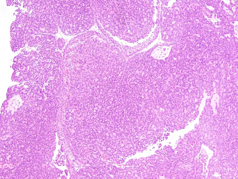
Mammary gland: the photomicrograph represents a portion of a densely cellular neoplasm. The neoplasm is composed of closely-packed lobules separated by a scant amount of fibrovascular stroma. Neoplastic lobules are mostly composed of solid areas with the formation of a few secondary lumens. |
Description |
Adenocarcinoma, solid, glandular, mammary gland |
Age at Necropsy |
unknown |
Contributor |
Ward JM (J:107304) |
Pathologist |
Mikaelian I (J:94320) |
Method |
H&E |
Model |
|
Strain |
|