Caption |
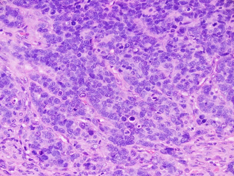
Trabecular mammary adenocarcinoma: neoplastic cells form interconnected trabeculae that are 2-3 cells thick and that are separated by a moderate amount of fibrovascular stroma with a prominent pleocellular infiltrate. Neoplastic cells show prominent nuclear palissading at the periphery of the trabeculae. Luminal spaces are not detected. Neoplastic cells are cuboidal to low columnar, with indistinct cell borders and a small to moderate amount of amphophilic cytoplasm. The nucleus is basal, oval, medium-size, normochromatic, with a punctate chromatin and 1-2 small basophilic nucleoli. Numerous areas of necrosis are present. |