Pathology Image Detail
Caption |
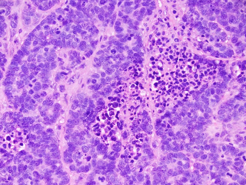
Trabecular mammary adenocarcinoma: this photomicrograph illustrates an area of coagulation necrosis (at the center of the photomicrograph) and nuclear palissading at the periphery of the trabeculae (in the upper left corner of the photomicrograph). Neoplastic cells occasionally form small acini. Luminal spaces are not formed. |
Description |
Adenocarcinoma, trabecular, solid, with prominent fibrosis and coagulation necrosis, mammary gland |
Age at Necropsy |
unknown |
Contributor |
Ward JM (J:107304) |
Pathologist |
Mikaelian I (J:94320) |
Method |
H&E |
Model |
|
Strain |
|