Pathology Image Detail

Caption

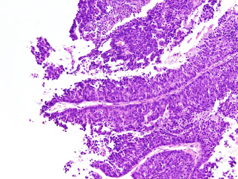
Mammary adenocarcinoma: this photomicrograph focuses on the papillary portions of the neoplasm. Neoplastic papillae have a slender fibrovascular core and are lined by a 2-15 cells-thick epithelium. Neoplastic cells are columnar to polygonal, with ill-defined cell borders and a small amount of amphophilic cytoplasm. The nucleus is oval to round, oriented in the long axis of the cell for columnar neoplastic cells, and slightly hyperchromatic. Numerous neoplastic cells undergo single cell necrosis and mitotic figures are numerous.

Description

Adenocarcinoma, papillary, solid, mammary gland

Age at Necropsy

unknown

Contributor

Ward JM (J:107304)

Pathologist

Mikaelian I (J:94320)

Method

H&E

Model

MMHC ID
MTB:29276
Tumor Name
Mammary gland • adenocarcinoma - mixed
Treatment Type
None (spontaneous)
Tumor Synonyms
Adenocarcinoma, papillary, solid, mammary gland
Organ Affected
Mammary gland
Frequency
observed
Reference
J:94320

Strain

Strain
STOCK Tg(Wap-BCL2)#Jag Tg(Wap-Tag)8Gmn
Strain Types
transgenic
General Note
This STOCK genetic background was a mixture of strains including at least NMRI, C57BL/6, and DBA/2.
Strain Synonyms
NMRI-Tg(WapSV40)(WapBcl2)#?
Strain Sex
Female
Reproductive Status
reproductive status not specified
Age of Detection
unknown