Pathology Image Detail
Caption |
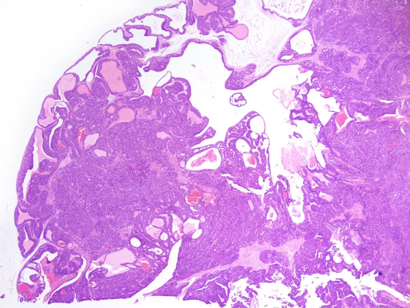
Mammary adenocarcinoma: this photomicrograph illustrates a portion of the neoplasm that is cystic and presents a few papillary projections (upper right corner). The other portions of the neoplasm is composed of closely-packed glands. |
Description |
Adenocarcinoma, glandular, acinar, cystic, solid with secondary lumens, mammary gland |
Age at Necropsy |
unknown |
Notes |
This tumor forms fronds that are reminiscent of fronds observed in type P tumors. However, this neoplasm differs from type P tumors in that the mitotic activity is not most important at the tip of the fronds and ductal differentiation with squamous metaplasia are absent. |
Contributor |
Ward JM (J:107304) |
Pathologist |
Mikaelian I (J:94320) |
Method |
H&E |
Model |
|
Strain |
|