Caption |
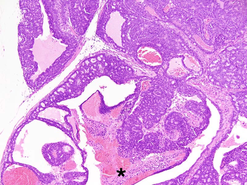
Mammary gland: the neoplasm is composed of coalescing lobules separated by thick fibrovascular septae. The center of the lobules is generally cystic and occasionally contains tissues that have undergone coagulation necrosis and hemorrhage (*). Neoplastic lobules are composed of closely-packed glands generally lined by a one cell-thick cuboidal epithelium. Neoplastic cell occasionally pile-up disorderly (up to 4-5 cells-thick) but retain the ability to form secondary lumens. |
Description |
Adenocarcinoma, glandular, acinar, cystic, solid with secondary lumens, mammary gland |
Age at Necropsy |
unknown |
Notes |
This tumor forms fronds that are reminiscent of fronds observed in type P tumors. However, this neoplasm differs from type P tumors in that the mitotic activity is not most important at the tip of the fronds and ductal differentiation with squamous metaplasia are absent. |
Contributor |
Ward JM (J:107304) |
Pathologist |
Mikaelian I (J:94320) |
Method |
H&E |