Caption |
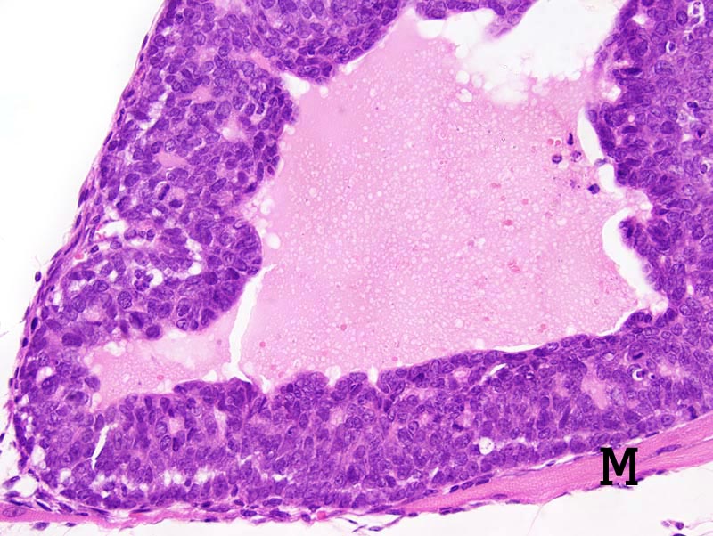
Mammary gland: this photomicrograph illustrates a cystic neoplastic lobule lined by a 5-10 cells thick epithelium that forms a few small secondary lumens. Neoplastic cells are cuboidal to polygonal, with indistinct cell borders and a small amount of strongly amphophilic cytoplasm. The nucleus is central, oval, normochromatic, with a clumped chromatin and occasionally a small basophilic nucleolus. Anisokaryosis and anisocytosis are mild. Mitoses are numerous. A skeletal muscle fiber (M) is entrapped in the neoplasm. |
Description |
Adenocarcinoma, glandular, acinar, cystic, solid with secondary lumens, mammary gland |
Age at Necropsy |
unknown |
Notes |
This tumor forms fronds that are reminiscent of fronds observed in type P tumors. However, this neoplasm differs from type P tumors in that the mitotic activity is not most important at the tip of the fronds and ductal differentiation with squamous metaplasia are absent. |
Contributor |
Ward JM (J:107304) |
Pathologist |
Mikaelian I (J:94320) |
Method |
H&E |