Pathology Image Detail
Caption |
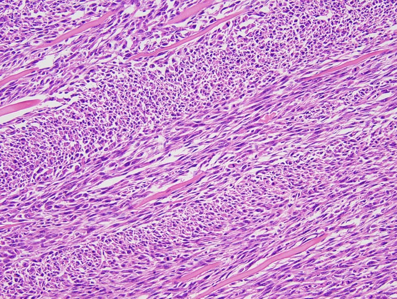
Mammary gland: this high magnification microphotograph focuses on the cells with an mesenchymal phenotype. These cells are organized in fascicles oriented at right angle one with another, are spindloid to fusifrom, with distinct cell borders, a small to moderate amount of acidophilic cytoplasm, and an elongated hyperchromatic nucleus. A moderate number of skeletal muscle fibers is entrapped in the neoplasm. |
Description |
Carcinoma, spindle cell, with epithelial areas, mammary gland |
Age at Necropsy |
unknown |
Contributor |
Ward JM (J:107304) |
Pathologist |
Mikaelian I (J:94320) |
Method |
H&E |
Model |
|
Strain |
|