Pathology Image Detail
Caption |
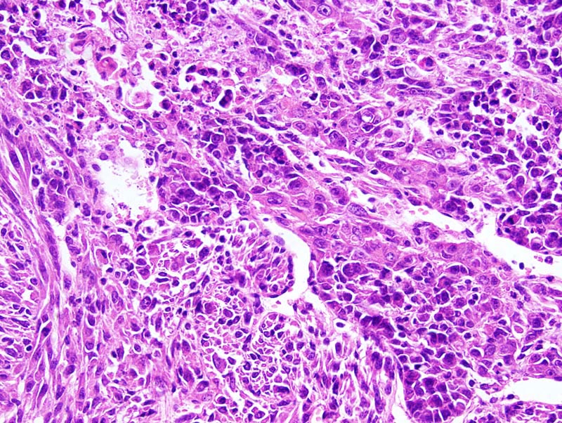
Spindle cell carcinoma: this high magnification photomicrograph illustrates a portion of the neoplasm where neoplastic cells have an epithelial phenotype and form trabeculae and gradually transform into cells with a mesenchymal phenotype forming fascicles. Dilated sinusoidal vessels are present in this portion of the neoplasm. |
Description |
Carcinoma, spindle cell, with epithelial areas, mammary gland |
Age at Necropsy |
unknown |
Contributor |
Ward JM (J:107304) |
Pathologist |
Mikaelian I (J:94320) |
Method |
H&E |
Model |
|
Strain |
|