Caption |
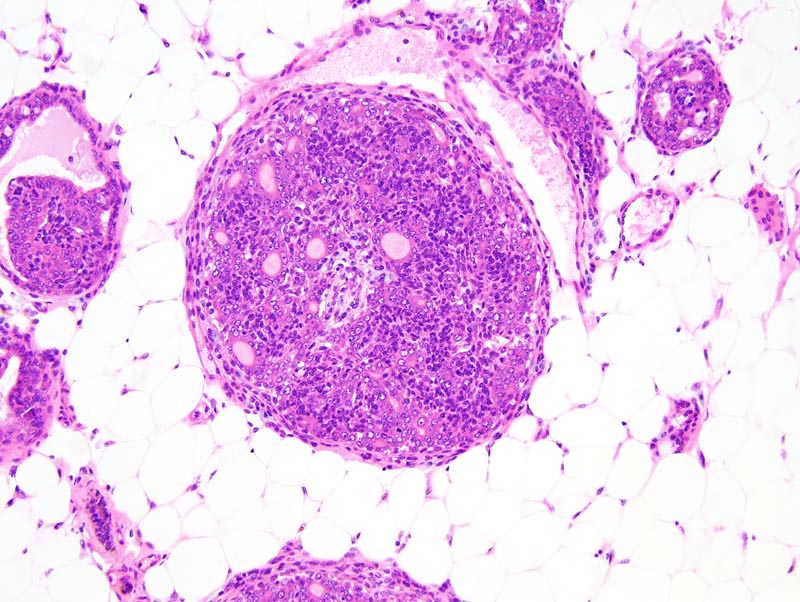
Mammary gland: an interlobular mammary duct is moderately expanded by a cauliflower-like proliferative process characterized by the formation of slender fibrovascular cores that support two populations of cells. First, there is a 1-5 cells-thick layer of oat-shaped cells with indistinct cell borders, a small amount of pale acidophilic cytoplasm and a small hyperchromatic oval nucleus. Second, there is a one cell-thick layer of cuboidal to low columnar cells that form conspicuous lumens filled with an amorphous proteinaceous material. These cells have indistinct cell borders, a moderate amount of acidophilic cytoplasm and a medium-size, central, normochromatic nucleus with a clumped chromatin. A few mast cells are present in the periductal connective tissue.
The presence of two layers of cells indicates that the cell that gave rise to these lesions is not committed to the luminal or to the basal lineage, and hence these lesions may be categorized as "complex" lesions. |