Caption |
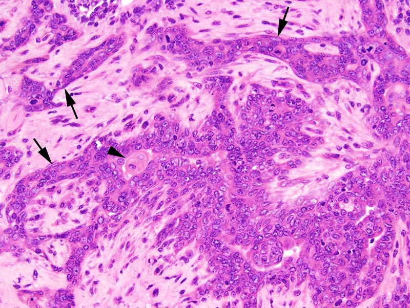
Mammary gland: a mammary lobule is greatly expanded. The ductal portions of the lobule are prominent and surrounded by prominent sclerosis. Cells in the ducts pile-up disorderly up to 3 cells-thick. Cells are cuboidal to polygonal, with indistinct cell borders and a moderate amount of granular and strongly acidophilic cytoplasm. The nucleus is central, oval, medium-size, normochromatic, with a punctate chromatin and one small basophilic nucleolus. Anisokaryosis and anisocytosis are mild. Mitoses are numerous. Myoepithelial differentiation is prominent (arrows). There is a focal area of squamous differentiation (arrowhead). |
Description |
Ductal carcinoma in situ, with sclerosis, mammary gland |
Age at Necropsy |
unknown |
Notes |
These lesions are very unique and are difficult to categorize. As indicated by the MMHCC panel of pathologists, these lesions have similarities with the human entity known as "sclerosing adenosis". However, they also differ from sclerosing adenosis of man in that the mitotic rate is higher, cells lining the ducts pile-up disorderly, there is squamous differentiation, and atypia is more prominent. |
Contributor |
Ward JM (J:107304) |
Pathologist |
Mikaelian I (J:94320) |
Method |
H&E |