Caption |
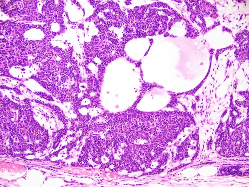
Mammary adenocarcinoma: this photomicrograph illustrates the morphology of the periphery of the neoplasm. The neoplasm in this area is composed of small nests, interconnected trabeculae, and areas of cystic degeneration separated by a moderate amount of loose fibrovascular stroma. There are two types of neoplastic cells: (1) there are large polygonal cells with a moderate amount of pale acidophilic cytoplasm and a large central hypochromatic round nucleus; (2) there are small polygonal cells with a small amount of amphophilic cytoplasm and a small hyperchromatic oval nucleus. Cells intermediate between these two phenotypes are also present. |