Pathology Image Detail
Caption |
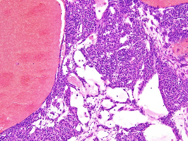
Mammary adenocarcinoma: this photomicrograph illustrates the morphology of the neoplasm towards its' center. Large blood-filled vascular spaces are present. Neoplastic cells form nests and trabeculae that often undergo cystic degeneration and are supported by a moderate amount of loose fibrovascular stroma. There are two types of neoplastic cells: (1) there are large polygonal cells with a moderate amount of pale acidophilic cytoplasm and a large central hypochromatic round to oval nucleus; (2) there are small polygonal cells with a small amount of amphophilic cytoplasm and a small hyperchromatic oval nucleus. Cells intermediate between these two phenotypes are also present. Anisokaryosis and anisocytosis are mild. Mitoses are rare. |
Description |
Adenocarcinoma, complex, mammary gland |
Age at Necropsy |
unknown |
Contributor |
Ward JM (J:107304) |
Pathologist |
Mikaelian I (J:94320) |
Method |
H&E |
Model |
|
Strain |
|