Pathology Image Detail
Caption |
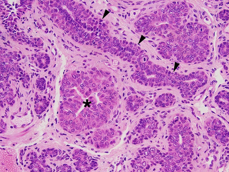
Atypical lobular hyperplasia: this high power photomicrograph illustrates an area of ductal differentiation (arrowheads) and an area of acinar differentiation (*). The myoepithelial layer is more prominent in the area of ductal differentiation than in the area of acinar differentiation. A few mitoses are noted. |
Description |
Lobular hyperplasia, atypical, mammary gland |
Age at Necropsy |
unknown |
Contributor |
Ward JM (J:107304) |
Pathologist |
Mikaelian I (J:94320) |
Method |
H&E |
Model |
|
Strain |
|