Caption |
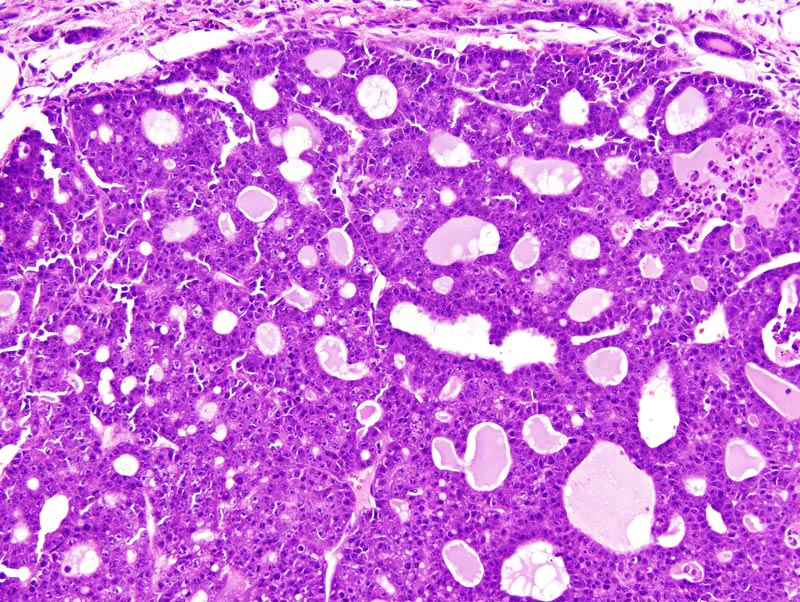
Mammary gland: neoplastic lobules are composed of closely-packed glands lined by a one cell-thick cuboidal epithelium. There are also a few solid areas with secondary lumens. Glands and secondary lumens show various degrees of ectasia, and some contain a small to moderate of amorphous acidophilic fluid and occasionally a few desquamated neoplastic cells. Neoplastic cells are cuboidal to polygonal, with distinct cell borders and a moderate amount of strongly amphophilic cytoplasm that occasionally contains small lipid vacuoles and hyaline droplets. The nucleus is central, medium-size to large, round to oval, slightly hypochromatic, with a finely stippled chromatin and 1-3 large basophilic nucleoli. Anisokaryosis and anisocytosis are moderate. A few mitoses are detected. Neoplastic cells occasionally pile-up disorderly. |