Pathology Image Detail
Caption |
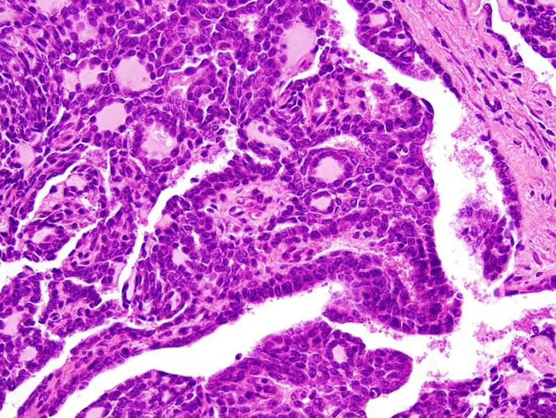
Mammary gland: neoplastic lobules are composed of closely-packed glands and acini supported by a small to moderate amount of loose fibrovascular stroma. The papillary pattern can be nicely appreciated at this magnification. Neoplastic papillae are massive and are lined by a two cells-thick cuboidal epithelium. The basal layer of cell is indicative of myoepithelial differentiation and is most easily identified in the papillary areas of the neoplasm (half lower right part of the photomicrographs), although it is also present in the glandular portions of the neoplasm (half upper left part of the photomicrograph). Anisokaryosis and anisocytosis are mild. |
Description |
Adenocarcinoma, papillary, glandular, complex, mammary gland |
Age at Necropsy |
unknown |
Contributor |
Ward JM (J:107304) |
Pathologist |
Mikaelian I (J:94320) |
Method |
H&E |
Model |
|
Strain |
|